. 2012 Jun 8;149(6):1257-68.
doi: 10.1016/j.cell.2012.03.050.

A thrombospondin-dependent pathway for a protective ER stress response

Affiliations

A thrombospondin-dependent pathway for a protective ER stress response

Jeffrey M Lynch et al. Cell. .

Abstract

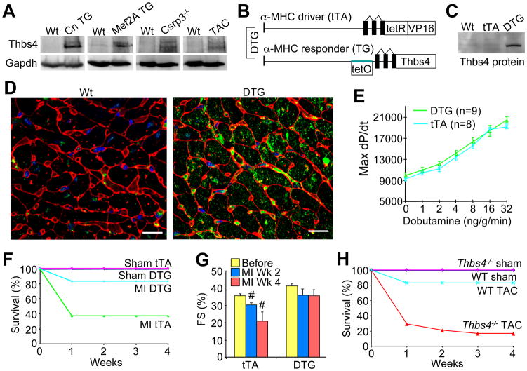
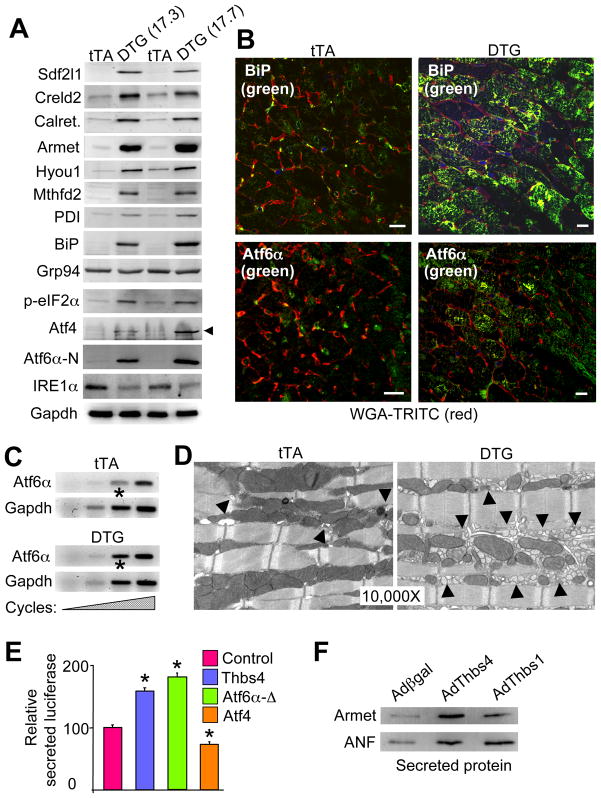
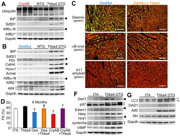
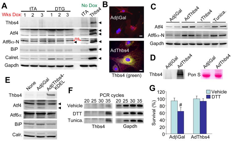
Thrombospondin (Thbs) proteins are induced in sites of tissue damage or active remodeling. The endoplasmic reticulum (ER) stress response is also prominently induced with disease where it regulates protein production and resolution of misfolded proteins. Here we describe a function for Thbs as ER-resident effectors of an adaptive ER stress response. Thbs4 cardiac-specific transgenic mice were protected from myocardial injury, whereas Thbs4(-/-) mice were sensitized to cardiac maladaptation. Thbs induction produced a unique profile of adaptive ER stress response factors and expansion of the ER and downstream vesicles. Thbs bind the ER lumenal domain of activating transcription factor 6α (Atf6α) to promote its nuclear shuttling. Thbs4(-/-) mice showed blunted activation of Atf6α and other ER stress-response factors with injury, and Thbs4-mediated protection was lost upon Atf6α deletion. Hence, Thbs can function inside the cell during disease remodeling to augment ER function and protect through a mechanism involving regulation of Atf6α.


Conflict of interest statement

Conflict of interest: None (no competing financial interests).

Figures

Figure 1
Figure 1
Thbs4 expression is up-regulated in diseased hearts and is protective. (A) Western blots for Thbs4 protein expression in different mouse models exhibiting cardiac disease. (B) Schematic diagram representing the binary transgenic system regulated by doxycycline to inducibly overexpress Thbs4 in the heart. (C) Representative western blot showing Thbs4 protein expression level in heart extracts from 4-month old DTG mice versus Wt and tTA only transgenic controls. (D) Immunohistochemistry for Thbs4 (green) in 4-month old mouse hearts. Red staining shows membranes and blue shows nuclei (scale bars = 10 μm). (E) Hemodynamic assessment of cardiac performance as maximal dP/dt in anesthetized, closed-chested mice in response to increasing doses of the β-adrenergic agonist dobutamine (measured in mmHg/s; +/− SD, N=6 mice each). (F) Survival plot for tTA and DTG mice following myocardial infarction (MI) injury (n = 10 male mice per group). (G) Cardiac ventricular fractional shortening (FS%) determined by echocardiography. At least 6 mice were analyzed in each group at 2 and 4 weeks after MI injury in the tTA control and DTG groups (#P<0.05 versus tTA before MI). (H) Survival plot in Thbs4−/− and Wt mice after pressure-overload by TAC (n = 10 male mice per group). Also See Figure S1 and S2
Figure 2
Figure 2
Unique ER stress induction profile associated with Thbs4 expression. (A) Western blots for expression of ER stress response proteins from the hearts of two lines of Thbs4 DTG mice versus tTA control hearts. (B) Immunohistochemistry for Atf6α and BiP (green) in heart sections from tTA controls or Thbs4 DTG mice. Membranes of the cardiomyocytes are shown in (scale bars = 10 μm). (C) Increasing RT-PCR cycles for Atf6α mRNA in hearts of tTA control or Thbs4 DTG mice. Gapdh mRNA was used as a loading/normalization control. The asterisk shows increased Aft6α mRNA in the DTG hearts at the most linear cycle number in the RT-PCR reaction. (D) Transmission electron microscopy (EM) of heart sections from tTA control and Thbs4 DTG mice shows ER/SR and vesicles (arrowheads) in control and Thbs4 DTG hearts. (E) Luciferase activity in the media of NIH 3T3 cells transfected with a plasmid encoding a secreted version of luciferase that was co-transfected with Thbs4, Atf6α-Δ (constitutively nuclear) or Atf4 (all internally normalized to co-transfected β-gal plasmid). *P<0.05 versus the luciferase plasmid only. (F) Western blotting for the ER processed and secreted proteins Armet and ANF from concentrated media of neonatal rat cardiomyocytes previously infected with adenoviruses encoding β-galactosidase (control), Thbs4 or Thbs1. Also see Figure S3 and S4.
Figure 3
Figure 3
Thbs4 induces a unique ER stress response signature that antagonizes protein aggregation and disease in the heart. (A) Western blots showing ER protein expression in αB-crystallin (CryAB) mutant or (B) desmin (DesMut) mutant hearts in comparison to non-transgenic (NTG) and Thbs4 DTG hearts. The hash marks or arrowheads show the different isoforms that were detected. (C) Immunohistochemistry of frozen heart sections from 6-month old mice of the indicated genotypes. Protein aggregation (green color) is dramatically reduced when Thbs4 is over-expressed. Red staining is for actin and shows the outline of cardiomyocytes (scale bars = 100 μm). (D) FS% as determined by echocardiography suggests that cardiac function is improved in both aggregation-prone cardiomyopathic transgenic mouse models when Thbs4 is overexpressed (N=6 or more mice in each group, *P<0.05 versus tTA; #P<0.05 versus DesMut; †P<0.05 versus CryAB). (F) Western blotting for proteins involved in ERAD from the hearts of Thbs4 DTG mice versus tTA control hearts. The arrowheads show the position of the relevant proteins, while the bracket shows 2 relevant bands. (G) Western blotting for proteins involved in autophagy (LC3, both I and II isoforms) or cellular protection. Also see Figure S5.
Figure 4
Figure 4
Thbs4 functions from within the cell to promote adaptive ER stress response signaling. (A) Western blots of ER stress response protein expression in heart extracts from 3-month old tTA and DTG mice fed chow containing doxycycline for the indicated periods of time to extinguish Thbs4 expression. “No dox” controls are also shown and were generated from extracts of 4-month old DTG mice. An N-terminal Atf6α antibody from ProSci was used. The arrowheads so the specific band that was detected; ns = non-specific. (B) Immunocytochemistry for Thbs4 in adenovirus infected neonatal cardiomyocytes showing that Thbs4 (green) has an ER and Golgi staining pattern. Red staining is for membranes and blue stains the nuclei (scale bars = 10 μm). (C) Western blotting for Atf4 and Atf6α protein expression in neonatal rat cardiomyocytes infected for 24 hrs with recombinant adenovirus or treated with recombinant Thbs4 (2 μg/μL) or tunicamycin (1μg/μL). (D) Western blotting from media for Thbs4 after AdThbs4 infection versus Adβgal control in neonatal cardiomyocytes. Ponceau staining is a control for protein loading between the samples. (E) Western blotting for ER stress response proteins from neonatal cardiomyocytes following infection with a control (Adβgal) adenovirus or an adenovirus expressing Thbs4 fused with a KDEL ER retention/relocalization signal. (F) RT-PCR for Thbs4 or Gapdh in neonatal cardiomyocytes treated for 5 hours with ER stress inducers DTT (5μg/μL) or tunicamycin (10μg/μL). (G) AlamarBlue survival assay in DTT treated (8-hrs, 4μg/μL) neonatal cardiomyocytes infected with AdThbs4 or Adβgal (assay was run in duplicate in three separate experiments; #P<0.05 versus vehicle).
Figure 5
Figure 5
Thbs proteins directly interact with Atf6α. (A) Western blot following GST-Thbs4 pull-down of the indicated proteins from cardiac protein extracts from Thbs4 DTG hearts. A C-terminal Atf6α antibody from ProSci was used. (B) Mouse Atf6α schematic diagram showing relative size of the luminal domains used for GST pull-down experiments (red bars). (C) Western blot for tagged truncations of two different Atf6α luminal domains following GST-Thbs4 pull-down. The ER luminal domain of Atf6β did not interact with GST-Thbs4. There is a non-specific band in the GST (Atf6α 471–656) only lane due to extreme loading of purified GST. (D) Western blot for a tagged Atf6α luminal domain (amino acids 361–656) following GST-Thbs1 pull-down. (E) Gal4-dependent luciferase reporter activity with the luminal fragments of Atf6α (amino acid numbers shown) as fusions to full-length Gal4 transiently transfected into COS cells with or without Thbs4. The basal activity of each construct was set to 100, from which Thbs co-expression was compared. *P<0.05 versus no Thbs4 control. Results are averaged from 3 separate experiments. (F) Schematic diagram of human Thbs4 with the relative size of the GST fragments indicated (red bars). A western blot for Atf6α luminal domain following GST pull-down with the different Thbs4 domains is shown below the schematic. (G) Western blot for Thbs4 and Atf6α (FL, full-length is shown detected with Abcam N-terminal antibody) after Atf6α immunoprecipitation (with C-terminal ProSci antibody) from cardiac extracts of control tTA or Thbs4 DTG hearts.
Figure 6
Figure 6
Thbs4 is necessary for Atf6α activation in the heart. (A) Immunocytochemistry for Atf6α in neonatal rat fibroblasts with Adβgal (control) or AdThbs4 infection, with our without the S1P inhibitor AEBSF. The arrows show prominent nuclear localization of Atf6α, while the arrowheads show Atf6α retention in the Golgi and ER. (scale bars = 10 μm). (B) Western blots showing ER stress response protein expression in hearts of Wt and Thbs4−/− mice at baseline (sham) or after 48-hrs of TAC stress stimulation. Arrowheads show the indicated protein. (C) Immunohistochemistry for Atf6α following 24-hrs TAC or a sham surgical procedure. Nuclear localized Atf6α (arrowheads) was much more prominent in Wt hearts after TAC relative to Thbs4−/− hearts with TAC. (scale bars = 10 μm). (D) Western blots showing ER stress response protein expression in hearts of Wt and Thbs4−/− mice at baseline (sham) or 3-days after MI injury. (E) Western blot for ER stress response protein expression from isolated adult myocytes in temporary culture from Wt or Thbs4−/− hearts. (F) AlamarBlue survival assay in tunicamycin treated adult cardiomyocytes isolated from Wt or Thbs4−/−hearts (assay was run in duplicate; *P<0.05 versus vehicle). Also see Figure S6.
Figure 7
Figure 7
Thbs4-Atf6α interaction is necessary for Atf6α function in ER stress induction in cardiomyocytes. (A) Schematic diagram of the region of mouse Atf6α that was used to make the dominant negative or decoy adenovirus (red bar). (B) Western blotting for the indicated proteins from neonatal cardiomyocytes infected with the indicated recombinant adenoviruses. The red-boxed areas show induction of ER stress response proteins by Thbs4 overexpression, which is attenuated with the Atf6α-dominant negative expressing adenovirus. (C) Schematic diagram of the region of Thbs4 that was used to make the dominant negative or decoy adenovirus (red bar). (D) Western blotting for the indicated proteins from neonatal cardiomyocytes infected with the indicated recombinant adenoviruses. The red-boxed areas show induction of ER stress response proteins by Thbs4 overexpression, which is attenuated with the Thbs4-dominant negative expressing adenovirus. (E) Immunocytochemistry for endogenous Atf6α in cultured neonatal rat fibroblasts 24 hrs after infection with the indicated adenoviruses. The arrows show prominent nuclear localization of endogenous Atf6α by AdThbs4 infection, which was blocked with either AdAtf6α(DN) or AdThbs4 (DN). (scale bars = 10 μm). (F) Gal4-dependent luciferase reporter assay with full-length human Atf6α-Gal4 that was co-transfected in HEK293 cells with combinations of plasmids encoding Thbs4 with either Atf6α(DN) or Thbs4(DN). Results were averaged from 3 independent experiments. *P<0.05 versus reporter alone (control); #P<0.05 versus Thbs4. (G) Western blotting for Atf6α (activated form) or Gapdh (control) in neonatal cardiomyocytes infected with the indicated adenoviruses with or without tunicamycin. The dominant negative Thbs4 construct prevents activation of endogenous Atf6α in response to tunicamycin treatment. (H) AlamarBlue survival assay in Wt or Atf6a−/− MEFs with or without AdThbs4 infection. Assay was performed in triplicate. *P<0.05 versus no drug; #P<0.05 versus Wt MEFs with AdThbs4. †P<0.05 versus Wt MEFs with Adβgal with same drug treatment. Also see Figure S6.

Comment in

Similar articles

Cited by

References

    1. Adachi Y, Yamamoto K, Okada T, Yoshida H, Harada A, Mori K. ATF6 is a transcription factor specializing in the regulation of quality control proteins in the endoplasmic reticulum. Cell Struc Func. 2008;33:75–89. - PubMed
    1. Belmont PJ, Tadimalla A, Chen WJ, Martindale JJ, Thuerauf DJ, Marcinko M, Gude N, Sussman MA, Glembotski CC. Coordination of growth and endoplasmic reticulum stress signaling by regulator of calcineurin 1 (RCAN1), a novel ATF6-inducible gene. J Biol Chem. 2008;283:14012–14021. - PMC - PubMed
    1. Beskow A, Grimberg KB, Bott LC, Salomons FA, Dantuma NP, Young P. A conserved unfoldase activity for the p97 AAA-ATPase in proteasomal degradation. J Mol Biol. 2009;394:732–746. - PubMed
    1. Bommiasamy H, Back SH, Fagone P, Lee K, Meshinchi S, Vink E, Sriburi R, Frank M, Jackowski S, Kaufman RJ, Brewer JW. ATF6alpha induces XBP1-independent expansion of the endoplasmic reticulum. J Cell Sci. 2009;122:1626–1636. - PMC - PubMed
    1. Briggs MD, Hoffman SM, King LM, Olsen AS, Mohrenweiser H, Leroy JG, Mortier GR, Rimoin DL, Lachman RS, Gaines ES, Cekleniak JA, Knowlton RG, Cohn DH. Pseudoachondroplasia and multiple epiphyseal dysplasia due to mutations in the cartilage oligomeric matrix protein gene. Nat Genet. 1995;10:330–336. - PubMed

Publication types