

Critical features for biosynthesis, stability, and functionality of a G protein-coupled receptor uncovered by all-versus-all mutations
- PMID: 22665811
- PMCID: PMC3382542
- DOI: 10.1073/pnas.1202107109
Critical features for biosynthesis, stability, and functionality of a G protein-coupled receptor uncovered by all-versus-all mutations
Abstract
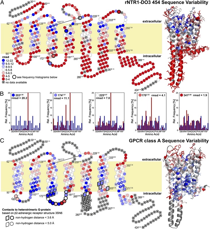
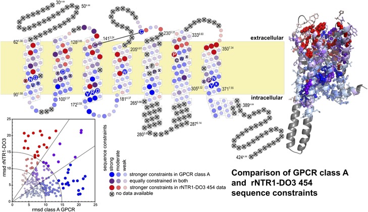
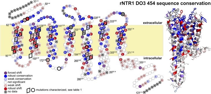
The structural features determining efficient biosynthesis, stability in the membrane and, after solubilization, in detergents are not well understood for integral membrane proteins such as G protein-coupled receptors (GPCRs). Starting from the rat neurotensin receptor 1, a class A GPCR, we generated a separate library comprising all 64 codons for each amino acid position. By combining a previously developed FACS-based selection system for functional expression [Sarkar C, et al. (2009) Proc Natl Acad Sci USA 105:14808-14813] with ultradeep 454 sequencing, we determined the amino acid preference in every position and identified several positions in the natural sequence that restrict functional expression. A strong accumulation of shifts, i.e., a residue preference different from wild type, is detected for helix 1, suggesting a key role in receptor biosynthesis. Furthermore, under selective pressure we observe a shift of the most conserved residues of the N-terminal helices. This unique data set allows us to compare the in vitro evolution of a GPCR to the natural evolution of the GPCR family and to observe how selective pressure shapes the sequence space covered by functional molecules. Under the applied selective pressure, several positions shift away from the wild-type sequence, and these improve the biophysical properties. We discuss possible structural reasons for conserved and shifted residues.
Conflict of interest statement
The authors declare no conflict of interest.
Figures
Similar articles
-
Maximizing detergent stability and functional expression of a GPCR by exhaustive recombination and evolution.J Mol Biol. 2012 Sep 21;422(3):414-28. doi: 10.1016/j.jmb.2012.05.039. Epub 2012 Jun 6. J Mol Biol. 2012. PMID: 22683350
-
Structure-Based Sequence Alignment of the Transmembrane Domains of All Human GPCRs: Phylogenetic, Structural and Functional Implications.PLoS Comput Biol. 2016 Mar 30;12(3):e1004805. doi: 10.1371/journal.pcbi.1004805. eCollection 2016 Mar. PLoS Comput Biol. 2016. PMID: 27028541 Free PMC article.
-
7TM Domain Structure of Adhesion GPCRs.Handb Exp Pharmacol. 2016;234:43-66. doi: 10.1007/978-3-319-41523-9_3. Handb Exp Pharmacol. 2016. PMID: 27832483 Review.
-
Evolution of three human GPCRs for higher expression and stability.J Mol Biol. 2011 May 13;408(4):599-615. doi: 10.1016/j.jmb.2011.02.051. Epub 2011 Mar 3. J Mol Biol. 2011. PMID: 21376730
-
Cryo-EM structures of GPCRs coupled to Gs, Gi and Go.Mol Cell Endocrinol. 2019 May 15;488:1-13. doi: 10.1016/j.mce.2019.02.006. Epub 2019 Mar 28. Mol Cell Endocrinol. 2019. PMID: 30930094 Review.
Cited by
-
Computational design of thermostabilizing point mutations for G protein-coupled receptors.Elife. 2018 Jun 21;7:e34729. doi: 10.7554/eLife.34729. Elife. 2018. PMID: 29927385 Free PMC article.
-
Comparison of fragments comprising the first two helices of the human Y4 and the yeast Ste2p G-protein-coupled receptors.Biophys J. 2012 Aug 22;103(4):817-26. doi: 10.1016/j.bpj.2012.07.012. Biophys J. 2012. PMID: 22947943 Free PMC article.
-
Structure of signaling-competent neurotensin receptor 1 obtained by directed evolution in Escherichia coli.Proc Natl Acad Sci U S A. 2014 Feb 11;111(6):E655-62. doi: 10.1073/pnas.1317903111. Epub 2014 Jan 22. Proc Natl Acad Sci U S A. 2014. PMID: 24453215 Free PMC article.
-
GPCR activation: protonation and membrane potential.Protein Cell. 2013 Oct;4(10):747-60. doi: 10.1007/s13238-013-3073-2. Epub 2013 Sep 20. Protein Cell. 2013. PMID: 24057762 Free PMC article. Review.
-
Advances in ligand-specific biosensing for structurally similar molecules.Cell Syst. 2023 Dec 20;14(12):1024-1043. doi: 10.1016/j.cels.2023.10.009. Cell Syst. 2023. PMID: 38128482 Free PMC article. Review.
References
Publication types
MeSH terms
Substances
LinkOut - more resources
Full Text Sources
Other Literature Sources
