. 2012 Jun 11;197(6):801-17.
doi: 10.1083/jcb.201108077. Epub 2012 Jun 4.

FGFR1 cleavage and nuclear translocation regulates breast cancer cell behavior

Affiliations

FGFR1 cleavage and nuclear translocation regulates breast cancer cell behavior

Athina-Myrto Chioni et al. J Cell Biol. .

Abstract

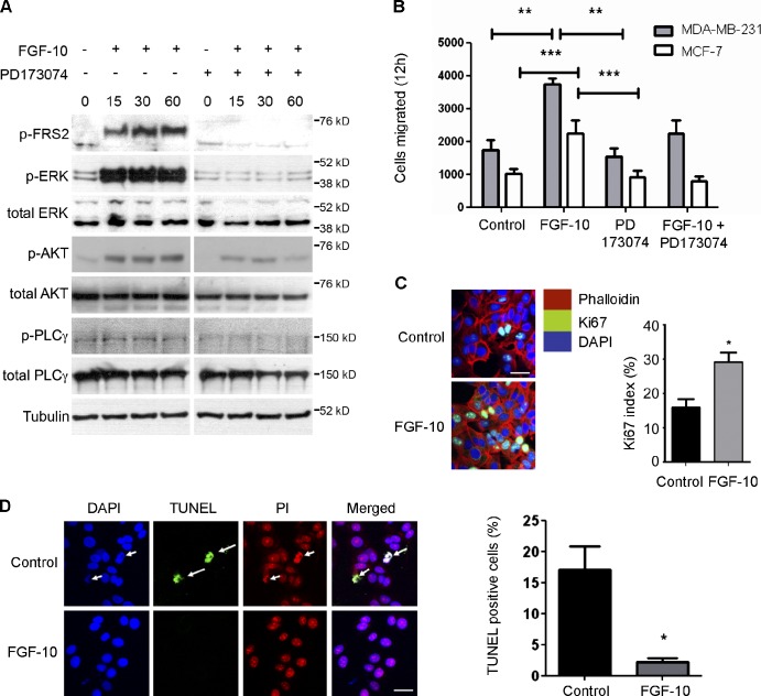
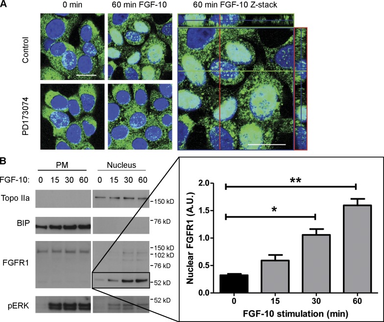
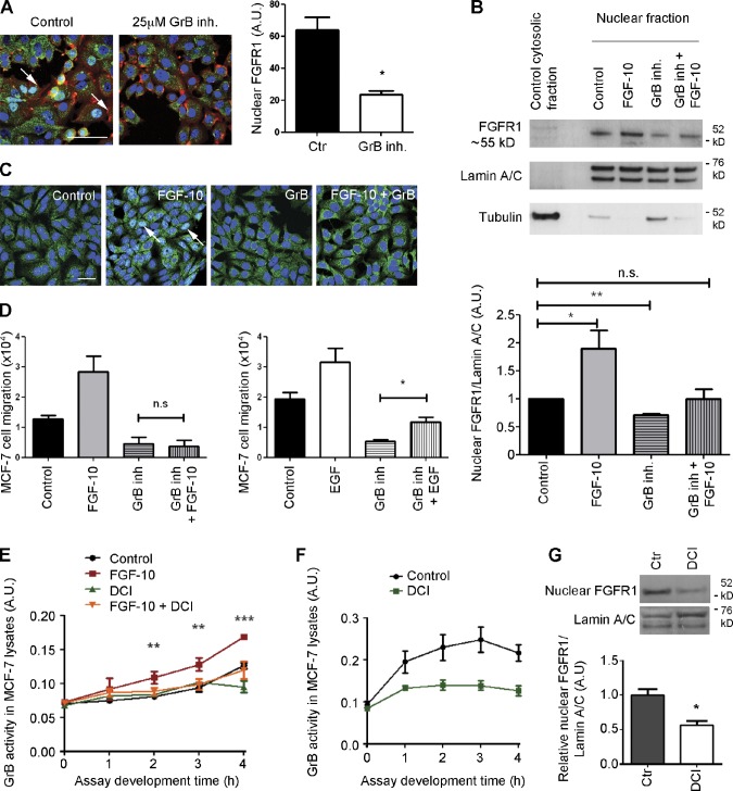
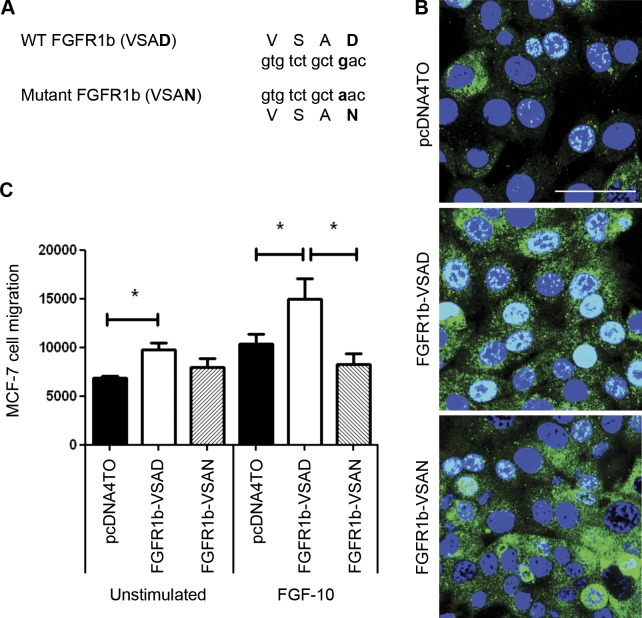
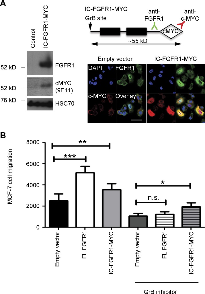
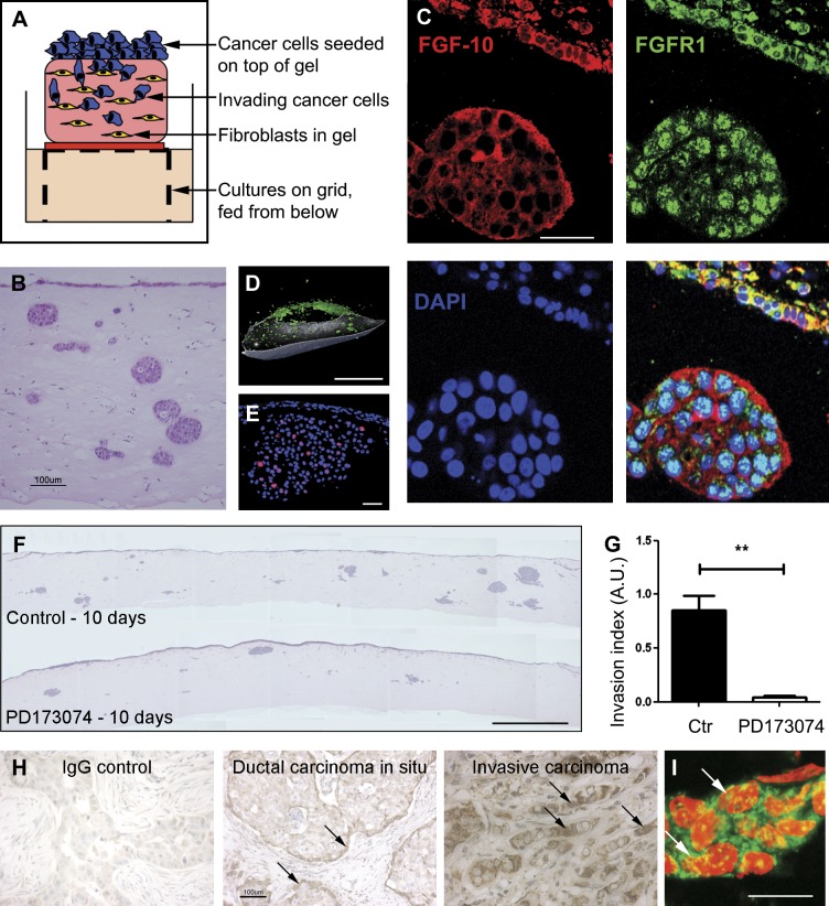
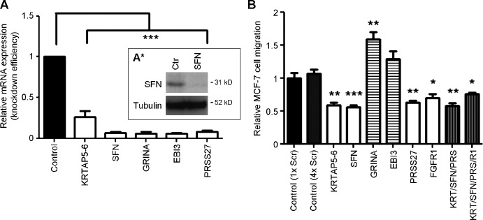
FGF-10 and its receptors, FGFR1 and FGFR2, have been implicated in breast cancer susceptibility and progression, suggesting that fibroblast growth factor (FGF) signaling may be co-opted by breast cancer cells. We identify a novel pathway downstream of FGFR1 activation, whereby the receptor is cleaved and traffics to the nucleus, where it can regulate specific target genes. We confirm Granzyme B (GrB) as the protease responsible for cleavage and show that blocking GrB activity stopped FGFR1 trafficking to the nucleus and abrogates the promigratory effect of FGF stimulation. We confirm the in vivo relevance of our findings, showing that FGFR1 localized to the nucleus specifically in invading cells in both clinical material and a three-dimensional model of breast cancer. We identify target genes for FGFR1, which exert significant effects on cell migration and may represent an invasive signature. Our experiments identify a novel mechanism by which FGF signaling can regulate cancer cell behavior and provide a novel therapeutic target for treatment of invasive breast cancer.


Figures

Figure 1.
Figure 1.
Cell signaling pathways activated upon FGF-10 stimulation and their impact on cell behavior. (A) Serum-starved MCF-7 cells were stimulated with 100 ng/ml FGF-10 in the presence of 7.5 µg/ml heparin in serum-free media for 15, 30, and 60 min. Where indicated, cells were pretreated with 2 µM PD173074 (1 h). Cell lysates were prepared, and Western blotting was performed using different primary antibodies. Stimulation with FGF-10 activated the FRS2–ERK pathway as well as the AKT pathway. FGF-10 treatment triggered rapid ERK phosphorylation, which was blocked by the FGFR inhibitor PD173074. Other pathways (PLC-γ and PI3K) were also investigated, but although FGF-10 induced AKT phosphorylation, this was not abrogated by treatment with PD173074. PLC-γ was not activated by FGF-10 treatment. (B) Migration of MCF-7 and MDA-MB-231 cells was monitored using 8-µm-pore Transwell migration filters in serum-free medium in the presence of FGF-10/heparin (100 ng/ml and 7.5 µg/ml, respectively) and/or 2 µM PD173074. Cells were allowed to migrate overnight. FGF-10 treatment increased Transwell migration significantly, and this effect was blocked in the presence of the PD173074. (C) Immunostaining with an anti-Ki67 antibody to quantify cell proliferation after 48 h revealed that treatment with FGF-10 resulted in a significant increase in the percentage of positive cell staining. (D) FGF-10 treatment reduced apoptosis in MCF-7 cells after 48-h treatment with FGF-10, as determined by TUNEL assay (PI, propidium iodide). *, P ≤ 0.05; **, P ≤ 0.01; ***, P ≤ 0.001 (ANOVA for B; Student’s t test for C and D). Bars, 25 µm. Error bars show means ± SEM.
Figure 2.
Figure 2.
Subcellular localization of FGFR1 in 2D culture. (A) FGF-10 stimulation (60 min) resulted in increased nuclear FGFR1 localization in serum-starved MCF-7 cells, confirmed by examining confocal z-stack sections. The effect was abolished in the presence of PD173074. The green, purple, and red boxes represent the x-z and y-z scan perspectives from the confocal z stack. Bars, 25 µm. (B) Subcellular fractionation revealed that FGF-10 treatment increased nuclear FGFR1 localization in MCF-7 cells. Over a time course of FGF-10 stimulation (0–60 min), there was a significant increase in accumulation of a truncated 55-kD C-terminal fragment of FGFR1 in the nuclear fraction (highlighted in black box), but there was no change in full-length FGFR1 levels. Immunoblotting with anti-TOPOIIa and anti-BIP antibodies confirmed the specificity of the cell fractionation protocol, and pERK confirmed the efficiency of FGF-10 stimulation. Nuclear FGFR1 was normalized to the nuclear marker TOPOIIa, confirming the significance of the accumulation (graph; *, P ≤ 0.05; **, P ≤ 0.01 [Student’s t test]). Error bars show means ± SEM. A.U., arbitrary unit; PM, plasma membrane.
Figure 3.
Figure 3.
Inhibition of GrB by RNAi in MCF-7 cells reduced nuclear FGFR1 accumulation. (A) Schematic representation of FGFR1 depicting the three IgG loops of the extracellular domain, the transmembrane region (TM), and the split tyrosine kinase domain. The arrow indicates GrB cleavage site at Asp-432. Numbers indicate amino acid residues. Adapted from Loeb et al. (2006). N, N terminus; C, C terminus. (B) Treatment of MCF-7 cells, growing in serum-containing medium, with a GrB inhibitor abolished nuclear FGFR1 after 24 h but did not change the total FGFR1 protein levels. Confocal analysis of MCF-7 cells growing in serum-containing medium treated with GrB RNAi for 72 h revealed efficient knockdown of GrB concomitant with decreased nuclear FGFR1 staining. **, P < 0.01; ***, P < 0.001 (Student’s t test). Bar, 25 µm. (C) Western blot analysis confirmed that GrB was expressed in MCF-7 cells and showed that 72-h treatment with RNAi to GrB reduced levels of nuclear FGFR1, despite an overall increase in full-length FGFR1. *, P < 0.05; **, P < 0.01 (Mann–Whitney test). Error bars show means ± SEM. A.U., arbitrary unit; Ctr, control.
Figure 4.
Figure 4.
Treatment of MCF-7 cells with a GrB inhibitor reduced nuclear FGFR1 accumulation and blocked FGF-10–dependent cell migration. (A) Treatment of MCF-7 cells, growing in serum-containing medium, with a GrB inhibitor (inh.) abolished nuclear FGFR1 (arrows) after 24 h but did not change the total FGFR1 protein levels. *, P < 0.05 (ANOVA). Bar, 100 µm. (B) Subcellular fractionation confirmed that FGF-10 treatment (1–2 h) increased nuclear FGFR1 (a truncated 55-kD C-terminal fragment of FGFR1) in serum-starved MCF-7 cells and decreased it when cells were treated with 25 µM GrB inhibitor (24 h). When cells were pretreated with GrB inhibitor and then treated with FGF-10, the translocation of the nuclear FGFR1 was partially blocked. Immunoblotting with Lamin A/C and tubulin antibodies confirmed the specificity of the cell fractionation protocol. Nuclear FGFR1 was normalized to the nuclear marker Lamin A/C, confirming the significance of the accumulation (graph). (C) MCF-7 cells were treated with 25 µM GrB inhibitor (24 h) in serum-free medium before stimulation with FGF-10/heparin (100 ng/ml and 7.5 µg/ml, respectively). Control cells showed increased staining for nuclear FGFR1 (arrows) after FGF-10 treatment, and this was blocked by GrB inhibition. Bar, 25 µm. (D) Transwell migration assays with MCF-7 cells showed that GrB inhibition abolished the promigratory effect of FGF-10. In contrast, cells were still able to migrate in response to EGF stimulation. *, P ≤ 0.05; **, P ≤ 0.01 (Student’s t test). (E) GrB activity in lysates from serum-starved MCF-7 cells with or without FGF-10/heparin treatment (100 ng/ml and 7.5 µg/ml, respectively) in the presence or absence of pretreatment with the serine protease inhibitor 3,4-dichloroisocoumarin (DCI; 50 µM; 1-h pretreatment). Activity was measured at 405-nm absorbance at hourly intervals after the addition of 200 µM Ac-IEPD-pNA substrate. FGF-10 treatment significantly increased GrB activity in MCF-7 cells, but this effect was abrogated by pretreatment with DCI. **, P ≤ 0.01; ***, P ≤ 0.001 (Student’s t test). (F) GrB activity in MCF-7 cells growing in full serum is markedly reduced by treatment with 50 µM DCI (2 h). (G) 5-h treatment with 50 µM DCI decreased nuclear FGFR1 protein significantly. *, P ≤ 0.05 (Student’s t test). Error bars show means ± SEM. A.U., arbitrary unit; Ctr, control.
Figure 5.
Figure 5.
Mutation of GrB cleavage site inhibited nuclear translocation of FGFR1. (A) Site-directed mutagenesis on wild-type (WT) FGFR1 changed the guanine nucleotide at position 1963 to adenine (g1963a; using sequence available from GenBank/EMBL/DDBJ under accession no. NM_023106.2) and thereby amino acid 432 from aspartic acid to alanine (D432N), abolishing the GrB cleavage site in Mutant FGFR1. (B) Confocal analysis of MCF-7 cells transiently transfected (48 h) with WT (VSAD) or mutant (VSAN) FGFR1b revealed that mutant (VSAN) FGFR1b did not translocate to the nucleus. Bar, 50 µm. (C) Transwell migration assays showed that, under control conditions, MCF-7 cells transfected with WT FGFR1b (VSAD) migrated more compared with empty vector (pcDNA4/TO)– and mutant FGFR1b (VSAN)–transfected cells. FGF-10 stimulation increased migration in cells transfected with WT FGFR1b (VSAD) but did not affect MCF-7 cells transfected with mutant FGFR1b (VSAN). *, P ≤ 0.05 (paired Student’s t test and ANOVA). Error bars show means ± SEM.
Figure 6.
Figure 6.
Expression of the intracellular domain of FGFR1 in MCF-7 cells increased cell migration and bypassed GrB inhibition. (A) Western blotting confirmed successful transfection of the intracellular C-terminal FGFR1 (IC-FGFR1-MYC) fragment in MCF-7 cells. A 55-kD band was present with both anti-FGFR1 antibody or an anti–c-MYC antibody. Similarly, immunofluorescence showed enhanced nuclear FGFR1 in the cells transfected with the IC-FGFR1-MYC construct. Bar, 25 µm. (B) Transwell migration assays showed that, under control conditions, MCF-7 cells transfected with both full-length FGFR1b and IC-FGFR1-MYC migrated more compared with empty vector (pcDNA4/TO) cells. GrB inhibition decreased migration in all cells transfected with empty vector, full-length (FL) FGFR1b, or IC-FGFR1-MYC compared with untreated cells, but there was a significant difference in the cells transfected with IC-FGFR1-MYC compared with empty vector, whether or not there was no difference between cell transfected with full-length FGFR1b. *, P ≤ 0.05; **, P < 0.01; ***, P < 0.001 (paired Student’s t test and ANOVA). Error bars show means ± SEM.
Figure 7.
Figure 7.
FGFR1 localizes to the nucleus in invading cells in 3D culture and in vivo. (A) A breast cancer organotypic model was designed using a collagen/Matrigel mix containing human foreskin fibroblasts as the stromal equivalent, overlaid with MDA-MB-231 cells. OCs were raised into an air–liquid interface and cultured for 10 d before fixation in 4% PFA. (B) Hematoxylin and eosin staining revealed invading particles (group of cells) in the stroma. (C) FGF-10 was highly expressed in both invading and noninvading cells. FGFR1 was localized mainly to the nucleus (DAPI stain) of invading cells and remained mainly in the plasma membrane and cytoplasm of noninvading cells. (D) Still optical projection tomography image of an OC stained with NESO antibody, a marker for metastatic breast cancer cells (Chioni et al., 2005), highlighted that the invading cells formed distinct groups within the stroma (Video 1). (E) Anti-Ki67 staining of transverse sections from OCs revealed that cell proliferation was restricted to invading cells and not those cells that remained at the air–liquid interface. (F and G) Breast cancer OCs growing for 10 d in the presence of PD173074 showed significantly less cancer cell invasion compared with control. The invasion index comprises the combined measurements of (a) depth of invasion (mean of several measurements from each OC, taken from the top layer of the noninvading cells to the middle of the invading particles). (b) number of invading particles, and (c) mean area of the invading particles (n = 4 OCs for each condition). Error bars show means ± SEM. (H) Immunohistochemistry of human tissues from patients with an anti-FGFR1 antibody revealed that, although the IgG negative control remained clear for brown DAB staining (left), specific FGFR1 staining was detected in the myoepithelial compartment of ductal carcinoma in situ (arrows in middle image) and even stronger staining was detected in invasive lobular carcinoma (arrows in right image), including nuclear staining in some invading cells (B). This is shown by confocal sectioning of immunofluorescently stained invasive carcinoma, in which FGFR1 (green) can be seen in the nuclei (red) as labeled by open arrows (colocalization in yellow). **, P ≤ 0.01 (ANOVA). Bars: (B, F, and H) 100 µm; (C, E, and I) 50 µm; (D) 500 µm.
Figure 8.
Figure 8.
Down-regulation of FGFR1 target genes by RNAi had an effect on cell migration. (A and B) Transwell migration assay showed that after 48-h RNAi treatment of KRTAP5-6, Stratifin (SFN), and PRSS27, MCF-7 cells migrated less compared with control cells (scrambled [Scr] RNAi treated). However, 48-h RNAi treatment of GRINA and EBI3 genes increased cell migration. *, P ≤ 0.05; **, P < 0.01; ***, P < 0.001 (paired Student’s t test). Error bars show means ± SEM. Ctr, control; KRT, keratin; PRS, PRSS27.

Similar articles

Cited by

References

    1. Adam R.M., Danciu T., McLellan D.L., Borer J.G., Lin J., Zurakowski D., Weinstein M.H., Rajjayabun P.H., Mellon J.K., Freeman M.R. 2003. A nuclear form of the heparin-binding epidermal growth factor-like growth factor precursor is a feature of aggressive transitional cell carcinoma. Cancer Res. 63:484–490 - PubMed
    1. Badea L., Herlea V., Dima S.O., Dumitrascu T., Popescu I. 2008. Combined gene expression analysis of whole-tissue and microdissected pancreatic ductal adenocarcinoma identifies genes specifically overexpressed in tumor epithelia. Hepatogastroenterology. 55:2016–2027 - PubMed
    1. Bray S.J. 2006. Notch signalling: a simple pathway becomes complex. Nat. Rev. Mol. Cell Biol. 7:678–689 10.1038/nrm2009 - DOI - PubMed
    1. Bryant D.M., Stow J.L. 2005. Nuclear translocation of cell-surface receptors: lessons from fibroblast growth factor. Traffic. 6:947–954 10.1111/j.1600-0854.2005.00332.x - DOI - PubMed
    1. Bryant D.M., Wylie F.G., Stow J.L. 2005. Regulation of endocytosis, nuclear translocation, and signaling of fibroblast growth factor receptor 1 by E-cadherin. Mol. Biol. Cell. 16:14–23 10.1091/mbc.E04-09-0845 - DOI - PMC - PubMed

Publication types

MeSH terms

Substances