

Collection protocol for human pancreas
- PMID: 22665046
- PMCID: PMC3466941
- DOI: 10.3791/4039
Collection protocol for human pancreas
Abstract
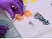
This dissection and sampling procedure was developed for the Network for Pancreatic Organ Donors with Diabetes (nPOD) program to standardize preparation of pancreas recovered from cadaveric organ donors. The pancreas is divided into 3 main regions (head, body, tail) followed by serial transverse sections throughout the medial to lateral axis. Alternating sections are used for fixed paraffin and fresh frozen blocks and remnant samples are minced for snap frozen sample preparations, either with or without RNAse inhibitors, for DNA, RNA, or protein isolation. The overall goal of the pancreas dissection procedure is to sample the entire pancreas while maintaining anatomical orientation. Endocrine cell heterogeneity in terms of islet composition, size, and numbers is reported for human islets compared to rodent islets. The majority of human islets from the pancreas head, body and tail regions are composed of insulin-containing β-cells followed by lower proportions of glucagon-containing α-cells and somatostatin-containing δ-cells. Pancreatic polypeptide-containing PP cells and ghrelin-containing epsilon cells are also present but in small numbers. In contrast, the uncinate region contains islets that are primarily composed of pancreatic polypeptide-containing PP cells. These regional islet variations arise from developmental differences. The pancreas develops from the ventral and dorsal pancreatic buds in the foregut and after rotation of the stomach and duodenum, the ventral lobe moves and fuses with the dorsal. The ventral lobe forms the posterior portion of the head including the uncinate process while the dorsal lobe gives rise to the rest of the organ. Regional pancreatic variation is also reported with the tail region having higher islet density compared to other regions and the dorsal lobe-derived components undergoing selective atrophy in type 1 diabetes. Additional organs and tissues are often recovered from the organ donors and include pancreatic lymph nodes, spleen and non-pancreatic lymph nodes. These samples are recovered with similar formats as for the pancreas with the addition of isolation of cryopreserved cells. When the proximal duodenum is included with the pancreas, duodenal mucosa may be collected for paraffin and frozen blocks and minced snap frozen preparations.
Similar articles
-
Staining protocols for human pancreatic islets.J Vis Exp. 2012 May 23;(63):e4068. doi: 10.3791/4068. J Vis Exp. 2012. PMID: 22665223 Free PMC article.
-
Quantitative analysis of pancreatic polypeptide cell distribution in the human pancreas.PLoS One. 2013;8(1):e55501. doi: 10.1371/journal.pone.0055501. Epub 2013 Jan 31. PLoS One. 2013. PMID: 23383206 Free PMC article.
-
Efficacy of human islet isolation from the tail section of the pancreas for the possibility of living donor islet transplantation.Transplantation. 2004 Sep 27;78(6):839-43. doi: 10.1097/01.tp.0000130177.03326.d5. Transplantation. 2004. PMID: 15385802
-
Network for Pancreatic Organ Donors with Diabetes (nPOD): developing a tissue biobank for type 1 diabetes.Diabetes Metab Res Rev. 2012 Oct;28(7):608-17. doi: 10.1002/dmrr.2316. Diabetes Metab Res Rev. 2012. PMID: 22585677 Free PMC article. Review.
-
Surgical aspects of human islet isolation.Islets. 2010 Sep-Oct;2(5):265-73. doi: 10.4161/isl.2.5.13019. Epub 2010 Sep 1. Islets. 2010. PMID: 21099323 Free PMC article. Review.
Cited by
-
The detection of glycosaminoglycans in pancreatic islets and lymphoid tissues.Methods Mol Biol. 2015;1229:413-30. doi: 10.1007/978-1-4939-1714-3_32. Methods Mol Biol. 2015. PMID: 25325969 Free PMC article.
-
Pancreas Optical Clearing and 3-D Microscopy in Health and Diabetes.Front Endocrinol (Lausanne). 2021 Apr 26;12:644826. doi: 10.3389/fendo.2021.644826. eCollection 2021. Front Endocrinol (Lausanne). 2021. PMID: 33981285 Free PMC article. Review.
-
Detection of enterovirus RNA in pancreas and lymphoid tissues of organ donors with type 1 diabetes.medRxiv [Preprint]. 2024 Sep 13:2024.09.11.24313112. doi: 10.1101/2024.09.11.24313112. medRxiv. 2024. PMID: 39314969 Free PMC article. Preprint.
-
Global Deletion of the Prolactin Receptor Aggravates Streptozotocin-Induced Diabetes in Mice.Front Endocrinol (Lausanne). 2021 Mar 5;12:619696. doi: 10.3389/fendo.2021.619696. eCollection 2021. Front Endocrinol (Lausanne). 2021. PMID: 33746901 Free PMC article.
-
Early prediction of autoimmune (type 1) diabetes.Diabetologia. 2017 Aug;60(8):1370-1381. doi: 10.1007/s00125-017-4308-1. Epub 2017 May 26. Diabetologia. 2017. PMID: 28550517 Free PMC article. Review.
References
-
- Brissova M. Assessment of human pancreatic islet architecture and composition by laser scanning confocal microscopy. J. Histochem. Cytochem. 2005;53:1087–1097. - PubMed
-
- Rahier J. The pancreatic polypeptide cells in the human pancreas: the effects of age and diabetes. J. Clin. Endocrinol. Metab. 1983;56:441–444. - PubMed
-
- Uchida T, Takada T, Ammori BJ, Suda K, Takahashi T. Three-dimensional reconstruction of the ventral and dorsal pancreas: a new insight into anatomy and embryonic development. J. Hepatobiliary Pancreat. Surg. 1999;6:176–180. - PubMed
-
- Rahier J, Goebbels R, Henquin J. Cellular composition of the human diabetic pancreas. Diabetologia. 1983;24:366–371. - PubMed
Publication types
MeSH terms
LinkOut - more resources
Full Text Sources
Miscellaneous
