. 2012;7(5):e37700.
doi: 10.1371/journal.pone.0037700. Epub 2012 May 25.

Hepatitis C virus sensitizes host cells to TRAIL-induced apoptosis by up-regulating DR4 and DR5 via a MEK1-dependent pathway

Affiliations

Hepatitis C virus sensitizes host cells to TRAIL-induced apoptosis by up-regulating DR4 and DR5 via a MEK1-dependent pathway

Zhongfan Deng et al. PLoS One. 2012.

Abstract

Background: Hepatitis C virus (HCV) is the leading cause of liver fibrosis, cirrhosis and hepatocellular carcinoma. It is believed that continuous liver cell apoptosis contributes to HCV pathogenesis. Recent studies have shown that HCV infection can sensitize host cells to TNF-related apoptosis-inducing ligand (TRAIL) induced apoptosis, but the mechanism by which HCV regulates the TRAIL pathway remains unclear.

Methods and results: Using a sub-genomic replicon and full length virus, JFH-1, we demonstrate that HCV can sensitize host cells to TRAIL-induced apoptosis by up-regulating two TRAIL receptors, death receptor 4 (DR4) and death receptor 5 (DR5). Furthermore, the HCV replicon enhanced transcription of DR5 via Sp1, and the HCV-mediated up-regulation of DR4 and DR5 required MEK1 activity. HCV infection also stimulated the activity of MEK1, and the inhibition of MEK1 activity or the knockdown of MEK1 increased the replication of HCV.

Conclusions: Our studies demonstrate that HCV replication sensitizes host cells to TRAIL-induced apoptosis by up-regulating DR4 and DR5 via a MEK1 dependent pathway. These findings may help to further understand the pathogenesis of HCV infection and provide a therapeutic target.


Conflict of interest statement

Competing Interests: The authors have declared that no competing interests exist.

Figures

Figure 1
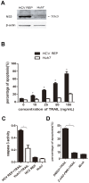
Figure 1. HCV replicon sensitizes host cells to TRAIL-induced and caspase-dependent apoptosis.
(A) Cell lysates from HCV replicon-containing cells (9–13) and Huh7 cells were subjected to western blot analyses using a rabbit polyclonal antibody against HCV NS3/4A. (B) Huh7 and 9–13 cells were treated with TRAIL at different concentrations (10 ng/mL, 20 ng/mL, 50 ng/mL or 100 ng/mL) for 2 hr and stained with annexin V and PI. The proportion of apoptotic cells was measured using flow cytometry. (C) Huh7 and 9–13 cells were treated with 50 ng/mL TRAIL for 2 hr, and caspase 3 activity was measured using a Caspase 3 Activity Assay Kit. (D) 9–13 cells were treated with 20 µM Z-VAD-FMK (a pan-caspase inhibitor) or DMSO for 1 hr and subsequently treated with 50 ng/mL TRAIL for 2 hr. The mock-treated samples were untreated 9–13 cells. The proportion of apoptotic cells was measured using flow cytometry after the cells were stained with annexin V and PI. The data are presented with the SD from three independent experiments, and statistical significance was calculated by t test or two-way ANOVA, * indicates a p value less than 0.05.
Figure 2
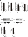
Figure 2. HCV replication up-regulates DR4 and DR5.
(A) The mRNA level of DR4, DR5, DcR1, and DcR2 in 9–13 and Huh7 cells was measured using real-time RT-PCR. (B) Huh7 and 9–13 cell lysates were subjected to western blot analyses using a rabbit polyclonal antibody against DR4 or DR5. (C) The DR4 reporter plasmid (DR4/−1156; 100 ng) or DR5 reporter plasmid (DR5/−1192; 100 ng) was co-transfected with the Renilla luciferase reporter plasmid (100 ng) into 9–13 or Huh7 cells cultured in a 24-well plate. After 2 days, the cells were harvested, and the luciferase activity was measured. (A and C) The data from the 9–13 cells were normalized to Huh7 cells to directly show the fold induction caused by HCV. The data are presented with the SD from three independent experiments, and statistical significance was calculated by t test, * indicates a p value less than 0.05.
Figure 3
Figure 3. Up-regulation of DR4 and DR5 is HCV replication-dependent.
(A) Lysates from Huh7, 9–13 and HCV-cured cells were subjected to western blot analysis using rabbit polyclonal antibodies against HCV NS3/4A, DR4 or DR5. (B) Huh7, 9–13 and HCV-cured cells were treated with 25 and 50 ng/mL TRAIL for 2 hr, and the proportion of apoptotic cells was measured using flow cytometry after the cells were stained with annexin V and PI. The data are presented with the SD from three independent experiments, and statistical significance was calculated by two-way ANOVA, * indicates a p value less than 0.05.
Figure 4
Figure 4. JFH-1 infection up-regulates the expression of DR4 and DR5.
(A) Western blot analysis was performed to measure the expression of DR4 and DR5 in Huh7.5.1 cells infected with JFH-1 1, 2, 3 days post-infection (MOI 0.02). (B) Real-time PCR was performed to measure the mRNA levels of DR4 and DR5 in Huh7.5.1 cells infected with JFH-1 (MOI 0.02) 3 days post-infection. (C) The DR4 reporter plasmid (DR4/−1156; 100 ng) or DR5 reporter plasmid (DR5/−1192; 100 ng) was co-transfected with the Renilla luciferase reporter plasmid (50 ng) into Huh7.5.1 cells, 6 hr later, cells were infected with JFH-1 (MOI 0.5). After 3 days, the cells were harvested, and the luciferase activity was measured. The data from the infected cells were normalized to Huh7.5.1 cells to directly show the fold induction caused by HCV. (D) Huh7.5.1 cells were infected with JFH-1 (MOI 0.5), 3 days later, cells were treated with indicated concentration of TRAIL for 2 hr, and stained with annexin V and PI. The proportion of apoptotic cells was measured using flow cytometry. The data are presented with the SD from three independent experiments, and statistical significance was calculated by t test or two-way ANOVA, * indicates a p value less than 0.05.
Figure 5
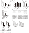
Figure 5. Transcriptional analysis of DR4 and DR5 in 9–13 cells.
(A and C) Luciferase reporter plasmids (100 ng) containing different regions of the (A) DR4 or (C) DR5 promoter and the Renilla luciferase reporter plasmid (100 ng) were co-transfected into 9–13 or Huh7 cells. 2 days post-transfection, the cells were harvested, and luciferase activity was measured. (B and D) The indicated reporter plasmids (100 ng) illustrated in (E) were co-transfected with the Renilla luciferase reporter plasmid (100 ng) into 9–13 cells. 2 days post-transfection, the cells were harvested, and the luciferase activity was measured. (E) The mutations introduced into the AP-1 binding sites in pDR4/−632 and Sp1-binding sites in pDR5/−560 are illustrated. (F) The Sp1-specific siRNA or the control siRNA (100 pmol) was transfected into 9–13 cells cultured in 6-well plates. 2 days post-transfection, the Sp1 and DR5 RNA and protein levels were measured using real-time PCR and western blot analyses, respectively. (G) A plasmid expressing HCV NS3/4A, NS4B, NS5A or NS5B (600 ng) was individually co-transfected with either DR4/−632 or DR5/−560 (100 ng) and the Renilla luciferase reporter plasmid (100 ng) into Huh7 cells. 2 days post-transfection, the cells were harvested, and luciferase activity was measured. The data are presented with the SD from three independent experiments, and statistical significance was calculated by t test or two-way ANOVA, * indicates a p value less than 0.05.
Figure 6
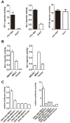
Figure 6. Over expression of MEK1 activates the DR4 and DR5 promoters.
(A) The AP-1, Sp1 or NF-κB luciferase reporter plasmid (100 ng) was co-transfected with the Renilla luciferase reporter plasmid (100 ng) into 9–13 or Huh7 cells cultured in 24-well plates. 2 days post-transfection, luciferase activity was measured. (B and C) pMEK1-pRK or pMEKK1-pRK (600 ng) and the Renilla luciferase reporter plasmid (100 ng) were co-transfected with the (B) AP-1 or Sp1 reporter plasmid (100 ng) or (C) the indicated DR4 or DR5 reporter plasmid (100 ng) into Huh7 cells cultured in 24-well plates. 2 days post-transfection, luciferase activity was measured. The data are presented with the SD from three independent experiments, and statistical significance was calculated by t test, * indicates a p value less than 0.05.
Figure 7
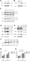
Figure 7. HCV-mediated increased DR4 and DR5 expression is MEK1 dependent.
Huh7 and 9–13 cell lysates (A) or Huh7.5.1 and JFH-1 infected Huh7.5.1 cell (MOI 0.02) lysates (B) were subjected to western blot analyses using antibodies against phospho-MEK1 or MEK1. (C) Untreated 9–13 cells or 9–13 cells treated with 100 µM PD98059, 100 µM SP600125 or DMSO for 2 days were harvested and subjected to western blot analyses using antibodies against DR4 or DR5. (D) The MEK1-specific siRNA or control siRNA (100 pmol) was transfected into 9–13 cells cultured in 6-well plates. 2 days post-transfection, the expression of MEK1, DR4 and DR5 was determined using western blot analyses. (E) Huh7.5.1 cells were transfected with the MEK1-specific siRNA or control siRNA and infected with JFH-1 (MOI 0.02) 6 hr post transfection. 3 days post infection, the expression of MEK1, DR4 and DR5 was determined using western blot analyses. (F) Huh7.5.1 cells were transfected with indicated siRNA, and the expression of MEK1 was measured by using western blot 2 days later. (G) 9–13 cells were transfected with MEK1 siRNA1, MEK1 siRNA2 or scramble RNA, 3 days post transfection, cells were treated indicated concentration of TRAIL for 2 hr, and stained with annexin V and PI. The proportion of apoptotic cells was analyzed by using flow cytometry. (H) Huh7.5.1 cells were transfected with MEK1 siRNA1, MEK1 siRNA2 or scramble RNA, 6 hr later, cells were infected with JFH-1 (MOI 0.5), 3 days post infection, cells were treated indicated concentration of TRAIL for 2 hr, and stained with annexin V and PI. The proportion of apoptotic cells was analyzed by using flow cytometry. The data are presented with the SD from three independent experiments, and statistical significance was calculated by two-way ANOVA, * indicates a p value less than 0.05.
Figure 8
Figure 8. Inhibition of MEK1 increases HCV replication.
Huh7.5.1 cells were transfected with a MEK1-specific siRNA (A) or treated with 10 µM PD98059 (B) and 6 hr post-transfection or treatment, infected with JFH-1 (MOI 0.02). 3 days post-infection, the expression of the HCV core protein was detected using western blot.

Similar articles

Cited by

References

    1. Wiley SR Schooley K, Smolak PJ, Din WS, Huang CP, et al. Identification and characterization of a new member of the TNF family that induces apoptosis. Immunity. 1995;3:673–682. - PubMed
    1. Walczak H, Miller RE, Ariail K, Gliniak B, Griffith TS, et al. Tumoricidal activity of tumor necrosis factor-related apoptosis-inducing ligand in vivo. Nat Med. 1999;5:157–163. - PubMed
    1. Liang X, Liu Y, Zhang Q, Gao L, Han L, et al. Hepatitis B virus sensitizes hepatocytes to TRAIL-induced apoptosis through Bax. J Immunol. 2007;178:503–510. - PubMed
    1. Lan L, Gorke S, Rau SJ, Zeisel MB, Hildt E, et al. Hepatitis C virus infection sensitizes human hepatocytes to TRAIL-induced apoptosis in a caspase 9-dependent manner. J Immunol. 2008;181:4926–4935. - PubMed
    1. Kotelkin A, Prikhod'ko EA, Cohen JI, Collins PL, Bukreyev A. Respiratory syncytial virus infection sensitizes cells to apoptosis mediated by tumor necrosis factor-related apoptosis-inducing ligand. J Virol. 2003;77:9156–9172. - PMC - PubMed

Publication types

MeSH terms

Substances