. 2012 Jul;8(7):646-54.
doi: 10.1038/nchembio.965. Epub 2012 Jun 3.

ATM and MET kinases are synthetic lethal with nongenotoxic activation of p53

Affiliations

ATM and MET kinases are synthetic lethal with nongenotoxic activation of p53

Kelly D Sullivan et al. Nat Chem Biol. 2012 Jul.

Abstract

The p53 tumor suppressor orchestrates alternative stress responses including cell cycle arrest and apoptosis, but the mechanisms defining cell fate upon p53 activation are poorly understood. Several small-molecule activators of p53 have been developed, including Nutlin-3, but their therapeutic potential is limited by the fact that they induce reversible cell cycle arrest in most cancer cell types. We report here the results of a genome-wide short hairpin RNA screen for genes that are lethal in combination with p53 activation by Nutlin-3, which showed that the ATM and MET kinases govern cell fate choice upon p53 activation. Genetic or pharmacological interference with ATM or MET activity converts the cellular response from cell cycle arrest into apoptosis in diverse cancer cell types without affecting expression of key p53 target genes. ATM and MET inhibitors also enable Nutlin-3 to kill tumor spheroids. These results identify new pathways controlling the cellular response to p53 activation and aid in the design of p53-based therapies.


Conflict of interest statement

Competing Financial Interest.

The authors declare no competing financial interests.

Figures
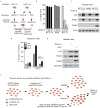
Figure 1
Figure 1. A genetic screen to identify modulators of the cellular response to p53 activation by Nutlin-3
(a) Experimental paradigms of cell type- and stimulus-specific p53-dependent cell fate choices used in this report. (b) Non-genotoxic activation of p53 by 20 μM Nutlin-3 leads to apoptosis only in select cell types. Relative viability of HCT116, A549 and BV173 cells as assessed by Annexin V and propidium iodide (PI) staining. (c) Nutlin-3 activates p53 and its pro-apoptotic target gene PUMA in all cell types tested, but activates executioner caspase 3 only in BV173 cells. Western blots for p53, PUMA and cleaved caspase 3 were performed on lysates prepared from cells treated with 20 μM Nutlin-3 or DMSO for 24 h. Actin serves as a loading control. Full blots can be found in Supplementary Fig. 7. (d) 5FU elicits a p53-dependent apoptotic response in HCT116 cells. Cells were treated with 5FU (375 μM) for the indicated period of time prior to Annexin V and PI analysis by flow cytometry. (e) Nutlin-3 (20 μM) and 5FU (375 μM) activate p53 and PUMA to similar levels, but only 5FU activates caspase 3. Lysates were prepared from cells treated with the indicated drug for 24 h and Western blots performed for p53, PUMA and cleaved caspase 3. Nucleolin serves as a loading control. (f) Schematic of genetic screen design.
Figure 2
Figure 2. Identification of ‘Synthetic Lethal with Nutlin-3’ genes
(a) Hierarchical clustering analysis of raw sequence counts of shRNAs from HCT116 cells. Heatmap of 19,411 shRNAs where the median count of one treatment is greater than the maximum count of the other treatment. (b) Actual read counts of top 10 candidate SLN shRNAs. (c) Distribution of fold change for each shRNA with a p-value <0.05 (total of 6,525) from HCT116 cells. Red diamonds indicate the position of the 30 shRNAs chosen for validation in e. Arrows indicate the position of 5 shRNAs highlighted throughout the figure. (d) Distribution of 1730 genes from HCT116 cells with p(wZ) values <0.05. Black diamonds indicate positions of the 30 genes tested in e. (e) Validation of 30 predicted SLNs. Cell lines stably expressing single hairpins targeting the indicated gene were treated with 20 μM Nutlin-3 for 72 hours and analyzed by SRB assay. Relative viability was calculated as the ratio of Nutlin-3 treatment to DMSO treatment and compared to a non-targeting control shRNA. Data shown are an average of three experiments ± standard deviation.
Figure 3
Figure 3. Pathway analysis points to ATM and MET as modulators of p53-dependent cell fate choice
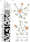
(a) Top, the highest scoring 505 SLNs from HCT116 cells were analyzed using IPA software. The top five canonical pathways identified are shown. Gray bars represent enrichment p-value for corresponding pathway. Black line represents absolute ratio of genes from a given pathway that are SLNs. Bottom, matrix of SLNs from each pathway. Listed genes were identified as SLNs in the genome-wide screen. Black boxes indicate that a gene is a component of a given pathway. (b) ATM module. Orange balls represent validated SLN and green balls represent SLNs predicted by BiNGS. Numbers represent fold change in abundance of shRNA with greatest change from screen. (c) MET module. Analyzed as in b.
Figure 4
Figure 4. ATM protects cells from p53-dependent apoptosis upon Nutlin-3 treatment
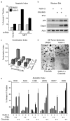
(a) ATM knockdown impairs cell viability in response to Nutlin-3 treatment. Cell lines stably expressing shRNAs targeting ATM were treated with 20 μM Nutlin-3 for 72 h and analyzed as in 2e. BCL2 serves as positive control. A non-targeting shRNA serves as a negative control. (b) ATM knockdown increases apoptosis upon Nutlin-3 treatment. Cell lines from a were treated with 20 μM Nutlin-3 or DMSO for 24 h prior to assessment of Annexin-V FITC and PI levels by flow cytometry. (c) Chemical inhibition of ATM activates executioner caspase 3 upon p53 activation by Nutlin-3. Lysates were prepared from cells treated with 10 μM ATMi, 20 μM Nutlin-3 and 375 μM 5FU for 24 h and Western blots performed for p53 and cleaved caspase 3. Actin serves as a loading control. Full blots can be found in Supplementary Fig. 7. (d) Treatment of cells with ATMi and Nutlin-3 results in robust apoptosis. Cells were treated with the indicated drugs as in c before Annexin V and PI levels were measured by flow cytometry. (e) The combination of ATMi and Nutlin-3 is highly synergistic. Cells were treated as in c for 24 h before SRB analysis. Combination index (CI) values were calculated using Calcusyn software. (f) Combination treatment is effective in a 3D culture system. HCT116 cells were plated on Matrigel pads for one week to allow tumor spheroid formation, then treated for 3 weeks before imaging. Scale bars, 1 mm.
Figure 5
Figure 5. MET protects against p53-dependent apoptosis upon Nutlin-3 treatment
(a) MET knockdown leads to increased apoptosis upon Nutlin-3 treatment. Cell lines expressing indicated shRNAs were treated with 20 μM Nutlin-3 or DMSO for 24 h prior to assessment of Annexin-V FITC and PI levels by flow cytometry. (b) Crizotinib converts the cellular response to Nutlin-3 from cell cycle arrest to apoptosis in HCT116 cells. Lysates were prepared from cells treated with 7 μM Crizotinib, 20 μM Nutlin-3 and 375 μM 5FU for 24 h and Western blots performed for p53 and cleaved caspase 3. Actin serves as a loading control. Full blots can be found in Supplementary Fig. 7. (c) The combination of Crizotinib and Nutlin-3 is highly synergistic. CI values were calculated as in Fig. 4e. (d) Crizotinib and Nutlin-3 clear tumor spheroids effectively. Tumor spheres were prepared and treated as in Fig. 4f for 12 days, at which point the combination treatment was devoid of viable cells. Scale bars, 1 mm. (e) Combinations of either ATMi or Crizotinib with Nutlin-3 increase the apoptotic index cancer cell types of different origins. Cells were treated with 10 μM ATMi, 7 μM Crizotinib and 20 μM Nutlin-3, for 24 h prior to analysis of Annexin V and PI by flow cytometry. Data shown are an average of three experiments ± standard deviation.
Figure 6
Figure 6. ATM and MET do not affect the ability of p53 to transactivate key genes in the cell cycle arrest and apoptosis modules
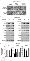
(a) PUMA is required for synthetic lethality of ATMi or Crizotinib in combination with Nutlin-3. Tumor spheres of HCT116 cells of different PUMA status were prepared as in Fig. 4f and treated with the indicated drug combinations for three weeks. Scale bars, 1 mm. (b) Neither ATMi nor Crizotinib affect protein expression of important p53-regulated cell cycle arrest or apoptotic target genes. Lysates were prepared from cells treated with 10 μM ATMi, 7 μM Crizotinib and 20 μM Nutlin-3, and expression of cell cycle arrest (p21 and 14-3-3σ), pro-apoptotic (PUMA and BAX), pro-survival (BCL2) and extrinsic apoptotic pathway (cleaved caspase 8 and tBID) proteins was analyzed by Western blot. Nucleolin serves as a loading control. Full blots can be found in Supplementary Fig. 7. (c) Drug combinations do not affect the transactivation ability of Nutlin-3-activated p53. RNA was harvested from cells treated with drugs as in b and Q-RT-PCR performed to test induction of p21, PUMA and MDM2 mRNAs.
Figure 7
Figure 7. 53BP1 is not required for ATM synthetic lethality
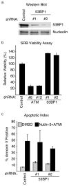
(a) Western blot of 53BP1 knockdown cell lines. Nucleolin serves as a loading control. Full blots can be found in Supplementary Fig. 7. (b) In contrast to ATM knockdown, 53BP1 knockdown increases cell viability in response to Nutlin-3 treatment. Cell lines stably expressing shRNAs targeting ATM or 53BP1 were treated with 20 μM Nutlin-3 for 72 hours prior to analysis by SRB staining. (c) 53BP1 knockdown does not block apoptosis in response to [Nutlin-3+ATMi]. Cell lines expressing either control or 53BP1 shRNAs were treated with the indicated drugs prior to assessment of Annexin-V-FITC staining by flow cytometry.

Similar articles

Cited by

References

    1. Vousden KH, Prives C. Blinded by the Light: The Growing Complexity of p53. Cell. 2009;137:413–31. - PubMed
    1. Brown CJ, Lain S, Verma CS, Fersht AR, Lane DP. Awakening guardian angels: drugging the p53 pathway. Nat Rev Cancer. 2009;9:862–73. - PubMed
    1. Levesque AA, Eastman A. p53-based cancer therapies: Is defective p53 the Achilles heel of the tumor? Carcinogenesis. 2007;28:13–20. - PubMed
    1. Mandinova A, Lee SW. The p53 pathway as a target in cancer therapeutics: obstacles and promise. Sci Transl Med. 2011;3:64rv1. - PMC - PubMed
    1. Vassilev LT, et al. In vivo activation of the p53 pathway by small-molecule antagonists of MDM2. Science. 2004;303:844–8. - PubMed

Publication types

MeSH terms