Cutting edge: TNF receptor-associated factor 4 restricts IL-17-mediated pathology and signaling processes
- PMID: 22649194
- PMCID: PMC3590847
- DOI: 10.4049/jimmunol.1200470
Cutting edge: TNF receptor-associated factor 4 restricts IL-17-mediated pathology and signaling processes
Abstract
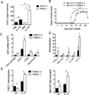
The effector T cell subset, Th17, plays a significant role in the pathogenesis of multiple sclerosis and of other autoimmune diseases. The signature cytokine, IL-17, engages the IL-17R and recruits the E3-ligase NF-κB activator 1 (Act1) upon stimulation. In this study, we examined the role of TNFR-associated factor (TRAF)4 in IL-17 signaling and Th17-mediated autoimmune encephalomyelitis. Primary cells from TRAF4-deficient mice displayed markedly enhanced IL-17-activated signaling pathways and induction of chemokine mRNA. Adoptive transfer of MOG35-55 specific wild-type Th17 cells into TRAF4-deficient recipient mice induced an earlier onset of disease. Mechanistically, we found that TRAF4 and TRAF6 used the same TRAF binding sites on Act1, allowing the competition of TRAF4 with TRAF6 for the interaction with Act1. Taken together, the results of this study reveal the necessity of a unique role of TRAF4 in restricting the effects of IL-17 signaling and Th17-mediated disease.
Figures


Similar articles
-
RKIP mediates autoimmune inflammation by positively regulating IL-17R signaling.EMBO Rep. 2018 Jun;19(6):e44951. doi: 10.15252/embr.201744951. Epub 2018 Apr 19. EMBO Rep. 2018. PMID: 29674348 Free PMC article.
-
IL-17 signaling for mRNA stabilization does not require TNF receptor-associated factor 6.J Immunol. 2009 Feb 1;182(3):1660-6. doi: 10.4049/jimmunol.182.3.1660. J Immunol. 2009. PMID: 19155515 Free PMC article.
-
TRAF4-SMURF2-mediated DAZAP2 degradation is critical for IL-25 signaling and allergic airway inflammation.J Immunol. 2015 Mar 15;194(6):2826-37. doi: 10.4049/jimmunol.1402647. Epub 2015 Feb 13. J Immunol. 2015. PMID: 25681341 Free PMC article.
-
IL-17 receptor signaling and T helper 17-mediated autoimmune demyelinating disease.Trends Immunol. 2011 May;32(5):232-9. doi: 10.1016/j.it.2011.02.007. Epub 2011 Apr 12. Trends Immunol. 2011. PMID: 21493143 Free PMC article. Review.
-
IL-17/IL-17 receptor system in autoimmune disease: mechanisms and therapeutic potential.Clin Sci (Lond). 2012 Jun;122(11):487-511. doi: 10.1042/CS20110496. Clin Sci (Lond). 2012. PMID: 22324470 Review.
Cited by
-
TRAF6-dependent Act1 phosphorylation by the IκB kinase-related kinases suppresses interleukin-17-induced NF-κB activation.Mol Cell Biol. 2012 Oct;32(19):3925-37. doi: 10.1128/MCB.00268-12. Epub 2012 Jul 30. Mol Cell Biol. 2012. PMID: 22851696 Free PMC article.
-
The Role of IL-17-Mediated Inflammatory Processes in the Pathogenesis of Intervertebral Disc Degeneration and Herniation: A Comprehensive Review.Front Cell Dev Biol. 2022 Mar 3;10:857164. doi: 10.3389/fcell.2022.857164. eCollection 2022. Front Cell Dev Biol. 2022. PMID: 35309927 Free PMC article. Review.
-
TRAF4 is a critical molecule for Akt activation in lung cancer.Cancer Res. 2013 Dec 1;73(23):6938-50. doi: 10.1158/0008-5472.CAN-13-0913. Epub 2013 Oct 23. Cancer Res. 2013. PMID: 24154876 Free PMC article.
-
Cytokine Imbalance as a Biomarker of Intervertebral Disk Degeneration.Int J Mol Sci. 2023 Jan 25;24(3):2360. doi: 10.3390/ijms24032360. Int J Mol Sci. 2023. PMID: 36768679 Free PMC article. Review.
-
Role of Interleukin-17A in the Pathomechanisms of Periodontitis and Related Systemic Chronic Inflammatory Diseases.Front Immunol. 2022 Mar 17;13:862415. doi: 10.3389/fimmu.2022.862415. eCollection 2022. Front Immunol. 2022. PMID: 35371044 Free PMC article. Review.
References
-
- Komiyama Y, Nakae S, Matsuki T, Nambu A, Ishigame H, Kakuta S, Sudo K, Iwakura Y. IL-17 plays an important role in the development of experimental autoimmune encephalomyelitis. J Immunol. 2006;177:566–573. - PubMed
-
- Nakae S, Nambu A, Sudo K, Iwakura Y. Suppression of immune induction of collagen-induced arthritis in IL-17-deficient mice. J Immunol. 2003;171:6173–6177. - PubMed
-
- Hu Y, Ota N, Peng I, Refino CJ, Danilenko DM, Caplazi P, Ouyang W. IL-17RC is required for IL-17A- and IL-17F-dependent signaling and the pathogenesis of experimental autoimmune encephalomyelitis. J Immunol. 2010;184:4307–4316. - PubMed
-
- Novatchkova M, Leibbrandt A, Werzowa J, Neubuser A, Eisenhaber F. The STIR-domain superfamily in signal transduction, development and immunity. Trends Biochem Sci. 2003;28:226–229. - PubMed
-
- Chang SH, Park H, Dong C. Act1 adaptor protein is an immediate and essential signaling component of interleukin-17 receptor. J Biol Chem. 2006;281:35603–35607. - PubMed
Publication types
MeSH terms
Substances
Grants and funding
LinkOut - more resources
Full Text Sources
Other Literature Sources
Molecular Biology Databases

