Polo-like kinase 1 is overexpressed in colorectal cancer and participates in the migration and invasion of colorectal cancer cells
- PMID: 22648245
- PMCID: PMC3560731
- DOI: 10.12659/msm.882900
Polo-like kinase 1 is overexpressed in colorectal cancer and participates in the migration and invasion of colorectal cancer cells
Abstract
Background: Polo-like kinase 1 (PLK1) is an important molecule in proliferation of many human cancers. The aim of study is to clarify the expression patterns and potential function of PLK1 in colorectal cancers.
Material/methods: Fifty-six colorectal cancers samples were collected and arranged onto a tissue array and the expression of PLK1 were detected by immunohistochemistry and correlated with clinico-pathological characteristics and expression of PCNA. Expression of PLK1 in 9 colorectal cancer cells lines was investigated by RT-PCR and Western blot, then SW1116 cells lines were treated with PLK1 siRNA and the efficiency was examined by Western blot. Transwell test was applied to detect the migration and invasion capability of cancer cells by counting the number of cells passing through the membranes. Cell proliferation and apoptosis were examined by Cell Counting Kit-8 (CCK-8) and Annexin-V Kit.
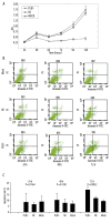
Results: PLK1 was positively expressed in 73.2% (41/56) of colorectal cancers tissues, but in only 3.6% (2/56) of normal tissues, and was associated with Duke's stage (P<0.01), tumor size (P<0.01), invasion extent (P<0.05) and lymphatic metastasis (P<0.01). The expression of PLK1 was correlated with expression of PCNA (R=0.553, P<0.01). PLK1 was inhibited in SW1116 cells by treating with PLK1 siRNA oligos, which resulted in a decreased number of cells passing through the membrane as compared with control groups (P<0.01) at 24 hours after transfection. Cell proliferation was inhibited from 48 hours after transfection, while cells apoptosis was induced from 72 hours after transfection.
Conclusions: PLK1 could be a progression marker for colorectal cancer patients and PLK1 depletion can inhibit migration and invasion capability of colorectal cancer cells SW1116, suggesting that PLK1 might be involved in metastasis and invasion of colorectal cancer. Therapeutic strategies targeting PLK1 may be a new approach to colorectal cancer.
Figures


Similar articles
-
[Influence of silencing Polo-like kinase 1 on migration and invasion of colorectal cancer cells].Zhonghua Wei Chang Wai Ke Za Zhi. 2011 Jan;14(1):61-4. Zhonghua Wei Chang Wai Ke Za Zhi. 2011. PMID: 21271384 Chinese.
-
Polo-like kinase 1 is overexpressed in renal cancer and participates in the proliferation and invasion of renal cancer cells.Tumour Biol. 2013 Jun;34(3):1887-94. doi: 10.1007/s13277-013-0732-0. Epub 2013 Mar 14. Tumour Biol. 2013. PMID: 23494182
-
Efficient inhibition of human colorectal carcinoma growth by RNA interference targeting polo-like kinase 1 in vitro and in vivo.Cancer Biother Radiopharm. 2011 Aug;26(4):427-36. doi: 10.1089/cbr.2010.0922. Epub 2011 Jul 28. Cancer Biother Radiopharm. 2011. PMID: 21797676
-
Targeting polo-like kinase 1 for cancer therapy.Nat Rev Cancer. 2006 Apr;6(4):321-30. doi: 10.1038/nrc1841. Nat Rev Cancer. 2006. PMID: 16557283 Review.
-
Polo-like kinase 1 (PLK1) signaling in cancer and beyond.Biochem Pharmacol. 2021 Nov;193:114747. doi: 10.1016/j.bcp.2021.114747. Epub 2021 Aug 26. Biochem Pharmacol. 2021. PMID: 34454931 Review.
Cited by
-
MicroRNA-200b Impacts Breast Cancer Cell Migration and Invasion by Regulating Ezrin-Radixin-Moesin.Med Sci Monit. 2016 Jun 8;22:1946-52. doi: 10.12659/msm.896551. Med Sci Monit. 2016. PMID: 27276064 Free PMC article.
-
Vimentin immunoexpression is associated with higher tumor grade, metastasis, and shorter survival in colorectal cancer.Int J Clin Exp Pathol. 2020 Mar 1;13(3):493-500. eCollection 2020. Int J Clin Exp Pathol. 2020. PMID: 32269687 Free PMC article.
-
The Emerging Role of Polo-Like Kinase 1 in Epithelial-Mesenchymal Transition and Tumor Metastasis.Cancers (Basel). 2017 Sep 27;9(10):131. doi: 10.3390/cancers9100131. Cancers (Basel). 2017. PMID: 28953239 Free PMC article. Review.
-
Role of Stress-Survival Pathways and Transcriptomic Alterations in Progression of Colorectal Cancer: A Health Disparities Perspective.Int J Environ Res Public Health. 2021 May 21;18(11):5525. doi: 10.3390/ijerph18115525. Int J Environ Res Public Health. 2021. PMID: 34063993 Free PMC article. Review.
-
Phase I trial of volasertib, a Polo-like kinase inhibitor, plus platinum agents in solid tumors: safety, pharmacokinetics and activity.Invest New Drugs. 2015 Jun;33(3):611-20. doi: 10.1007/s10637-015-0223-9. Epub 2015 Mar 22. Invest New Drugs. 2015. PMID: 25794535 Free PMC article. Clinical Trial.
References
-
- Jemal A, Siegel R, Xu J, Ward E. Cancer statistics, 2010. CA Cancer J Clin. 2010;60:277–300. - PubMed
-
- Sung JJ, Lau JY, Goh KL, Leung WK Asia Pacific Working Group on Colorectal Cancer. Increasing incidence of colorectal cancer in Asia: implications for screening. Lancet Oncol. 2005;6:871–76. - PubMed
-
- Boxberger F, Albrecht H, Konturek PC, et al. Neoadjuvant treatment with weekly high-dose 5-fluorouracil as a 24h-infusion, folinic acid and biweekly oxaliplatin in patients with primary resectable liver metastases of colorectal cancer: long-term results of a phase II trial. Med Sci Monit. 2010;16(2):CR49–55. - PubMed
-
- Gennari L, Russo A, Rossetti C. Colorectal cancer: what has changed in diagnosis and treatment over the last 50 years? Tumori. 2007;93:235–41. - PubMed
-
- Michor F, Iwasa Y, Vogelstein B, et al. Can chromosomal instability initiate tumorigenesis? Semin Cancer Biol. 2005;15:43–49. - PubMed
Publication types
MeSH terms
Substances
LinkOut - more resources
Full Text Sources
Medical
Miscellaneous

