. 2012 May 30:9:111.
doi: 10.1186/1742-2094-9-111.

Heterotrimeric G protein-dependent WNT-5A signaling to ERK1/2 mediates distinct aspects of microglia proinflammatory transformation

Affiliations

Heterotrimeric G protein-dependent WNT-5A signaling to ERK1/2 mediates distinct aspects of microglia proinflammatory transformation

Carina Halleskog et al. J Neuroinflammation. .

Abstract

Background: WNT-5A signaling in the central nervous system is important for morphogenesis, neurogenesis and establishment of functional connectivity; the source of WNT-5A and its importance for cellular communication in the adult brain, however, are mainly unknown. We have previously investigated the inflammatory effects of WNT/β-catenin signaling in microglia in Alzheimer's disease. WNT-5A, however, generally recruits β-catenin-independent signaling. Thus, we aim here to characterize the role of WNT-5A and downstream signaling pathways for the inflammatory transformation of the brain's macrophages, the microglia.

Methods: Mouse brain sections were used for immunohistochemistry. Primary isolated microglia and astrocytes were employed to characterize the WNT-induced inflammatory transformation and underlying intracellular signaling pathways by immunoblotting, quantitative mRNA analysis, proliferation and invasion assays. Further, measurements of G protein activation by [γ-(35)S]GTP binding, examination of calcium fluxes and cyclic AMP production were used to define intracellular signaling pathways.

Results: Astrocytes in the adult mouse brain express high levels of WNT-5A, which could serve as a novel astroglia-microglia communication pathway. The WNT-5A-induced proinflammatory microglia response is characterized by increased expression of inducible nitric oxide synthase, cyclooxygenase-2, cytokines, chemokines, enhanced invasive capacity and proliferation. Mapping of intracellular transduction pathways reveals that WNT-5A activates heterotrimeric G(i/o) proteins to reduce cyclic AMP levels and to activate a G(i/o) protein/phospholipase C/calcium-dependent protein kinase/extracellular signal-regulated kinase 1/2 (ERK1/2) axis. We show further that WNT-5A-induced ERK1/2 signaling is responsible for distinct aspects of the proinflammatory transformation, such as matrix metalloprotease 9/13 expression, invasion and proliferation.

Conclusions: Thus, WNT-5A-induced and G protein-dependent signaling to ERK1/2 is important for the regulation of proinflammatory responses in mouse primary microglia cells. We show for the first time that WNT-5A/G protein signaling mediates physiologically important processes in primary mammalian cells with natural receptor and G protein stochiometry. Consequently, WNT-5A emerges as an important means of astrocyte-microglia communication and we, therefore, suggest WNT-5A as a new player in neuroinflammatory conditions, such as neurodegenerative disease, hypoxia, stroke, injury and infection.


Figures

Figure 1
Figure 1
WNT-5A expression in mouse GFAP+astrocytes. (A) Immunohistochemistry was performed on adult mouse brain sections using an anti-WNT-5A antibody in combination with anti-glial fibrillary acidic protein (GFAP) and ionized calcium-binding adaptor protein1 (IBA1) antibodies as astrocyte and microglia marker, respectively. Merge presents the overlay of IBA1, GFAP, WNT-5A. Size bar −2 μm. The images represent a maximum intensity projection of a Z-stack of 5 μm thickness. The white square marked ‘B’ indicates the area magnified in B: (B) Close up of a GFAP+ astrocyte reveals the expression of WNT-5A in this cell type. (C) shows immunoblot detection of recombinant WNT-5A (rWNT-5A; 375 ng/lane) in comparison to lysates from mouse primary microglia and mixed astrocyte cultures. β-actin serves as a loading control. (D) The bar graph depicts expression levels of WNT-5A mRNA in mouse primary microglia and mixed astrocyte cultures measured by QPCR. Data are normalized to GAPDH expression and analyzed with a non-parametric Mann–Whitney test. *, P < 0.05; **, P < 0.01; ***, P < 0.001. n = 4 to 8. (E) shows indirect immunocytochemistry of mixed astrocyte cultures employing anti-GFAP as astrocyte and anti-CD11b as microglia markers. DAPI is used as nuclear counterstain. Image represents a maximum image projection of an 8 μm Z-stack. Size bar 20 μm. The frame shows 63 cells in total and 10 CD11b-positive microglia (arrows). Routinely 10% to 18% microglia were observed (n = 4). DAPI, 4',6-diamidino-2-phenylindole; GADPH, glyceraldehyde 3-phosphate dehydrogenase; n, number.
Figure 2
Figure 2
WNT-5A induced a transient phosphorylation of ERK1/2. WNT-5A stimulation of mouse primary microglia induced ERK1/2 phosphorylation in a time- (A; 300 ng/ml WNT-5A) and dose-dependent (B; 30 minutes WNT-5A) manner. Bar graphs provide a summary of densitometric analysis of three independent experiments. Data were normalized to the response in ctrl-stimulated microglia. Variation is shown as s.e.m. (n ≥ 3). *, P < 0.05; **, P < 0.01; ***, P < 0.001. ctrl, control; ERK1/2, extracellular signal-regulated kinase 1/2; n, number; s.e.m, standard error of the mean.
Figure 3
Figure 3
WNT-5A-induced ERK1/2 phosphorylation requires activation of Gαi/o. (A) Primary microglia, pretreated with PTX (100 ng/ml, overnight), were stimulated with 300 ng/ml WNT-5A for 30 minutes and their total cell lysate was analyzed for P-ERK1/2 by immunoblotting. β-actin serves as a loading control. Densitometric analysis of three independent experiments is summarized in the bar graph (error bars - s.e.m.). **, P < 0.01. (B) In a similar stimulation paradigm, cells were lysed after two hours and the lysate was analyzed for the formation of PS-DVL3 (filled triangle: DVL3, open triangle: PS-DVL3). n = 3. (C) Indirect immunocytochemistry and confocal microscopy were used to detect and localize P-ERK1/2 in ctrl, WNT-5A (300 ng/ml, 30 minutes) and WNT-5A/pertussis toxin (PTX, 100 ng/ml)-treated mouse primary microglia. IBA1 was used as a microglia marker and DAPI as nuclear counterstain. Arrows mark WNT-5A-responsive microglia with increased P-ERK1/2 levels (n = 3). Size bar = 50 μm. ctrl, control; DAPI, 4',6-diamidino-2-phenylindole; DVL, disheveled; ERK1/2, extracellular signal-regulated kinase 1/2; IBA1, ionized calcium-binding adaptor protein1; n, number; P-ERK1/2, ERK1/2 phosphorylation; PS-DVL, phosphorylated and shifted DVL; s.e.m., standard error of the mean.
Figure 4
Figure 4
WNT-5A-induced G protein activation mediates decrease in cAMP and mobilization of [Ca2+]i. (A) Isolated plasma membranes from mouse primary microglia were stimulated with WNT-5A (300 ng/ml) as described in the Methods section to assess activation of heterotrimeric G proteins. Incorporation of [γ-35 S]GTP, a measure for activation of G proteins, was quantified by scintillation counting. Data from three independent experiments are shown in the bar graph. ***, P < 0.001. Error bars give s.e.m.. (B) cDNA from mouse primary microglia was analyzed for expression of PTX-sensitive variants of the Gi/o family of Gα subunits by RT-PCR. cDNA synthesis was performed in the absence and presence of RT (+/− RT) to control for the purity of the preparation. (C) Detection of intracellular cAMP levels reveals that WNT-5A reduces forskolin-induces cAMP production in a dose-dependent manner (error bars: s.e.m.; n = 3; **, P < 0.01; ***, P < 0.001.). (D-F) Stimulation of Fluo-3-loaded primary microglia with WNT-5A induced fast and transient elevation of [Ca2+]i. ATP was used as a positive control. The [Ca2+]i trace shown in D originates from a single cell. Typically 15% to 30% of the cultured microglia responded to WNT-5A. (E) shows a representative view of Fluo-3-loaded cells at baseline and upon WNT-5A (300 ng/ml) exposure. The images are pseudocolored with warm colors presenting high [Ca2+]i and cold colors low [Ca2+]i. Size bar 20 μm. The bar graph summarizes data from five different experiments with ATP and WNT-5A in combination with PTX. Error bar gives s.e.m. n, number; PTX, pertussis toxin; s.e.m., standard error of the mean.
Figure 5
Figure 5
Mapping of the WNT-5A-induced signaling pathway leading to ERK1/2 phosphorylation in primary microglia. (A) Cells were treated with pharmacological inhibitors 10 minutes prior to WNT-5A challenge (300 ng/ml, 20 minutes). Immunoblotting analysis of P-ERK1/2 and P-MEK1/2 is shown. β-actin serves as loading control. Representative data from at least three experiments are shown and data are quantified in the bar graphs next to each inhibitor (error bars - s.e.m.; *, P < 0.05; **, P < 0.01; ***, P < 0.001). (B) Lysates from microglia treated with WNT-5A in the absence or presence of D4479 were analyzed for the formation of PS-DVL3. (C) Table summarizes concentrations, targets and effects of the inhibitors used. Pharmacological inhibitors: D4476, CK1 inhibitor; M119, βγ inhibitor; U73122, PLC inhibitor; BIS,(bisindolmaleimide VIII), PKC inhibitor; wortmannin/LY294002, phosphatidylinositol-3'-kinase inhibitor; BAPTA-AM, Ca2+-chelator; SL327, MEK1/2 inhibitor. DVL3, disheveled 3; ERK1/2, extracellular signal-regulated kinase 1/2; MEK1/2, MAPK/ERK kinase 1/2; PKC, calcium-dependent protein kinase; PLC, phospholipase C; PS-DVL3, phosphorylated and shifted DVL3; s.e.m, standard error of the mean.
Figure 6
Figure 6
WNT-5A induces a proinflammatory transformation in mouse microglia. (A, B) iNOS, COX-2 and TNFα were detected by immunoblotting in lysates from mouse primary microglia after WNT-5A stimulation (ctrl, 300 ng/ml, 6 hours). (B’) shows TNFα levels in the supernatant of primary microglia under ctrl conditions and upon WNT-5A stimulation (ctrl, 300 ng/ml, 24 hours; n = 4). At least three experiments are summarized in the bar graphs. Data are normalized to ctrl. Error bars give s.e.m. (C) Microglial proliferation was assessed by an MTT assay monitoring mitochondrial activity, which is proportional to cell number [see Additional file 1: Figure S5]. Stimulation with WNT-5A (300 ng/ml, 24 hours) increased MTT, which was blocked by PTX (100 ng/ml, overnight) or the MEK1/2 (10 μM) inhibitor, SL327. (D). Experiments were done in triplicate and data from three independent experiments are shown. *, P < 0.01; ***, P < 0.001: Error bars show s.e.m.. (E) Cell tracker (red)-stained primary microglia were seeded on top of a collagen matrix in 35 mm glass bottom dishes. One day after ctrl or 300 ng/ml WNT-5A stimulation, invasion was observed by confocal microscopy and Z-stacking using a Zeiss LSM710 microscope and subsequent analysis with the Bitplane Imaris software. The size of the collagen cube shown is 1,000 (Z) x 1,400 (Y) x 1,400 (X) μm. Three invasion experiments in the absence and presence of the MEK1/2 inhibitor SL327 were quantified. Data are presented in a bar graph (F). *, P < 0.05; error bars show s.e.m.. (G) cDNA of ctrl stimulated (−) and WNT-5A stimulated (+) primary microglia was analyzed by QPCR for expression of inflammatory genes. At least three independent experiments are summarized. Gene expression is normalized to the housekeeping gene GAPDH and expressed as arbitrary units (2-Δct). *, P < 0.05; **, P < 0.01. COX-2, cyclooxygenase 2; iNOS, inducible nitric oxide synthase; MTT, 3-(4,5-dimethylthiazol-2-yl)-2,5-diphenyltetrazolium bromide; n, number; s.e.m., standard error of the mean.
Figure 7
Figure 7
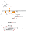
Schematic overview of the results from the present study indicating WNT-5A secretion from astrocytes and the WNT-5A-induced signaling pathway towards ERK1/2 regulating distinct aspects of the proinflammatory transformation of microglia. Pharmacological inhibitors used in the study: D4476, pertussis toxin (PTX), M119, BAPTA-AM, U73122, bisindolaleiimide (BIS), SL327. Abbreviations: AC, adenylyl cyclase; cAMP, cyclic AMP; DAG, diacylglycerol; DVL, disheveled; ERK1/2, extracellular signal-regulated kinases 1/2; FZD, Frizzled; IP3, inositoltriphosphate; MEK1/2, MAPK/ERK kinase 1/2; MMP, matrix metalloprotease; PLC, phospholipase C; PKC, Ca2+-dependent protein kinase; Gi, heterotrimeric G proteins of the Gi/o family; WNT, Wingless/int-1.

Similar articles

Cited by

References

    1. van Amerongen R, Nusse R. Towards an integrated view of Wnt signaling in development. Development. 2009;136:3205–3214. doi: 10.1242/dev.033910. - DOI - PubMed
    1. Nusse R. Wnts and Hedgehogs: lipid-modified proteins and similarities in signaling mechanisms at the cell surface. Development. 2003;130:5297–5305. doi: 10.1242/dev.00821. - DOI - PubMed
    1. Kikuchi A, Yamamoto H, Sato A, Matsumoto S. Wnt5a: its signalling, functions and implication in diseases. Acta Physiol (Oxf) 2012;204:17–33. doi: 10.1111/j.1748-1716.2011.02294.x. - DOI - PubMed
    1. Freese JL, Pino D, Pleasure SJ. Wnt signaling in development and disease. Neurobiol Dis. 2010;38:148–153. doi: 10.1016/j.nbd.2009.09.003. - DOI - PMC - PubMed
    1. Malaterre J, Ramsay RG, Mantamadiotis T. Wnt-Frizzled signalling and the many paths to neural development and adult brain homeostasis. Front Biosci. 2007;12:492–506. doi: 10.2741/2077. - DOI - PubMed

Publication types