. 2012 Aug 15;446(1):113-23.
doi: 10.1042/BJ20120112.

The RNA-binding protein CUG-BP1 increases survivin expression in oesophageal cancer cells through enhanced mRNA stability

Affiliations

The RNA-binding protein CUG-BP1 increases survivin expression in oesophageal cancer cells through enhanced mRNA stability

Elizabeth T Chang et al. Biochem J. .

Abstract

Survivin, a member of the IAP (inhibitor of apoptosis protein) family, plays important roles in maintaining cellular homoeostasis and regulating cell-cycle progression. This IAP is overexpressed in oesophageal cancer cells, leading to uncontrolled cell growth and resistance to apoptosis. CUG-BP1 (CUG-binding protein 1) is an RNA-binding protein that regulates the stability and translational efficiency of target mRNAs. In the present paper, we report that CUG-BP1 is overexpressed in oesophageal cancer cell lines and human oesophageal cancer specimens. CUG-BP1 associates with the 3'-untranslated region of survivin mRNA, thereby stabilizing the transcript and elevating its expression in oesophageal cancer cells. Our results show that overexpression of CUG-BP1 in oesophageal epithelial cells results in increased survivin mRNA stability and consequently survivin protein expression. Conversely, silencing CUG-BP1 in oesophageal cancer cells destabilizes survivin mRNA, lowering the level of survivin protein. In addition, we have found that altering CUG-BP1 expression modulates susceptibility to chemotherapy-induced apoptosis. Overexpression of CUG-BP1 in oesophageal epithelial cells increases resistance to apoptosis, whereas silencing CUG-BP1 makes oesophageal cancer cells more susceptible to chemotherapy-induced apoptosis. Co-transfection experiments with small interfering RNA directed against survivin suggest that the anti-apoptotic role for CUG-BP1 is not entirely dependent on its effect on survivin expression.


Figures

Figure 1
Figure 1
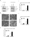
A: A representative esophagectomy specimen containing both normal and malignant tissue was examined after H&E staining and IHC staining for CUG-BP1 and survivin. a. Representative photomicrograph (100×) of H&E stained benign squamous mucosa (top) and malignant and invasive glandular tissue (bottom). b. IHC staining for CUB-GP1 (100×). The benign squamous mucosa shows expression in the basal zone (small arrowheads), with loss of expression at the luminal surface (large arrowhead). The adjacent malignant glandular tissue shows diffuse staining (arrow) of CUG-BP1. c. IHC staining for survivin (100×). The benign squamous mucosa shows marked staining for survivin in the basal zone (small arrowheads), which is reduced at the luminal surface (large arrowhead). The adjacent malignant glandular tissue shows strong diffuse staining for survivin (arrow). B: Baseline levels of CUG-BP1 and survivin protein in nhESO cells, and two esophageal cancer cell lines, TE7 and TE10. Twenty-five micrograms of total protein were loaded into each lane, and immunoblots were probed with either a CUG-BP1 or survivin-specific antibody. Actin hybridization was performed as an internal control to ensure equal loading. The approximate sizes of CUG-BP1, survivin, and actin are 60kDa, 17kDa and 42kDa, respectively. C: Western blot analysis from (B) was quantified by densitometry and graphed relative to TE10 cells. *p < 0.05 compared with nhESO.
Figure 2
Figure 2
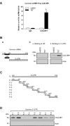
A: Levels of association of endogenous CUG-BP1 with survivin mRNA in nhESO and TE7 cell lines. Whole cell lysates from nhESO and TE7 cells were used for immunoprecipitation (IP) in the presence of anti-CUG-BP1 antibody, or non-specific IgG. RNA in the IP materials was used in qRT-PCR reactions to detect the presence of survivin mRNA. *p < 0.05 compared nhESO cells immunoprecipitated with CUG-BP1. B: a. Schematic representation of survivin mRNA coding region (CR) and 3′ untranslated region (UTR). b. Binding of CUG-BP1 to the CR (a) and 3′ UTR (b) of survivin mRNA in biotin pull-down experiments. Cytoplasmic lysates from nhESO and TE7 cell lines were isolated and incubated with 6μg of biotinylated survivin CR or 3′-UTR. Streptavidin-coated beads were used to pull-down the resultant RNP complexes. The presence of CUG-BP1 in the pull-down material was assayed by Western blotting. Actin in the pull-down material was also examined and served as a negative control. c. Levels of input CUG-BP1 and actin proteins in each cell line are shown by Western blot. C: Schematic representation of the fragmented survivin 3′-UTR biotinylated transcripts used in this series of experiments. D: Representative CUG-BP1 immunoblots of the material pulled down by the different biotinylated fragments of the survivin mRNA 3′-UTR (F-1 to F-8), using the same methods as described in (B).
Figure 3
Figure 3
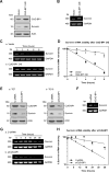
Changes in survivin mRNA stability following modulation of CUG-BP1. A: Representative immunoblots of CUG-BP1 and survivin after overexpression of CUG-BP1 in nhESO cells. Following transfection with control vector or a CUG-BP1 overexpressing vector whole cell lysates were harvested for Western blot analysis. Actin was used as a loading control. B: Levels of survivin mRNA in cells that were processed as described in (A). Total cellular RNA was extracted and levels of survivin mRNA were measured by RT-PCR. GAPDH was concurrently amplified to serve as a control for equal loading. Survivin and GAPDH mRNA sizes are as indicated on the left. C: Half-life of survivin mRNA in cells described in (A). Total cellular RNA was isolated at indicated times after administration of actinomycin D (5μg/μl), and the remaining levels of survivin and GAPDH mRNA were measured by qRT-PCR analysis. D: Percentage of survivin mRNA remaining in cells that were processed as described in (C). Relative levels of survivin mRNA were normalized to the amount of GAPDH mRNA (optical density of survivin mRNA/optical density of GAPDH mRNA). *p < 0.001 compared with nhESO cells transfected with vector control. E: Representative immunoblots of CUG-BP1 and survivin protein in CUG-BP1-silenced cells. After TE7 and TE10 cells were transfected with either siRNA targeting CUG-BP1 (siCUG-BP1) or C-siRNA for 48 hours, whole cell lysates were isolated for Western blot analysis. Actin was used as an internal loading control. CDK4 levels were also determined after silencing CUG-BP1 in TE7 cells. F: Levels of survivin mRNA in TE7 cells that were processed as described in (E). G: Half-life of survivin mRNA in TE7 cells described in (E), following procedure described in (C). H: Percentage of survivin mRNA remaining in TE7 cells that were processed as described in (G). *p < 0.001 compared with TE7 cells transfected with C-siRNA.
Figure 4
Figure 4
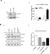
Silencing CUG-BP1 in esophageal cancer cells increases association of survivin mRNA with P-body components. A: a. Western blots confirming the successful silencing of CUG-BP1 by siRNA in TE7 cells. b. Levels of association of endogenous Ago2 with survivin mRNA in CUG-BP1-silenced TE7 cell lines. Whole cell lysates from TE7 cells treated with either C-siRNA or siCUG-BP1 were used for immunoprecipitation (IP) in the presence of anti-Ago2 antibody, or non-specific IgG. RNA in the IP materials was used in qRT-PCR reactions to detect the presence of survivin mRNA. B: Effect on survivin protein level after silencing both CUG-BP1 and Lsm4 in TE7 cells. a. Western blots confirming the silencing of CUG-BP1 and Lsm4, as well as the change in survivin protein levels. b. Densitometry analysis of survivin protein levels. *p < 0.05 compared with TE7 cells transfected with siLsm4 alone or in combination with siCUG-BP1.
Figure 5
Figure 5
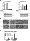
Overexpression of CUG-BP1 enhances resistance to apoptosis in nhESO cells. A: nhESO cells were transfected with either control vector (Vector) or the CUG-BP1 cDNA (CUG-BP1 O/E). Following transfection, cells were either not induced or exposed to camptothecin (1μM) for 6 hours and whole cell lysates were obtained. a. Western blot analysis of un-induced (Panel a) or camptothecin-induced (Panel b) levels of CUG-BP1, procaspase-3, caspase-3, and actin. b. Densitometry analysis quantifying caspase-3 protein levels relative to levels in un-induced samples. *p < 0.0001 compared to vector control. B: Apoptosis was measured in transfected cells as described in (A). a. Representative morphological analysis of treated cells. b. Percentage of apoptotic cells. *p < 0.01 compared to vector control. C: Early apoptosis was detected by caspase-3 colorimetric assay in cells prepared as described in (A). Whole cell lysates were collected and lysed by Lysis Buffer, then incubated with appropriate reaction buffers, and the caspase-3 colorimetric substrate (DEVD-pNA) at 37°C. Enzymatic activities for each condition were then read and recorded on a microplate reader using a 405 nm wavelength light after 1 hour. *p < 0.05 compared to vector control.
Figure 6
Figure 6
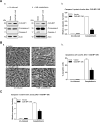
Down-regulation of CUG-BP1 in esophageal cancer cells sensitizes cells to apoptosis. A: a. Representative immunoblots of CUG-BP1, procaspase-3, caspase-3, and actin in CUG-BP1-silenced TE7 cells following no induction (Panel a) or exposure to camptothecin (10μM) for 6 hours (Panel b). b. Densitometry analysis of caspase-3 protein levels. *p < 0.0001 compared to C-siRNA. B: a. Representative morphologic analysis of treated cells. Cells were prepared as described in (A). b. Percentage of apoptotic cells. *p < 0.01 compared to C-siRNA. C: Caspase-3 colorimetric assay was performed as described in Figure 5D. *p < 0.05 compared to C-siRNA.
Figure 7
Figure 7
Overexpression of CUG-BP1 in esophageal cancer cells abrogates increased sensitivity to apoptosis following down-regulation of survivin. A: Representative immunoblots and densitometry analyses of survivin and CUG-BP1 after the co-transfection of siSurvivin plus CUG-BP1 cDNA in TE7 cells. B: Representative morphologic analyses of un-induced (top panels) and camptothecin-induced (bottom panels) TE7 cells after a. C-siRNA + vector, b. C-siRNA + CUG-BP1 O/E, c. siSurvivin + vector, and d. siSurvivin + CUG-BP1 O/E. Cells were transfected with appropriate siRNA and cDNA for 48 hours and then exposed to camptothecin for 6 hours. C: Percentage of apoptotic cells after co-transfection. Cells were prepared as described in (B) and apoptotic cells were counted and graphed as a percentage of total cells. *p < 0.05 compared to C-siRNA + CUG-BP1 O/E and siSurvivin + CUG-BP1 O/E; +p < 0.001 compared to C-siRNA + CUG-BP1 O/E and siSurvivin + CUG-BP1 O/E.

Similar articles

Cited by

References

    1. Siegel R, Ward E, Brawley O, Jemal A. Cancer statistics, 2011: The impact of eliminating socioeconomic and racial disparities on premature cancer deaths. CA Cancer J Clin. 2011;61:212–236. - PubMed
    1. Chang E, Donahue J, Smith A, Hornick J, Rao JN, Wang JY, Battafarano RJ. Loss of p53, rather than beta-catenin overexpression, induces survivin-mediated resistance to apoptosis in an esophageal cancer cell line. J Thorac Cardiovasc Surg. 2010;140:225–232. - PubMed
    1. Li F, Ambrosini G, Chu EY, Plescia J, Tognin S, Marchisio PC, Altieri DC. Control of apoptosis and mitotic spindle checkpoint by survivin. Nature. 1998;396:580–584. - PubMed
    1. Grabowski P, Kuhnel T, Muhr-Wilkenshoff F, Heine B, Stein H, Hopfner M, Germer CT, Scherubl H. Prognostic value of nuclear survivin expression in oesophageal squamous cell carcinoma. Br J Cancer. 2003;88:115–119. - PMC - PubMed
    1. Ikeguchi M, Yamaguchi K, Kaibara N. Survivin gene expression positively correlates with proliferative activity of cancer cells in esophageal cancer. Tumour Biol. 2003;24:40–45. - PubMed

Publication types

MeSH terms