. 2012 May 23;32(21):7178-90.
doi: 10.1523/JNEUROSCI.0008-12.2012.

Identification of two functionally distinct endosomal recycling pathways for dopamine D₂ receptor

Affiliations

Identification of two functionally distinct endosomal recycling pathways for dopamine D₂ receptor

Yun Li et al. J Neurosci. .

Abstract

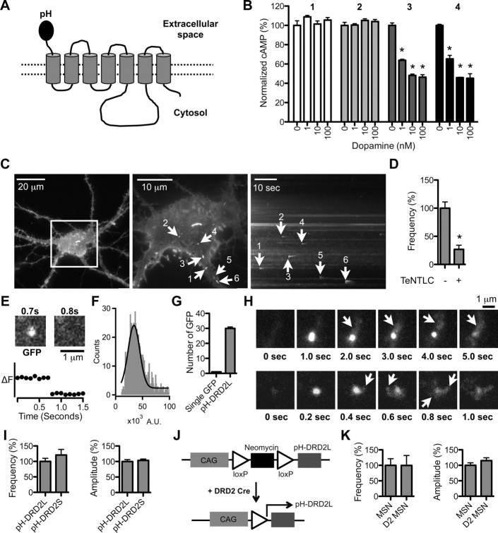
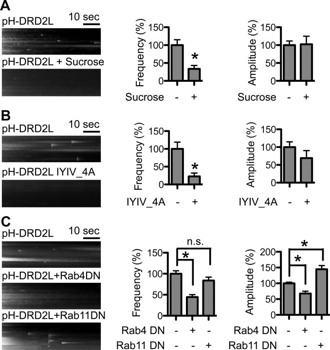
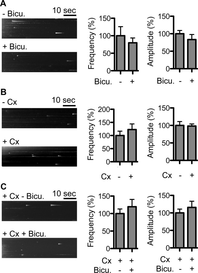
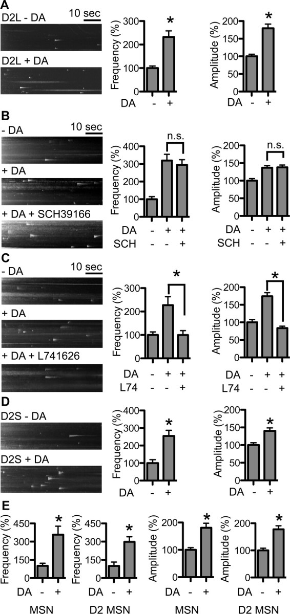
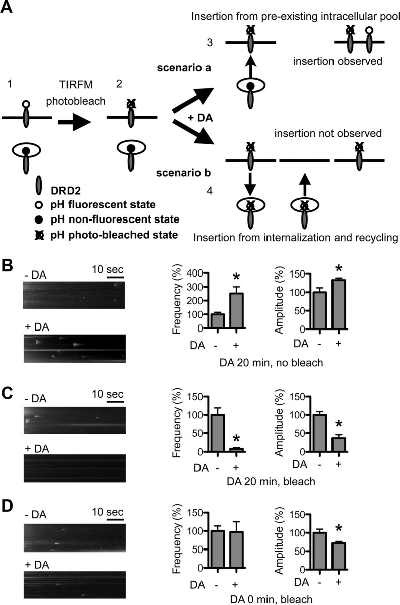
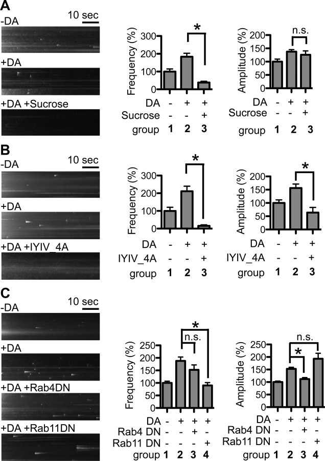
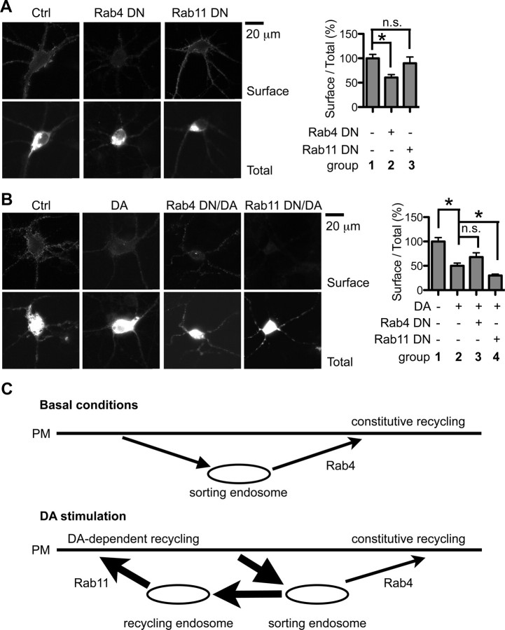
Dopamine D₂ receptor (DRD2) is important for normal function of the brain reward circuit. Lower DRD2 function in the brain increases the risk for substance abuse, obesity, attention deficit/hyperactivity disorder, and depression. Moreover, DRD2 is the target of most antipsychotics currently in use. It is well known that dopamine-induced DRD2 endocytosis is important for its desensitization. However, it remains controversial whether DRD2 is recycled back to the plasma membrane or targeted for degradation following dopamine stimulation. Here, we used total internal reflection fluorescent microscopy (TIRFM) to image DRD2 with a superecliptic pHluorin tagged to its N terminus. With these technical advances, we were able to directly visualize vesicular insertion events of DRD2 in cultured mouse striatal medium spiny neurons. We showed that insertion of DRD2 occurs on neuronal somatic and dendritic surfaces. Lateral diffusion of DRD2 was observed following its insertion. Most importantly, using our new approach, we uncovered two functionally distinct recycling pathways for DRD2: a constitutive recycling pathway and a dopamine activity-dependent recycling pathway. We further demonstrated that Rab4 plays an important role in constitutive DRD2 recycling, while Rab11 is required for dopamine activity-dependent DRD2 recycling. Finally, we demonstrated that the two DRD2 recycling pathways play distinct roles in determining DRD2 function: the Rab4-sensitive constitutive DRD2 recycling pathway determines steady-state surface expression levels of DRD2, whereas the Rab11-sensitive dopamine activity-dependent DRD2 recycling pathway is important for functional resensitization of DRD2. Our findings underscore the significance of endosomal recycling in regulation of DRD2 function.


Figures

Figure 1.
Figure 1.
Visualization of pH-DRD2 insertion in cultured MSNs. A, Schematic drawing of the pH-DRD2 construct. B, cAMP accumulation assay to determine DRD2 function in HEK 293 cells: untransfected cells (1), vector-transfected cells (2), DRD2 (nontagged) transfected cells (3), and pH-DRD2 transfected cells (4). Activation of DRD2 inhibits cAMP accumulation. Untransfected and vector-transfected cells do not respond to DA stimulation, while DRD2 (nontagged) and pH-DRD2 display similar sensitivities to DA at three different concentrations: 1, 10, and 100 nm. C, Left, pH-DRD2L in a cultured MSN visualized under epifluorescent mode using a 40×, 1.30 NA objective. The white square in the middle represents the field of view under TIRF imaging mode using a 100×, 1.46 NA objective, as seen in the middle. Middle, Maximum-intensity projection image of pH-DRD2L fluorescence of the same cultured MSN visualized under TIRF imaging mode; white spots with arrowheads represent individual pH-DRD2L insertion events as visualized under TIRFM. Right, y–t maximum-intensity projection image of pH-DRD2L fluorescence in the same cultured MSN visualized under TIRF imaging mode; arrowheads indicate individual pH-DRD2 insertion events as indicated in the middle panel. D, Quantification of pH-DRD2L insertion frequency with or without TeNTLC. E, The two images show a single GFP fluorescent spot before (left) and after (right) the single-step photobleaching; the plot underneath the images shows fluorescence changes over time at this fluorescent spot. Single-step photobleaching occurs between 0.7 and 0.8 s. F, Fitting the primary peak of single GFP fluorescence intensity to a Gaussian function results in a Gaussian profile with the center of the Gaussian function representing a single GFP fluorescence level. G, By normalizing the average total fluorescence level of pH-DRD2 to single GFP fluorescence, the number of pH-DRD2L molecules in individual insertion events can be derived. H, Two representative insertion events of pH-DRD2 followed by lateral diffusion. The arrows indicate diffusion of pH-DRD2L from the initial insertion site. I, Comparison of pH-DRD2L and pH-DRD2S in MSNs. J, Experimental design for examination of pH-DRD2L insertion properties in D2-type MSNs. In pCALNL-pH-DRD2L, the translation of pH-DRD2L is prevented by a floxed neomycin cassette. Transfecting this pCALNL-pH-DRD2L construct into MSNs cultured from DRD2 Cre embryos will result in specific expression of pH-DRD2L in D2-type MSNs. K, Comparison of pH-DRD2L insertion between MSNs and D2-type MSNs. MSN, pH-DRD2L insertion without discrimination of D1-type versus D2-type MSNs, using pRK5-pH-DRD2L construct; D2 MSN, pH-DRD2L insertion in D2-type MSNs using pCALNL-pH-DRD2L construct. Asterisks denote statistical significance compared to the control group.
Figure 2.
Figure 2.
Observed DRD2 insertion represents constitutive DRD2 recycling. Left, y–t maximum-intensity projection images of pH-DRD2 insertion events in each experimental group. A, Hypertonic sucrose inhibits DRD2 insertion. −Sucrose, Without hypertonic sucrose treatment; +Sucrose: with hypertonic sucrose treatment. B, Arrestin-binding mutant of DRD2 displays reduced insertion. −IYIV_4A, Wild-type pH-DRD2L; + IYIV_4A, pH-DRD2L mutant with arrestin-binding site abolished. C, Rab4 regulates constitutive pH-DRD2L recycling. pH-DRD2L, Control group; pH-DRD2L+Rab4DN, coexpression of pH-DRD2L with dominant-negative mutant of Rab4; pH-DRD2L+Rab11DN, coexpression of pH-DRD2L with dominant-negative mutant of Rab11. Asterisks denote statistical significance compared to the control group. n.s., Not significant.
Figure 3.
Figure 3.
Glutamatergic or GABAergic activity does not affect pH-DRD2 insertion. Left, y–t maximum-intensity projection images of pH-DRD2 insertion events in each experimental group. A, Inhibition of GABAergic activity does not affect pH-DRD2 insertion in MSN. Bicu., 20 μm bicuculline; −Bicu., pH-DRD2 insertion in the absence of bicuculline; +Bicu., pH-DRD2 insertion in the presence of bicuculline. B, Coculture of cortical neurons with MSN does not affect pH-DRD2 insertion. C, Inhibition of GABAergic activity in coculture of MSN and Cx does not affect pH-DRD2 insertion.
Figure 4.
Figure 4.
DA activity-dependent pH-DRD2 insertion. Left, y–t maximum-intensity projection images of pH-DRD2 insertion events in each experimental group. A, Twenty minute treatment with 1 μm DA enhances the frequency and amplitude of pH-DRD2 insertion in MSN. B, Dopamine D1 receptor antagonist SCH does not affect DA activity-dependent pH-DRD2 insertion. C, DRD2 antagonist L74 abolishes DA activity-dependent pH-DRD2 insertion. D, pH-DRD2S also showed DA activity-dependent insertion. E, Comparison of DA activity-dependent pH-DRD2L insertion in MSNs versus D2-type MSNs. MSN, pH-DRD2L insertion in MSNs without discrimination of D1- versus D2-type MSNs; D2 MSNs, pH-DRD2L insertion in DRD2 Cre-labeled D2-type MSNs. −DA, pH-DRD2 insertion in the absence of DA stimulation; +DA, pH-DRD2 insertion following DA stimulation; +DA +SCH39166, pH-DRD2L with DA stimulation in the presence of DRD1 antagonist SCH39166; +DA +L741626, pH-DRD2L with DA stimulation in the presence of DRD2 antagonist L741626. Asterisks denote statistical significance compared to the control group. n.s., Not significant.
Figure 5.
Figure 5.
DA activity facilitates recycling of internalized DRD2L. A, Rationale and design for the photobleaching experiments: (1) Before photobleaching, pH-DRD2L on the PM is fluorescent, and pH-DRD2L in intracellular pools is nonfluorescent. (2) Photobleach is performed in TIRFM mode; consequently, pH-DRD2L on the PM is photobleached, but due to the nonfluorescent nature of intracellular pH-DRD2L and photobleach in TIRFM mode, pH-DRD2L in intracellular pools is protected from photobleach. (3) Scenario a, If DA stimulation enhances the insertion of pH-DRD2L that originated from intracellular pools, then DA stimulation should still be able to increase pH-DRD2L insertion after photobleaching, as intracellular pools of pH-DRD2L would be protected from photobleach. (4) Scenario b, If DA stimulation enhances the recycling of internalized pH-DRD2L, then photobleaching should result in a photobleached pHluorin attached to the recycled DRD2L; hence, insertion of DRD2L should be undetectable to our imaging system, and we would expect to observe a significant reduction in DRD2L insertion. B, Without photobleaching, 20 min of DA stimulation consistently enhances the frequency and amplitude of pH-DRD2L insertion. C, Following photobleaching, the same protocol of 20 min of DA stimulation almost completely abolishes pH-DRD2L insertion, as predicted by scenario b, suggesting that following DA stimulation, the observed pH-DRD2L insertion originates primarily from recycling of endocytosed DRD2L. D, Following photobleaching, and immediately after the onset of DA stimulation, pH-DRD2L insertion is clearly visible but the frequency is not increased, and the amplitude is slightly reduced. Asterisks denote statistical significance compared to the control group.
Figure 6.
Figure 6.
DA activity-dependent increase in DRD2L insertion depends on DRD2L endocytosis and Rab11-dependent recycling endosomes. A, Hypertonic sucrose abolishes DA activity-dependent DRD2L recycling. B, Arrestin-binding mutant of DRD2 abolishes DA activity-dependent DRD2 recycling. C, DA stimulation enhances DRD2L recycling through Rab11-dependent recycling endosomes. −DA, pH-DRD2L without DA stimulation; +DA, pH-DRD2L with DA stimulation; +DA +Sucrose, pH-DRD2L with DA stimulation in the presence of hypertonic sucrose; +DA +IYIV_4A, pH-DRD2L IYIV_4A mutant with DA stimulation; +DA +Rab4DN, pH-DRD2L with DA stimulation in the presence of dominant-negative mutant of Rab4; +DA +Rab11DN, pH-DRD2L with DA stimulation in the presence of dominant-negative mutant of Rab11. Asterisks denote statistical significance compared to the control group. n.s., Not significant.
Figure 7.
Figure 7.
Rab4 and Rab11 differentially regulate DRD2L function. A, Rab4DN reduces steady-state surface expression of DRD2L. Surface, Surface staining of pH-DRD2L; Total, total level of pH-DRD2L; group 1, control; group 2, Rab4DN; group 3, Rab11DN. B, Rab11DN inhibits DRD2L re-sensitization following DA stimulation. Group 1, Control; group 2, control plus DA; group 3, Rab4DN plus DA; group 4, Rab11DN plus DA. C, A model for the roles of Rab4 and Rab11 in regulation of DRD2L recycling. Under basal conditions, internalized DRD2L constitutively recycles from sorting endosomes to the PM via the Rab4-dependent route. Rab11 contributes little to constitutive DRD2L recycling. In contrast, when stimulated with DA, the Rab4-dependent fast recycling remains fully functional, but DRD2L recycling and functional resensitization occur primarily via the Rab11-dependent recycling pathway. Asterisks denote statistical significance compared to the control group. n.s., Not significant.

Similar articles

Cited by

References

    1. Bartlett SE, Enquist J, Hopf FW, Lee JH, Gladher F, Kharazia V, Waldhoer M, Mailliard WS, Armstrong R, Bonci A, Whistler JL. Dopamine responsiveness is regulated by targeted sorting of D2 receptors. Proc Natl Acad Sci U S A. 2005;102:11521–11526. - PMC - PubMed
    1. Binda AV, Kabbani N, Lin R, Levenson R. D2 and D3 dopamine receptor cell surface localization mediated by interaction with protein 4.1N. Mol Pharmacol. 2002;62:507–513. - PubMed
    1. Blum K, Sheridan PJ, Wood RC, Braverman ER, Chen TJ, Cull JG, Comings DE. The D2 dopamine receptor gene as a determinant of reward deficiency syndrome. J R Soc Med. 1996;89:396–400. - PMC - PubMed
    1. Bunzow JR, Van Tol HH, Grandy DK, Albert P, Salon J, Christie M, Machida CA, Neve KA, Civelli O. Cloning and expression of a rat D2 dopamine receptor cDNA. Nature. 1988;336:783–787. - PubMed
    1. Carlsson A. The current status of the dopamine hypothesis of schizophrenia. Neuropsychopharmacology. 1988;1:179–186. - PubMed

Publication types

MeSH terms

LinkOut - more resources