

Interleukin-8 and its receptor CXCR2 in the tumour microenvironment promote colon cancer growth, progression and metastasis
- PMID: 22617157
- PMCID: PMC3364111
- DOI: 10.1038/bjc.2012.177
Interleukin-8 and its receptor CXCR2 in the tumour microenvironment promote colon cancer growth, progression and metastasis
Abstract
Background: Colorectal cancer (CRC) is a leading cause of death in the United States. Increased level of interleukin-8 (IL-8) and CXCR2 on tumours and in the tumour microenvironment has been associated with CRC growth, progression and recurrence in patients. Here, we aimed to evaluate the effects of tissue microenvironment-encoded IL-8 and CXCR2 on colon cancer progression and metastasis.
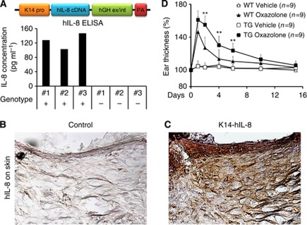
Methods: A novel immunodeficient, skin-specific IL-8-expressing transgenic model was generated to evaluate colon cancer growth and metastasis. Syngeneic mouse colon cancer cells were grafted in CXCR2 knockout (KO) mice to study the contribution of CXCR2 in the microenvironment to cancer growth.
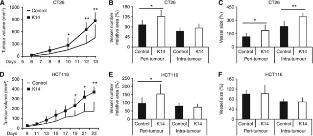
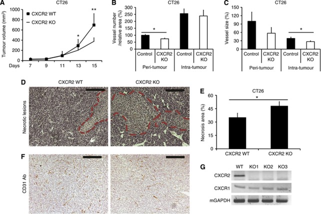
Results: Elevated levels of IL-8 in the serum and tumour microenvironment profoundly enhanced the growth of human and mouse colon cancer cells with increased peri-tumoural angiogenesis, and also promoted the extravasation of the cancer cells into the lung and liver. The tumour growth was inhibited in CXCR2 KO mice with significantly reduced tumour angiogenesis and increased tumour necrosis.
Conclusion: Increased expression of IL-8 in the tumour microenvironment enhanced colon cancer growth and metastasis. Moreover, the absence of its receptor CXCR2 in the tumour microenvironment prevented colon cancer cell growth. Together, our study demonstrates the critical roles of the tumour microenvironment-encoded IL-8/CXCR2 in colon cancer pathogenesis, validating the pathway as an important therapeutic target.
© 2012 Cancer Research UK
Conflict of interest statement
The authors declare no conflict of interest.
Figures


Similar articles
-
Expression of interleukin 8 and its receptors in human colon carcinoma cells with different metastatic potentials.Clin Cancer Res. 2001 Oct;7(10):3298-304. Clin Cancer Res. 2001. PMID: 11595728
-
The CXCR2 antagonist, SCH-527123, shows antitumor activity and sensitizes cells to oxaliplatin in preclinical colon cancer models.Mol Cancer Ther. 2012 Jun;11(6):1353-64. doi: 10.1158/1535-7163.MCT-11-0915. Epub 2012 Mar 5. Mol Cancer Ther. 2012. PMID: 22391039
-
GRO-α and IL-8 enhance ovarian cancer metastatic potential via the CXCR2-mediated TAK1/NFκB signaling cascade.Theranostics. 2018 Feb 2;8(5):1270-1285. doi: 10.7150/thno.22536. eCollection 2018. Theranostics. 2018. PMID: 29507619 Free PMC article.
-
Targeting IL-8 in colorectal cancer.Expert Opin Ther Targets. 2012 May;16(5):491-7. doi: 10.1517/14728222.2012.677440. Epub 2012 Apr 12. Expert Opin Ther Targets. 2012. PMID: 22494524 Review.
-
The CXCL8-CXCR1/2 pathways in cancer.Cytokine Growth Factor Rev. 2016 Oct;31:61-71. doi: 10.1016/j.cytogfr.2016.08.002. Epub 2016 Aug 25. Cytokine Growth Factor Rev. 2016. PMID: 27578214 Free PMC article. Review.
Cited by
-
The Role of Chemokines in Promoting Colorectal Cancer Invasion/Metastasis.Int J Mol Sci. 2016 Apr 28;17(5):643. doi: 10.3390/ijms17050643. Int J Mol Sci. 2016. PMID: 27136535 Free PMC article. Review.
-
Extracellular matrix gene expression profiling using microfluidics for colorectal carcinoma stratification.Biomicrofluidics. 2016 Oct 31;10(5):054124. doi: 10.1063/1.4966245. eCollection 2016 Sep. Biomicrofluidics. 2016. PMID: 27822332 Free PMC article.
-
Insights on CXC chemokine receptor 2 in breast cancer: An emerging target for oncotherapy.Oncol Lett. 2019 Dec;18(6):5699-5708. doi: 10.3892/ol.2019.10957. Epub 2019 Oct 3. Oncol Lett. 2019. PMID: 31788042 Free PMC article. Review.
-
A Review of Gut Microbiota-Derived Metabolites in Tumor Progression and Cancer Therapy.Adv Sci (Weinh). 2023 May;10(15):e2207366. doi: 10.1002/advs.202207366. Epub 2023 Mar 23. Adv Sci (Weinh). 2023. PMID: 36951547 Free PMC article. Review.
-
Astragalus mongholicus Bunge and Curcuma aromatica Salisb. inhibits liver metastasis of colon cancer by regulating EMT via the CXCL8/CXCR2 axis and PI3K/AKT/mTOR signaling pathway.Chin Med. 2022 Aug 3;17(1):91. doi: 10.1186/s13020-022-00641-4. Chin Med. 2022. PMID: 35922850 Free PMC article.
References
-
- Araki S, Omori Y, Lyn D, Singh RK, Meinbach DM, Sandman Y, Lokeshwar VB, Lokeshwar BL (2007) Interleukin-8 is a molecular determinant of androgen independence and progression in prostate cancer. Cancer Res 67(14): 6854–6862 - PubMed
-
- Baier PK, Eggstein S, Wolff-Vorbeck G, Baumgartner U, Hopt UT (2005) Chemokines in human colorectal carcinoma. Anticancer Res 25(5): 3581–3584 - PubMed
-
- Chapman RW, Minnicozzi M, Celly CS, Phillips JE, Kung TT, Hipkin RW, Fan X, Rindgen D, Deno G, Bond R, Gonsiorek W, Billah MM, Fine JS, Hey JA (2007) A novel, orally active CXCR1/2 receptor antagonist, Sch527123, inhibits neutrophil recruitment, mucus production, and goblet cell hyperplasia in animal models of pulmonary inflammation. J Pharmacol Exp Ther 322(2): 486–493 - PubMed
Publication types
MeSH terms
Substances
LinkOut - more resources
Full Text Sources
Other Literature Sources
Research Materials
