. 2012;8(5):e1002708.
doi: 10.1371/journal.ppat.1002708. Epub 2012 May 17.

Induction of GADD34 is necessary for dsRNA-dependent interferon-β production and participates in the control of Chikungunya virus infection

Affiliations

Induction of GADD34 is necessary for dsRNA-dependent interferon-β production and participates in the control of Chikungunya virus infection

Giovanna Clavarino et al. PLoS Pathog. 2012.

Abstract

Nucleic acid sensing by cells is a key feature of antiviral responses, which generally result in type-I Interferon production and tissue protection. However, detection of double-stranded RNAs in virus-infected cells promotes two concomitant and apparently conflicting events. The dsRNA-dependent protein kinase (PKR) phosphorylates translation initiation factor 2-alpha (eIF2α) and inhibits protein synthesis, whereas cytosolic DExD/H box RNA helicases induce expression of type I-IFN and other cytokines. We demonstrate that the phosphatase-1 cofactor, growth arrest and DNA damage-inducible protein 34 (GADD34/Ppp1r15a), an important component of the unfolded protein response (UPR), is absolutely required for type I-IFN and IL-6 production by mouse embryonic fibroblasts (MEFs) in response to dsRNA. GADD34 expression in MEFs is dependent on PKR activation, linking cytosolic microbial sensing with the ATF4 branch of the UPR. The importance of this link for anti-viral immunity is underlined by the extreme susceptibility of GADD34-deficient fibroblasts and neonate mice to Chikungunya virus infection.


Conflict of interest statement

The authors have declared that no competing interests exist.

Figures
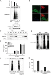
Figure 1
Figure 1. Translation inhibition and IFN-β production are induced by poly I:C in MEFs.
A) Protein synthesis was monitored in poly I:C(pI:C)-stimulated MEFs using puromycin labelling followed by immunoblot with the anti-puromycin mAb 12D10. Controls are cells not treated with puromycin (No puro) and cells treated with cycloheximide (chx) 5 min prior puromycin incorporation. β-actin immunoblot is shown for equal loading control. Quantification of puromycin signal was quantified with ImageJ software and is represented above the immunoblot. Phosphorylation of eIF2α (P-eIF2α) was assessed in the same MEFs extracts. B) Immunofluorescence staining for puromycin, P-eIF2α and dsRNA of MEFs treated with poly I:C for 4 h and labeled with puromycin for 1 h. Scale bar, 10 µm. C) WT and PKR−/− MEFs were stimulated for 8 h with poly I:C (pI:C), thapsigargin (th) or arsenite (as). PKR and P-eIF2α were detected by immunoblot. D) WT and PKR−/− MEFs were stimulated for 8 h with poly I:C and protein synthesis was monitored like in (A). β-actin immunoblot is shown for equal loading control. E) IFN-β levels were measured, by ELISA, in cell culture supernatants of WT, PKR−/−, eIF2αA/A and control eIF2αS/S MEFs after 4 and 8 h of poly I:C stimulation. Data are mean ± standard deviation of 3 independent experiments. F) Protein synthesis was measured in NIH3T3 cells by puromycin incorporation after 7 h of poly I:C treatment. Where indicated, a chase of 1 h with fresh media was performed prior to puromycin labeling and immunoblotting. Samples with cycloheximide (chx) and arsenite (as) added respectively 5 min and 30 min before the puromycin pulse are shown as controls. G) IFN-β was quantified by ELISA in culture supernatants in the conditions described above after 7 h of poly I:C stimulation or 7 h of poly I:C stimulation followed by 1 h with fresh media (chase). Data are mean ± standard deviation of 4 independent experiments.
Figure 2
Figure 2. PKR is required for ATF4 and GADD34 expression in response to poly I:C.
A) WT and PKR−/− MEFs were stimulated for 8 h with poly I:C (pI:C), or the UPR-inducing drug, thapsigargin (th) for 6 h. ATF4 protein expression was detected by immunoblot on nuclear extracts. Nuclear HDAC1 immunoblot is shown for equal loading control. * indicates unspecific band. B) GADD34 mRNA levels were quantified by qPCR after 6 h of poly I:C (pI:C) treatment in WT and PKR−/− MEFs. For the same cell extracts, immunoblots of GADD34 (middle panel), PKR and P-eIF2α (right panel) were performed. C) The same analysis was performed in eIF2α A/A and control eIF2α S/S MEFs. Treatment with thapsigargin (th), for 6 h was used as control to induce GADD34 and P-eIF2α. eIF2α and β-actin immunoblots are shown for equal loading control. Quantitative PCR data are the mean ± standard deviation of 3 independent experiments.
Figure 3
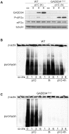
Figure 3. GADD34 mediates eIF2α dephosphorylation but not global translation recovery in response to poly I:C.
A) After treatment with poly I:C, protein extracts of WT and GADD34ΔC/ΔC MEFs were immunobloted for GADD34 and P-eIF2α. B) Protein synthesis was analyzed in WT cells treated for 1 to 6 hours with poly I:C (pI:C) alone or together with thapsigargin (th). Controls are cells not treated with puromycin (co) and cells treated with cycloheximide (chx) 5 min before puromycin incorporation. C) Protein synthesis was analyzed in GADD34ΔC/ΔC cells treated for 1 to 6 hours with poly I:C (pI:C) alone or together with thapsigargin (th). Tubulin or β-actin immunoblot are shown for equal loading control. In GADD34ΔC/ΔC cells translation is strongly impacted by thapsigargin, but not poly I:C.
Figure 4
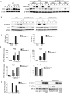
Figure 4. GADD34 is required for cytokine production in poly I:C-stimulated MEFs.
A) Left Panel, immunoblot for cystatin C after treatment or not with brefeldin A (BFA) in poly I:C-stimulated WT MEFs. Arrow indicates N-glycosylated-Cystatin C. Right panel, WT and GADD34ΔC/ΔC MEFs were treated with poly I:C (pI:C), thapsigargin (th) or tunicamycin (tun) for the indicated times. Levels of GADD34 and Cystatin C (CysC) were examined by immunoblot. β-actin immunoblot is shown as equal loading control. B) Immunoblots for GADD34 and PKR in WT and GADD34-inactivated cells treated with poly I:C for the indicated periods of time. The UPR-inducing drugs, Thapsigargin (th) and tunicamycin (tun), were used as controls to induce GADD34. Immunoblot of tubulin is shown as equal loading control. C) Amount of IFN-β (left panel) and IL-6 (right panel) in cell culture supernatants of WT and GADD34ΔC/ΔC MEFs after 6 h of poly I:C stimulation. Mock are samples treated with lipofectamine alone. Data are mean ± standard deviation of five (IFN-β) and three (IL-6) independent experiments. D) Transcription of IFN-β, IL-6, PKR and Cystatin C was analyzed by qPCR in samples of WT and GADD34ΔC/ΔC MEFs treated with poly I:C (pI:C). Mock represent samples treated with lipofectamine alone. E) WT and GADD34ΔC/ΔC MEFs were transfected overnight with an expression plasmid carrying the murine GADD34 (G34) cDNA and then treated with poly I:C for 6 h. IFN-β production was quantified by ELISA, left panel, in cell culture supernatants and plotted as a ratio of IFN-β to total cell proteins to compensate for different cell mortality levels induced by the transfection. In the right panel immunoblots for GADD34 and P-eIF2α in the same experimental conditions are shown. One representative analysis of 3 independent experiments is shown.
Figure 5
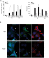
Figure 5. PKR is required to control CHIKV infection and IFN-β production in MEFs.
A) WT and PKR−/− MEFs were infected with CHIKV-GFP at an MOI of 10 or 50, for 24 h and 48 h. The amount of infected cells was determined by GFP expression, left panel. Interferon β present in the cell culture supernatants was measured by ELISA, right panel. Data represented are mean ± standard deviation from 3 experiments. B) WT (top panel) and PKR−/− MEFs (bottom panel) were infected for 24 h with CHIKV-GFP, then labeled with puromycin for 1 h prior fixation. GFP-CHIKV positive (green) were visualized by confocal microscopy after staining with specific antibodies for puromycin (cyan) and phospho-eIF2α (red). Cell Nuclei are stained with Hoechst 33258 (blue). Infection by CHIKV inhibits protein synthesis (visualized by puromycin incorporation) in WT, but not in PKR-deficient cells (arrows). In WT MEFs, eIF2α phosphorylation levels correlate with translation inhibition, although variability is observed among different infected cells, presumably due to GADD34 activity and time of infection. Non-infected WT and PKR−/− cells serve as a reference for normal translation activity and are indicated by an arrowhead. Scale bar 10 µm.
Figure 6
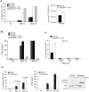
Figure 6. CHIKV infection and IFN-β production are controlled by GADD34 in MEFs.
A) WT and GADD34ΔC/ΔC cells were infected with CHIKV-GFP for a period of 24 and 48 h. The percentage of infected, GFP positive, cells and viral titers were analyzed. B) Levels of IFN-β in cell culture supernatants of WT and GADD34ΔC/ΔC MEFs infected with CHIKV-GFP for 24 h and 48 h. C) Murine IFN-β was added 3 h before infection of WT and GADD34ΔC/ΔC MEFs with CHIKV-GFP (10 MOI). Productive infection was estimated by GFP expression 24 h after CHIKV exposure. D) Cells were treated with guanabenz 3 h before and during infection (24 h) with CHIKV-GFP, percentage of infected cells (left) and corresponding IFN-β production (middle), are shown. Immunoblot of GADD34 and P-eIF2α are shown on the right. NI stands for non-infected. Percentage of infected cells, viral titers and IFN-β measurements data represent mean ± standard deviation of 3 experiments. p values shown in (D) were obtained applying a Student's t test.
Figure 7
Figure 7. CHIKV infection in mouse neonates.
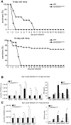
A) Kaplan–Meier plots representing the survival of FVB (WT) and GADD34ΔC/ΔC mouse neonates 9-day-old (n = 11 per group) (upper panel) or 12-day-old (n = 14 per group) (lower panel) after intradermal inoculation with 106 PFU of CHIKV and observed for 21 days. B) Left panel, viral titers in different tissues and serum of 12-day-old mice inoculated with 106 PFU of CHIKV via the intradermal route. Mice were sacrificed 5 days after infection and the amount of infectious virus in serum and tissues quantified by TCID50 (see methods) (n = 5). In addition of considerably increased levels of viral replication in CHIKV target tissues, GADD34ΔC/ΔC neonates also display signs of heart infection. Right panel, Quantification of IFN-β for the same different tissues, CHIKV-infected target tissues of GADD34ΔC/ΔC mice produced less IFN-β than WT. C) 17-day-old mice were infected with 106 PFU of CHIKV via the intradermal route, and sacrificed 72 h later. Quantification of viral titers and IFN-β/viral titers ratio is presented for different tissues. A broken line indicates the detection threshold. In B and C represented data are arithmetic mean ± standard deviation, n = 5. In B and C p values were calculated using a Student's t test, *p≤0.1, **p≤0.05.
Figure 8
Figure 8. CHIKV infection causes severe myocarditis in mouse neonates.
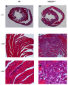
Histological appearance of horizontal sections of the heart through left and right ventricles of 12-day FVB (A, C and E) and GADD34ΔC/ΔC mice at D5 pi (B, D and F). Normal appearance of heart of FVB infected mice, at low magnification (A, ×10) with normal cardiomyocytes (C, ×100) and exceptional small foci of lymphocytes (E, ×400). Numerous foci of necrosis in the heart of GADD34ΔC/ΔC infected mice, at low magnification (B, ×10) and extensive through the ventricular wall (D, ×100). Higher magnification shows few residual cardiomyocytes (arrow head) and inflammation mainly composed of monocytes as well as extensive deposition of calcium (F, ×400). The mice were inoculated with 106 PFU of CHIKV via the intradermal route.

Similar articles

Cited by

References

    1. Kawai T, Akira S. Innate immune recognition of viral infection. Nat Immunol. 2006;7:131–137. - PubMed
    1. Kawai T, Akira S. The roles of TLRs, RLRs and NLRs in pathogen recognition. Int Immunol. 2009;21:317–337. - PMC - PubMed
    1. Akira S, Uematsu S, Takeuchi O. Pathogen recognition and innate immunity. Cell. 2006;124:783–801. - PubMed
    1. Garcia MA, Meurs EF, Esteban M. The dsRNA protein kinase PKR: virus and cell control. Biochimie. 2007;89:799–811. - PubMed
    1. Malathi K, Dong B, Gale M, Jr, Silverman RH. Small self-RNA generated by RNase L amplifies antiviral innate immunity. Nature. 2007;448:816–819. - PMC - PubMed

Publication types

MeSH terms