. 2012 Jul 19;120(3):626-35.
doi: 10.1182/blood-2012-02-409235. Epub 2012 May 21.

The origin and function of platelet glycosyltransferases

Affiliations

The origin and function of platelet glycosyltransferases

Hans H Wandall et al. Blood. .

Abstract

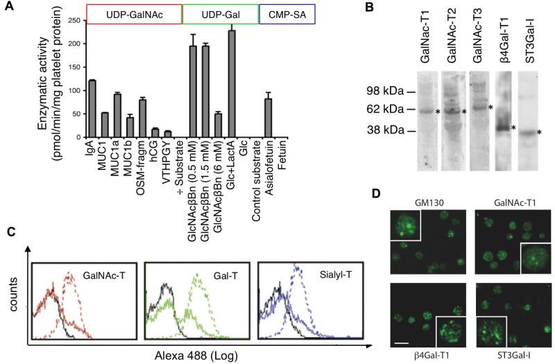
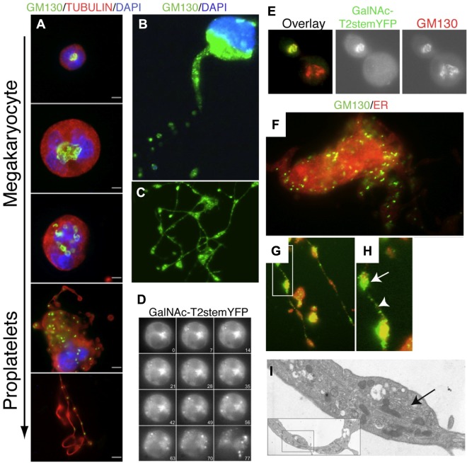
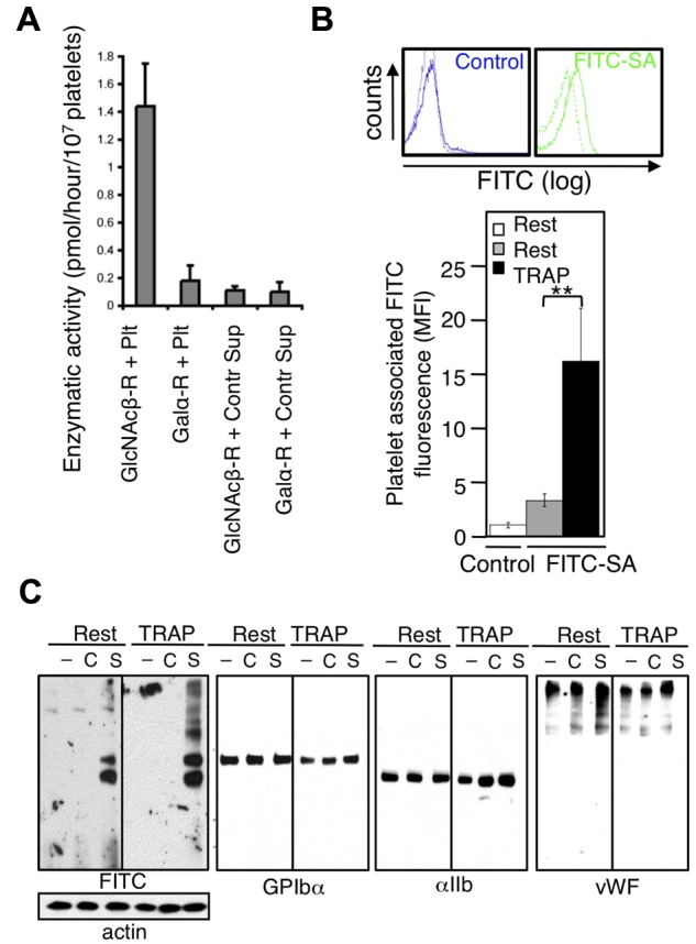
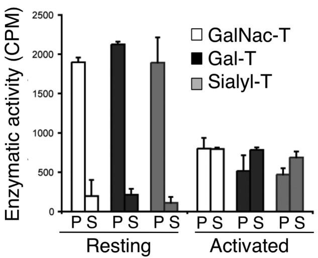
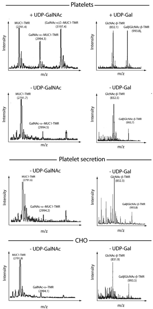
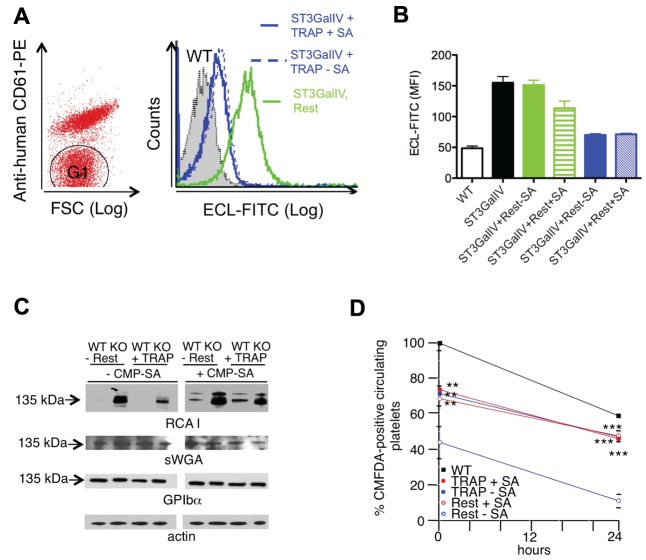
Platelets are megakaryocyte subfragments that participate in hemostatic and host defense reactions and deliver pro- and antiangiogenic factors throughout the vascular system. Although they are anucleated cells that lack a complex secretory apparatus with distinct Golgi/endoplasmic reticulum compartments, past studies have shown that platelets have glycosyltransferase activities. In the present study, we show that members of 3 distinct glycosyltransferase families are found within and on the surface of platelets. Immunocytology and flow cytometry results indicated that megakaryocytes package these Golgi-derived glycosyltransferases into vesicles that are sent via proplatelets to nascent platelets, where they accumulate. These glycosyltransferases are active, and intact platelets glycosylate large exogenous substrates. Furthermore, we show that activation of platelets results in the release of soluble glycosyltransferase activities and that platelets contain sufficient levels of sugar nucleotides for detection of glycosylation of exogenously added substrates. Therefore, the results of the present study show that blood platelets are a rich source of both glycosyltransferases and donor sugar substrates that can be released to function in the extracellular space. This platelet-glycosylation machinery offers a pathway to a simple glycoengineering strategy improving storage of platelets and may serve hitherto unknown biologic functions.


Figures

Figure 1
Figure 1
Human platelets contain functional surface and intracellular glycosyltransferases. (A) GalNAc-transferase, galactosyltransferase, and sialyltransferase activities in platelets. Detergent-extracted platelets were incubated with UDP-14C-GalNAc, UDP-14C-Gal, or CMP-14C-sialic acid (SA) and acceptor substrates. See supplemental Methods for details on acceptor substrates. Shown are the means ± SD of 3 experiments. (B) Immunoblots of platelet lysates subjected to SDS-PAGE using mAbs specific for GalNAc-T1, GalNAc-T2, GalNAc-T3, β4Gal-T1, and ST3Gal-I glycosyltransferases. Asterisks indicate bands with molecular weights corresponding to the glycosyltransferases (n = 3). (C) Flow cytometry histograms of GalNAc-T1 (red), β4Gal-T1 (green), and ST3Gal-I (blue) expression in nonpermeabilized (solid line) and permeabilized (dotted line) resting platelets. Isotype control mAbs (black line; n = 3). (D) Galactosyltransferase and Golgi marker (GM130) distribution in resting platelets determined using anti-GM130 and anti–GalNAc-T1, β4Gal-T1, and ST3Gal-I mAbs (n > 5). Insets show platelets at a higher magnification. Scale bars indicate 5 μm.
Figure 2
Figure 2
Golgi redistribution during thrombogenesis. (A) Immunofluorescence micrographs of immature and proplatelet-producing megakaryocytes showing Golgi remodeling into condensed vesicles that move into proplatelets during megakaryocyte maturation. Tubulin is visualized with an antitubulin mAb (red); nuclei by DAPI staining (blue); Golgi by anti-GM130 Abs (green). The micrographs are representative of > 10 experiments. Scale bar indicates10 μm. (B-C) Location of GM130 in proplatelets. Golgi is fragmented and condensed into small vesicular structures (green). Scale bar indicates 10 μm. (D) Kinetics of the Golgi disassembly in a mouse megakaryocyte transfected with the Golgi marker GalNAc-T2-stem-YFP. Panels of micrographs selected from 77 minutes of real-time observation. (E) Double immunofluorescence microscopy of transfected megakaryocyte with GalNAc-T2-stem-YFP using anti-YFP (green) and anti-GM130 (red) Ab. Colocalization was seen in cells expressing GalNAc-T2-stem-YFP regardless of the maturation level (not shown). (F) Immunofluorescence microscopy of differentiated megakaryocytes using the Golgi marker anti-GM130 Ab (green) and the endoplasmic reticulum (ER) marker Calnexin (red). (G-H) Arrow shows GM130 accumulation in a proplatelet swelling. Between swellings, small GM130-containing vesicles are stained. (I) Electron micrograph of a section of a proplatelet extension/swelling containing a small Golgi-like vesicular stack (arrow).
Figure 3
Figure 3
Platelets glycosylate exogenous platelet and plasma acceptors. (A) Surface galactosyltransferase activity measured by incubating platelets with GlcNAc-βGlcNAc-PAA 2.8 μm (GlcNAcβ-R + Plt) or α-Gal-Dynabead conjugates (Galα-R + Plt) and UDP-14C-galactose. Galactosyltransferase activity released into the supernatant during incubation with Dynabead conjugates (GlcNAcβ-R + Sup and Galα-R + Sup). (B) Surface sialyltransferase incorporated FITC-conjugated CMP-SA (FITC-SA) into resting (dotted line) or TRAP-activated platelets. FITC alone (Control) was added to resting (dotted line) or TRAP-activated platelets (solid line). Quantification of resting and TRAP-activated platelet-associated mean fluorescence intensity (MFI); n = 3. (C) Immunoblots of lysates from resting (Rest) or TRAP-activated platelets (TRAP), treated with FITC (C) or CMP-FITC-SA (S) or left untreated (-) with Abs to FITC, GPIbα, αIIb, and VWF. Actin is shown as a loading control (n = 3).
Figure 4
Figure 4
Activated platelets release soluble glycosyltransferases. GalNAc-, Gal-, and sialyltransferase activities in ultracentrifuged supernatant (S) from TRAP-activated platelets and detergent lysates of resting platelets (P). Glycosyltransferase activities were measured using C14-labeled donor sugars and enzyme-specific acceptor substrates (n = 3-4).
Figure 5
Figure 5
Platelets glycosylate exogenous acceptors. Matrix-assisted laser desorption/ionization–time of flight mass spectrometry of glycosylation products in detergent extracts from resting platelets (Platelets), secretion from TRAP-activated platelets (Plt. secretion), Chinese hamster ovary cell lysates after addition of exogenous GalNAc-transferase acceptor TMR-PEG-MUC1 (left), or Gal-transferase acceptor TMR-βGlcNAc (right). No exogenous donor UDP-GalNAc or UDP-Gal was added, with the exception of the top panels, in which 100μM UDP-GalNAc and UDP-Gal were added, respectively (n = 3).
Figure 6
Figure 6
Platelets sialylate specific surface glycoproteins in neighboring cells. (A) Sialylation of GPIbα in ST3GalIV−/− mouse platelets by activated human platelets. Human platelets were identified and separated from mouse platelets (G1) by flow cytometry using PE-conjugated anti–human CD61. Flow cytometric analysis of terminal β-galactose using ECL-FITC-labeled lectin on mouse ST3GalIV−/− platelets (G1) incubated with TRAP-activated human platelets (TRAP) or resting platelets (Rest) in the absence of CMP-SA. Relative binding of ECL-FITC to ST3GalIV+/+ (WT) and ST3GalIV−/− (KO) platelets, and to ST3GalIV−/− platelets incubated with medium from resting (Rest) or TRAP-activated human platelets (TRAP) with (+SA) and without CMP-SA (-SA). (B) Histograms report the mean ± SEM. SA indicates CMP-SA (n = 3). (C) Incorporation of sialic acid in GPIbα. ST3GalIV+/+ (WT) and ST3GalIV−/− (KO) mouse platelets were incubated with supernatants from resting (Rest) or TRAP (TRAP)–activated human platelets with (+CMP-SA) or without (-CMP-SA) CMP-SA. Lysates were subjected to SDS-PAGE and immunoblotted with sWGA, RCA-I, or Abs to GPIbα and actin (n = 3). (D) Survival of ST3GalIV+/+ (WT) or ST3GalIV−/− mouse platelets incubated with resting (Rest) or TRAP-activated human platelet supernatants (TRAP) in the presence (+CMP-SA) or absence (-CMP-SA) of CMP-SA. The y-axis reflects the platelet recovery defined by the number of platelets circulating at the first sample point after injection, expressed as the percentage of circulating nonactivated WT platelets. Shown are means ± SEM for 6 recipients. **P < .01; ***P < .001.
Figure 7
Figure 7
Sialic acid incorporation decreases platelet activation in vitro and in vivo. (A) Platelet-rich plasma from mice was incubated with FITC-conjugated CMP-SA or FITC (Control) at a 40μM final concentration for the indicated time points. At the indicated times, platelets in plasma were activated with 25μM TRAP for 5 minutes (TRAP) or left untreated (Rest), and platelet-associated FITC fluorescence was measured by flow cytometry (n = 2). (B) Representative side and forward scatter plots of platelets isolated from mice 60 minutes after infusion of FITC-SA or FITC (Control) and activated with 25μM TRAP or left untreated (Resting; n = 3). (C) The ratio of side to forward and side scatter platelets isolated from mice injected with FITC-SA (■) and FITC (□, Control) was determined. Unstimulated platelets isolated at 0 minutes were set as 1, and platelet shapes of isolates at 30 and 60 minutes activated with 25μM TRAP were compared. Data are means ± SD (n = 3). n.s. indicates not significant. *P < .05. (D) P-selectin expression was determined after TRAP activation by flow cytometry using a PE-conjugated anti–mouse P-selectin mAb (n = 2). (E) Activation status of platelets isolated from peripheral (Peripheral) or blood shed from a tail wound (Shed blood) 60 minutes after infusion of FITC (Control) or FITC-SA as assessed by light scatter characteristics. In the left panel, platelets are identified by PE–anti-CD61 mAb binding. The FITC mean fluorescence associated with platelets is plotted in the middle panel (n = 3; **P < .01. The right panel shows the ratio of side versus forward scatter of platelets in shed blood obtained from control mice (Control) or mice injected with FITC-SA (FITC-SA; n = 3; **P < .01).

Similar articles

Cited by

References

    1. Varki A. Biological roles of oligosaccharides: all of the theories are correct. Glycobiology. 1993;3(2):97–130. - PMC - PubMed
    1. Colley KJ, Lee EU, Paulson JC. The signal anchor and stem regions of the beta-galactoside alpha 2,6-sialyltransferase may each act to localize the enzyme to the Golgi apparatus. J Biol Chem. 1992;267(11):7784–7793. - PubMed
    1. Nilsson T, Au CE, Bergeron JJ. Sorting out glycosylation enzymes in the Golgi apparatus. FEBS Lett. 2009;583(23):3764–3769. - PubMed
    1. Paulson JC, Colley KJ. Glycosyltransferases. Structure, localization, and control of cell type-specific glycosylation. J Biol Chem. 1989;264(30):17615–17618. - PubMed
    1. Berger EG. Ectopic localizations of Golgi glycosyltransferases. Glycobiology. 2002;12(2):29R–36R. - PubMed

Publication types

MeSH terms