. 2012 Jul;32(14):2823-36.
doi: 10.1128/MCB.00496-12. Epub 2012 May 14.

SIRT1 negatively regulates the activities, functions, and protein levels of hMOF and TIP60

Affiliations

SIRT1 negatively regulates the activities, functions, and protein levels of hMOF and TIP60

Lirong Peng et al. Mol Cell Biol. 2012 Jul.

Abstract

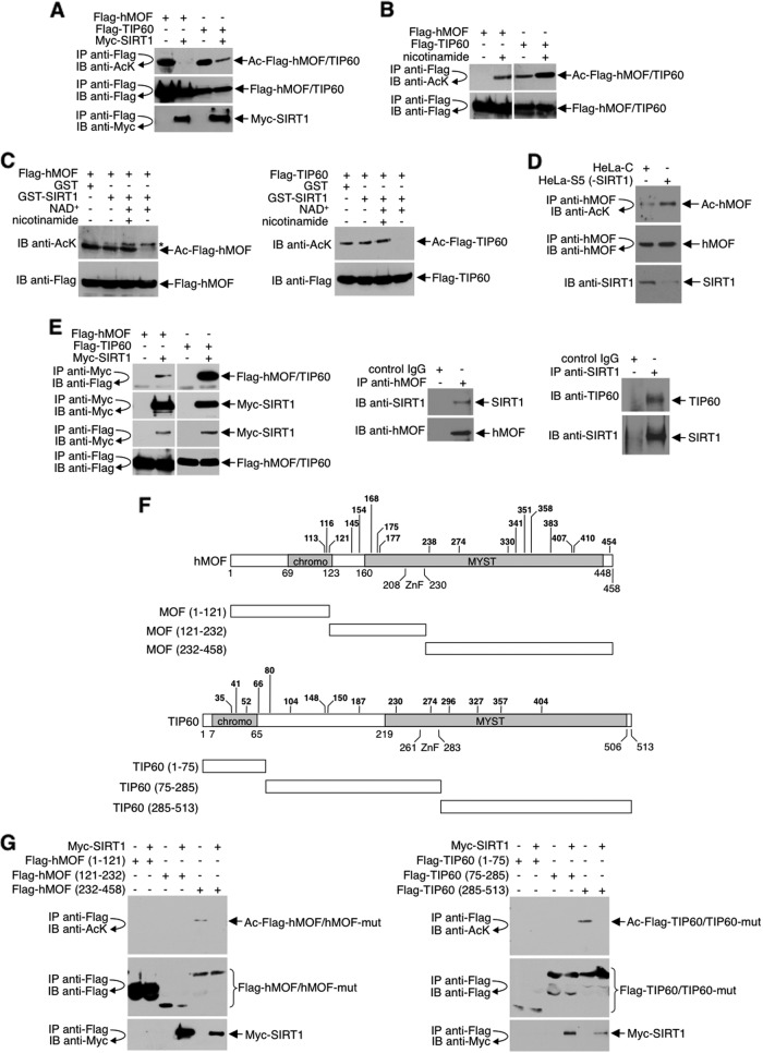
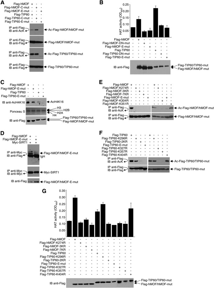
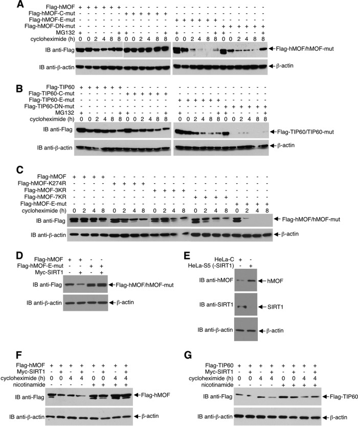
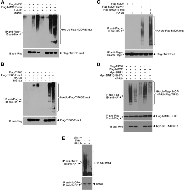
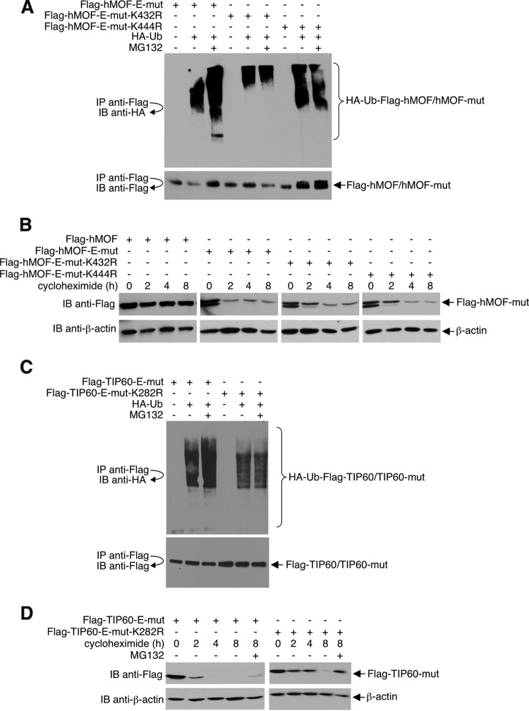
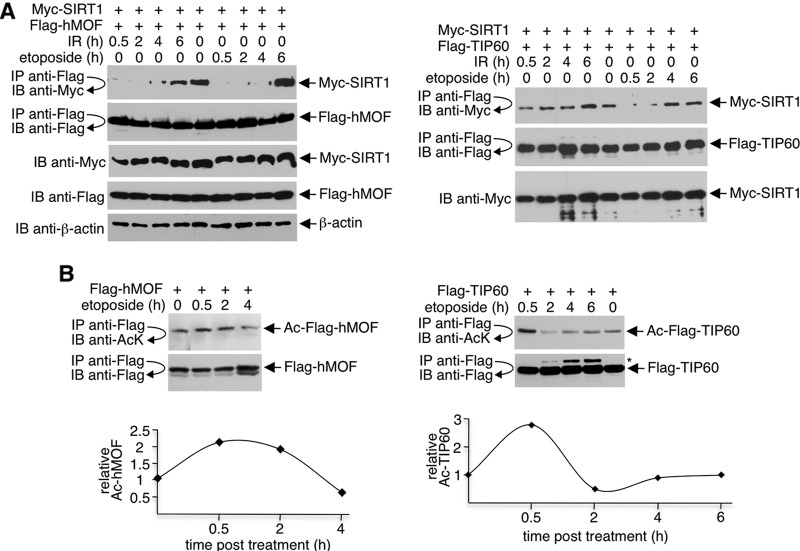
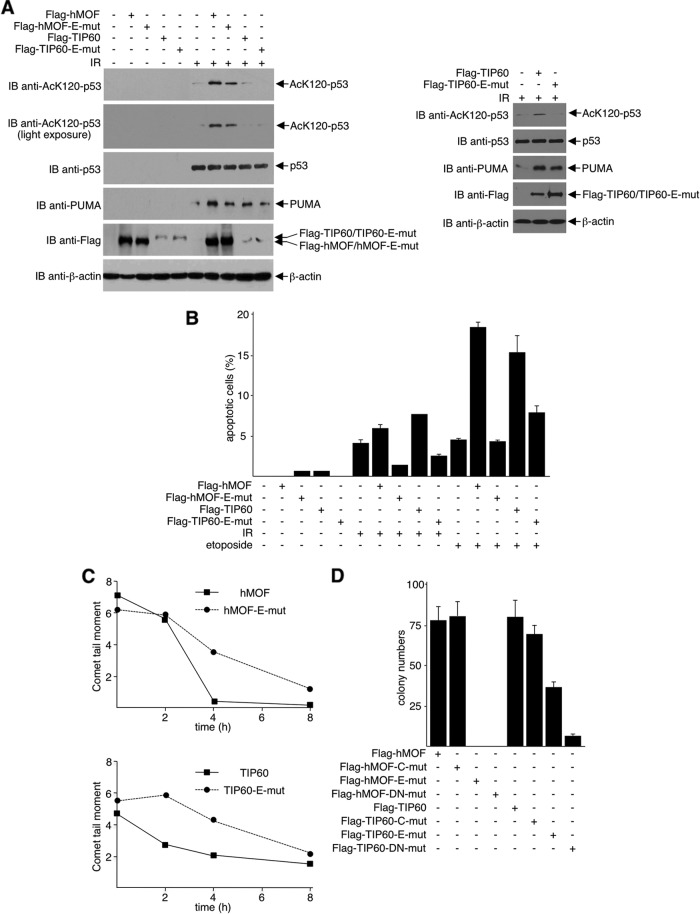
SIRT1 is a NAD(+)-dependent histone H4K16 deacetylase that controls several different normal physiologic and disease processes. Like most histone deacetylases, SIRT1 also deacetylates nonhistone proteins. Here, we show that two members of the MYST (MOZ, Ybf2/Sas3, Sas2, and TIP60) acetyltransferase family, hMOF and TIP60, are SIRT1 substrates. SIRT1 deacetylation of the enzymatic domains of hMOF and TIP60 inhibits their acetyltransferase activity and promotes ubiquitination-dependent degradation of these proteins. Importantly, immediately following DNA damage, the binding of SIRT1 to hMOF and TIP60 is transiently interrupted, with corresponding hMOF/TIP60 hyperacetylation. Lysine-to-arginine mutations in SIRT1-targeted lysines on hMOF and TIP60 repress DNA double-strand break repair and inhibit the ability of hMOF/TIP60 to induce apoptosis in response to DNA double-strand break. Together, these findings uncover novel pathways in which SIRT1 dynamically interacts with and regulates hMOF and TIP60 through deacetylation and provide additional mechanistic insights by which SIRT1 regulates DNA damage response.


Figures

Fig 1
Fig 1
SIRT1 binds to and deacetylates the enzymatic domains of hMOF and TIP60. Cell lysates were prepared either from 293T cells transfected with plasmids expressing the indicated proteins (A, E, left, and G), HeLa cells (E, middle), or MEFs (E, right) and immunoprecipitated (IP) with the indicated antibodies. The resulting immunoprecipitates were subjected to immunoblotting (IB) with the indicated antibodies. (B) Thirty-six hours posttransfection, cells were left untreated or were treated overnight with 10 mM nicotinamide. Cell lysates were subjected to immunoprecipitation and immunoblotting as before. (C) In vitro deacetylation assays were performed with Flag-hMOF/TIP60 substrates and SIRT1 enzyme. Reaction products were subjected to Western blot analysis with antiacetyllysine (anti-AcK) and reprobed with anti-Flag. (D) Lysates prepared from HeLa cells depleted of SIRT1 (HeLa-S5) or stably expressing a scrambled control shRNA (HeLa-C) were immunoprecipitated with anti-hMOF and immunoblotted with anti-AcK or anti-hMOF antibodies. Immunoblotting was also performed to assess SIRT1 expression. (F) Schematic diagram of hMOF/TIP60 and deletion mutants (not drawn to scale). Acetylated lysines identified by mass spectrometry are indicated in bold type. Chromo, chromatin organization modifier (chromo) domain; MYST, conserved MYST domain; Znf, C2HC zinc fingers. Ac, acetyl.
Fig 2
Fig 2
Deacetylation of hMOF and TIP60 inhibits their enzymatic activities. (A, D, E, and F) 293T cells were transfected with plasmids expressing the indicated proteins. Cell lysates were prepared and immunoprecipitated with the indicated antibodies, and the resulting immunoprecipitates were subjected to immunoblotting with the indicated antibodies. (B and G) Nuclear lysates prepared from transfected cells were subjected to HAT activity assays. Data are represented as mean ± standard error of the mean from three independent experiments. A representative blot of proteins used in the assays is shown. (C) 293T cells were transfected with plasmids expressing the indicated proteins. Histones were isolated, resolved by SDS-PAGE, and subjected to Western blot analysis with the indicated antibodies. The quality and quantity of histones were determined by Ponceau S staining of the membrane before Western blotting. All transfections were optimized to ensure equal expression levels of wild-type and mutant proteins. For example, in the experiment shown in panel A, three times more Flag-hMOF-E-mut and 1.5 times more Flag-TIP60-E-mut expression plasmids were transfected into the cells for each experiment than Flag-hMOF and Flag-TIP60.
Fig 3
Fig 3
SIRT1 deacetylation of hMOF and TIP60 promotes protein degradation. (A to D, F, and G) 293T cells were transfected with plasmids expressing the indicated proteins. Thirty-six hours posttransfection, cells were treated with 50 μg/ml cycloheximide for the indicated time, or left untreated, in combination with or without 10 μM MG132. Some reaction mixtures in the experiments shown in panels F and G included 10 mM nicotinamide. Specific protein levels were examined by Western blotting with the indicated antibodies. All transfections were adjusted according to the expression levels of Flag-hMOF or Flag-TIP60. For example, three times more plasmids were used for hMOF-E-mut and hMOF-DN-mut than for Flag-hMOF and Flag-hMOF-C-mut, and 1.5 times more TIP60-E-mut and TIP60-DN-mut was used than Flag-TIP60 and Flag-TIP60-C-mut. (E) Lysates prepared from HeLa cells depleted of SIRT1 (HeLa-S5) or stably expressing a scrambled control shRNA (HeLa-C) were immunoblotted with the indicated antibodies to assess protein expressions.
Fig 4
Fig 4
SIRT1 deacetylation of hMOF and TIP60 promotes protein ubiquitination. 293T cells (A to D) and MEFs (E) were transfected with plasmids expressing the indicated proteins. In some experiments, cells were left untreated or were treated with 10 μM MG132 for 8 h. Cell lysates were prepared and immunoprecipitated with the indicated antibodies, followed by immunoblotting with the indicated antibodies. Western blotting was also performed to assess protein expression levels. All transfections (A to D) were adjusted to ensure equal expression of wild-type and mutant proteins. For comparison of ubiquitinated hMOF, equal amounts of immunopurified hMOF proteins were loaded onto each lane (E). Ub, ubiquitin.
Fig 5
Fig 5
hMOF-K432/K444 and TIP60-K282 are ubiquitinated. 293T cells were transfected with plasmids expressing the indicated proteins. Thirty-six hours posttransfection, cells were treated with 50 μg/ml of cycloheximide for the indicated time, or left untreated, in combination with or without 10 μM MG132. Cell lysates were prepared and immunoprecipitated with the indicated antibodies, followed by immunoblotting with the indicated antibodies. Direct Western blotting was also performed to assess protein expression levels. Ub, ubiquitin.
Fig 6
Fig 6
SIRT1-hMOF/TIP60 interactions are dynamically regulated upon the DNA damage response. 293T cells were transfected with plasmids expressing the indicated proteins and left untreated or treated with IR (10 Gy) or etoposide (10 μM) for the indicated time. Flag-hMOF/TIP60 was immunoprecipitated with Flag M2 agarose. Cell lysates were prepared and immunoprecipitated with the indicated antibodies, and the resulting immunoprecipitates were subjected to immunoblotting with the indicated antibodies. Relative amounts of acetylated hMOF and TIP60 were determined using densitometry of Western blots.
Fig 7
Fig 7
Deacetylation inhibits hMOF- or TIP60-mediated apoptosis and DNA repair in response to DNA damage. (A) U2OS cells were transfected with plasmids expressing the indicated proteins and then treated with 10 Gy of ionizing radiation. Two hours after treatment, cell lysates were prepared and subjected to Western blot analysis with the indicated antibodies. (B) U2OS cells were transfected with plasmids expressing the indicated proteins. After selection with G418, cells were seeded and subjected to 10 Gy of IR or 50 μM etoposide (12 h) or left untreated. Twenty-four hours after treatment, cells were fixed, stained with DAPI, and analyzed for apoptosis. Data are represented as mean ± standard error of the mean from three separate experiments. (C) Pools of U2OS cells stably expressing the indicated proteins were treated with 1 Gy of ionizing radiation or left untreated for the indicated time, harvested, and analyzed with the neutral comet assay. (D) U2OS cells were transfected with plasmids expressing the indicated proteins. Twenty-four hours later, 104 cells from each transfectant were seeded and cultured for 2 weeks. Cell colonies were stained with Giemsa and counted. Data are represented as mean ± standard error of the mean from three separate experiments.

Similar articles

Cited by

References

    1. Akhtar A, Becker PB. 2000. Activation of transcription through histone H4 acetylation by MOF, an acetyltransferase essential for dosage compensation in Drosophila. Mol. Cell 5:367–375 - PubMed
    1. Avvakumov N, Cote J. 2007. The MYST family of histone acetyltransferases and their intimate links to cancer. Oncogene 26:5395–5407 - PubMed
    1. Blander G, Guarente L. 2004. The Sir2 family of protein deacetylases. Annu. Rev. Biochem. 73:417–435 - PubMed
    1. Bouras T, et al. 2005. SIRT1 deacetylation and repression of p300 involves lysine residues 1020/1024 within the cell cycle regulatory domain 1. J. Biol. Chem. 280:10264–10276 - PubMed
    1. Brunet A, et al. 2004. Stress-dependent regulation of FOXO transcription factors by the SIRT1 deacetylase. Science 303:2011–2015 - PubMed

Publication types

LinkOut - more resources