. 2012 Jun 29;287(27):22889-99.
doi: 10.1074/jbc.M112.374447. Epub 2012 May 9.

Mechanism of release and fate of excised oligonucleotides during nucleotide excision repair

Affiliations

Mechanism of release and fate of excised oligonucleotides during nucleotide excision repair

Michael G Kemp et al. J Biol Chem. .

Abstract

A wide range of environmental and carcinogenic agents form bulky lesions on DNA that are removed from the human genome in the form of short, ∼30-nucleotide oligonucleotides by the process of nucleotide excision repair. Although significant insights have been made regarding the mechanisms of damage recognition, dual incisions, and repair resynthesis during nucleotide excision repair, the fate of the dual incision/excision product is unknown. Using excision assays with both mammalian cell-free extract and purified proteins, we unexpectedly discovered that lesion-containing oligonucleotides are released from duplex DNA in complex with the general transcription and repair factor, Transcription Factor IIH (TFIIH). Release of excision products from TFIIH requires ATP but not ATP hydrolysis, and release occurs slowly, with a t(1/2) of 3.3 h. Excised oligonucleotides released from TFIIH then become bound by the single-stranded binding protein Replication Protein A or are targeted by cellular nucleases. These results provide a mechanism for release and an understanding of the initial fate of excised oligonucleotides during nucleotide excision repair.


Figures

FIGURE 1.
FIGURE 1.
Excised oligonucleotides become partially degraded in cell-free extract. A, time course of excision repair in reactions with CHO AA8 cell-free extract. Locations of the full-length (6-4) photoproduct-containing substrate, the small (<30 nt) excision products, and oligonucleotide size makers (27 and 22 nt) are indicated. B, quantification of excision repair assays. The results indicate the averages and standard deviations from three independent experiments. C, distribution of excision product lengths during excision assay time course experiments. The results indicate the average from three independent experiments.
FIGURE 2.
FIGURE 2.
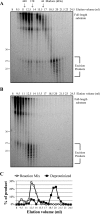
The excised oligonucleotides are protein-bound. A, products of an excision repair reaction (50 μl, 60 min) with cell-free extract were deproteinized as described under “Experimental Procedures” and analyzed by gel filtration chromatography on a Superdex 200 column. The fractions were then electrophoresed in a denaturing urea gel. Locations of the gel filtration protein standard size markers (ferritin, aldolase, and ovalbumin) are indicated. B, excision repair reaction mixture (50 μl, 60 min) containing both protein and DNA were directly analyzed by gel filtration chromatography. C, excision products from each of the fractions in A and B were quantified as a percentage of the total excision product signal across the column for each experiment and plotted. Reaction Mix indicates direct loading onto the column, and Deproteinized indicates deproteinization prior to gel filtration chromatography.
FIGURE 3.
FIGURE 3.
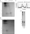
The excised oligonucleotides associate with TFIIH and RPA. A, the indicated proteins were immunoprecipitated from excision repair reactions (90 min) with specific antibodies. DNA from the immunoprecipitations were purified and then analyzed in a denaturing urea gel (top panel). Immunoprecipitates were also analyzed by SDS-PAGE and Western blotting. B, the unbound material (flow-through) from the immunoprecipitations in A were similarly processed and analyzed by denaturing gel electrophoresis and SDS-PAGE. C, quantification of the percentage of 25–30-mer oligonucleotides depleted from the excision assay immunoprecipitations in B. The results show the averages and standard deviations from three independent experiments. D, an excision repair reaction (100 μl, 90 min) was analyzed by gel filtration chromatography. Fractions were electrophoresed in both urea and SDS gels. E, fractions 10 and 13 from D, which represent peak fractions for two excision repair product species, were immunoprecipitated with either anti-mouse IgG or antibodies against RPA and XPB. Purified DNA from the IP was electrophoresed in a urea gel. F, time course experiments were performed, and then XPB and RPA were immunoprecipitated. DNA from the IPs were analyzed by urea-PAGE.
FIGURE 4.
FIGURE 4.
Excised oligonucleotides generated with a reconstituted excision reaction associate with TFIIH and RPA. A, deproteinized excision repair products from a reconstituted excision assay were analyzed by Superdex 200 chromatography and urea gel electrophoresis. B, a reconstituted repair reaction was directly fractionated by Superdex 200 chromatography followed by urea gel electrophoresis. C, quantification of excision product distribution in A and B. Reaction Mix indicates direct loading onto the column, and Deproteinized indicates deproteinization prior to gel filtration chromatography. D, a reconstituted reaction was subjected to immunoprecipitation with the indicated antibodies. DNA was purified and electrophoresed in a urea gel.
FIGURE 5.
FIGURE 5.
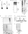
TFIIH and RPA associate with excised oligonucleotides released from duplex DNA. A, schematic for excision repair reaction with immobilized substrate DNA. Internally 32P-labeled T-T (6-4) photoproduct-containing substrate was prepared as described under “Experimental Procedures” with one oligonucleotide containing a 5′-biotin, which allowed for immobilization on Dynabeads M280 streptavidin magnetic beads. An asterisk represents the radiolabel, and the triangle indicates the (6-4) photoproduct. B, DNA from excision assays with CHO CFE was collected on a magnet after the indicated periods of time (immediately or following 0.5 h of incubation). Bound (lanes B) and unbound (lanes U) DNA were purified and electrophoresed in a urea gel. C, excision products released from the immobilized DNA during a 90-min excision reaction were subjected to immunoprecipitation with the indicated antibodies and then analyzed by urea- and SDS-PAGE. D, excision products released from immobilized DNA during a 90-min excision reaction were purified and subjected to gel filtration chromatography and urea gel electrophoresis (top panel). A portion of the purified excision products were reincubated in a standard excision reaction for 30 min and then fractionated by gel filtration chromatography (middle panel). The fractions were analyzed by urea-PAGE (top and middle panels) and SDS-PAGE (bottom panels). E, quantification of excision product distribution in D. Reaction indicates direct loading of the excision products reincubated in CHO AA8 CFE onto the column, and Deproteinized indicates deproteinization prior to gel filtration chromatography. F, purified excision products reincubated in CHO CFE-containing reaction mixtures for 30 min were subjected to immunoprecipitation with the indicated antibodies.
FIGURE 6.
FIGURE 6.
The slow release of excision products from TFIIH requires ATP but not ATP hydrolysis. A, a TFIIH immunoprecipitate from an excision reaction with CHO CFE and immobilized, radiolabeled substrate DNA was incubated in reaction buffer with either no ATP or with 2 mm ATP. At the indicated time points, the TFIIH immunoprecipitates were pelleted by centrifugation. Excision products that were released from TFIIH and that remained bound to TFIIH were purified and analyzed by denaturing urea-PAGE and phosphorimaging. B, quantification of results in A. The results show the averages and standard deviations from three independent experiments. C, TFIIH excision product immunoprecipitates described in A were incubated for 7 h in the presence of no ATP, ATP, ATPγS, or AMP-PCP. The plotted data are the averages and standard deviations from three independent experiments. D, TFIIH excision product immunoprecipitates described in A were incubated for 7 h in the presence of no ATP, ATP, ADP, or AMP. Lanes B, bound DNA; lanes U, unbound DNA.
FIGURE 7.
FIGURE 7.
RPA binds excision products released from TFIIH and limits excision product degradation. A, TFIIH immunoprecipitates from an excision assay with CHO CFE and immobilized, radiolabeled substrate were incubated in reactions with or without RPA (50 nm) and ATP (2 mm). After the 7-h incubation, the TFIIH immunoprecipitates were pelleted by centrifugation, and the unbound and bound DNAs were purified and analyzed by urea-PAGE and phosphorimaging. B, a portion of the unbound fractions shown in A were subjected to a second round of immunoprecipitation with anti-RPA antibody. DNA associating with the RPA IP was analyzed by urea-PAGE and phosphorimaging. C, HeLa CFE was incubated with IgG or anti-RPA34 antibodies to yield mock and RPA-depleted CFE. Depleted CFE was analyzed by Western blotting with antibodies against the indicated proteins. D, excision products deproteinized and purified from a TFIIH immunoprecipitate from an excision reaction with CHO CFE and immobilized, radiolabeled DNA were incubated in new reactions containing RPA-depleted HeLa CFE supplemented or not with recombinant RPA (50 nm). At the indicated time points, the reactions were stopped, and the excision products were purified for analysis by denaturing urea-PAGE.
FIGURE 8.
FIGURE 8.
Model for the mechanism of release and fate of excised oligonucleotides during nucleotide excision repair. Double-stranded DNA exposed to UV light generates photoproducts in DNA, as shown by the T<>T dimer. The nucleotide excision repair factors TFIIH-XPC, XPA, and RPA assemble on the damaged DNA in a random and cooperative manner that involves the kinetic proofreading and helicase activities of TFIIH (PIC1, preincision complex 1). XPG enters and stabilizes the preincision complex, which coincides with XPC leaving the damaged DNA (PIC2, preincision complex 2). XPF associates with the other repair factors at the site of damage, followed by incision events by the XPF and XPG nucleases (PIC3, preincision complex 3). RPA remains on the gapped DNA to promote the subsequent repair resynthesis and ligation steps of nucleotide excision repair. The excision product is released in complex with TFIIH after the dual incision events. The ATP-dependent release of the excision product from TFIIH recycles TFIIH for use in additional repair events and leads to either oligonucleotide degradation and/or RPA binding to the excision product.

Similar articles

Cited by

References

    1. Huang J. C., Svoboda D. L., Reardon J. T., Sancar A. (1992) Human nucleotide excision nuclease removes thymine dimers from DNA by incising the 22nd phosphodiester bond 5′ and the 6th phosphodiester bond 3′ to the photodimer. Proc. Natl. Acad. Sci. U.S.A. 89, 3664–3668 - PMC - PubMed
    1. Huang J. C., Sancar A. (1994) Determination of minimum substrate size for human excinuclease. J. Biol. Chem. 269, 19034–19040 - PubMed
    1. Svoboda D. L., Taylor J. S., Hearst J. E., Sancar A. (1993) DNA repair by eukaryotic nucleotide excision nuclease. Removal of thymine dimer and psoralen monoadduct by HeLa cell-free extract and of thymine dimer by Xenopus laevis oocytes. J. Biol. Chem. 268, 1931–1936 - PubMed
    1. Pathania S., Nguyen J., Hill S. J., Scully R., Adelmant G. O., Marto J. A., Feunteun J., Livingston D. M. (2011) BRCA1 is required for postreplication repair after UV-induced DNA damage. Mol. Cell 44, 235–251 - PMC - PubMed
    1. Reardon J. T., Thompson L. H., Sancar A. (1997) Rodent UV-sensitive mutant cell lines in complementation groups 6–10 have normal general excision repair activity. Nucleic Acids Res. 25, 1015–1021 - PMC - PubMed

Publication types