

Transient nuclear envelope rupturing during interphase in human cancer cells
- PMID: 22567193
- PMCID: PMC3342953
- DOI: 10.4161/nucl.18954
Transient nuclear envelope rupturing during interphase in human cancer cells
Abstract
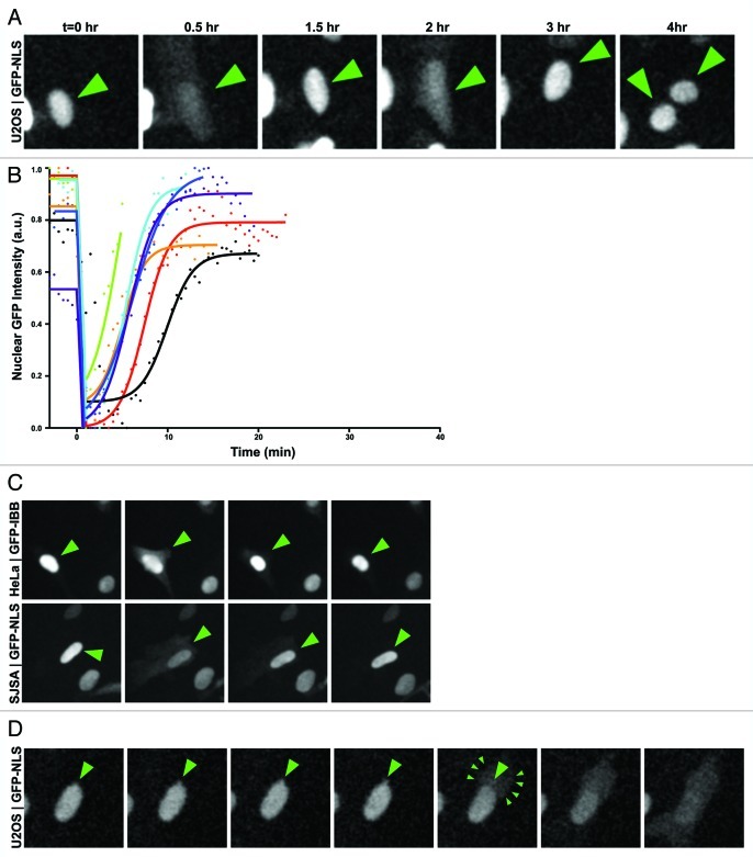
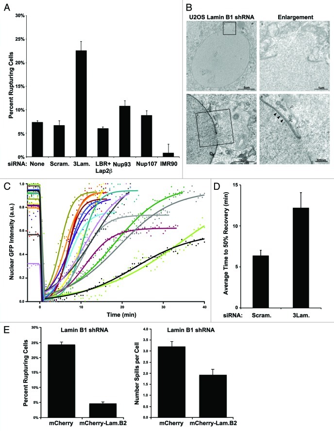
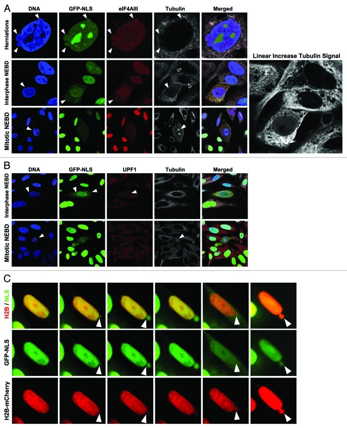
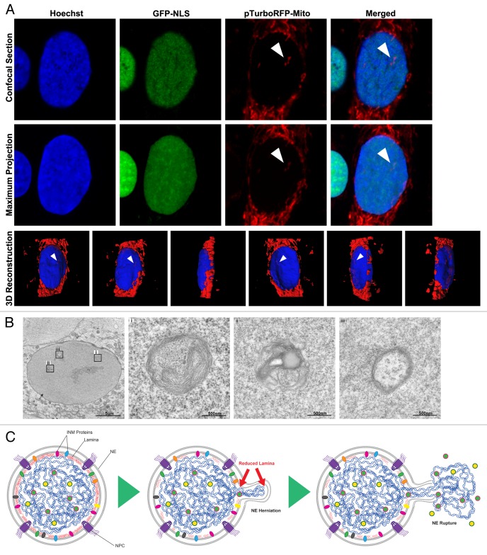
Neoplastic cells are often characterized by specific morphological abnormalities of the nuclear envelope (NE), which have been used for cancer diagnosis for more than a century. The NE is a double phospholipid bilayer that encapsulates the nuclear genome, regulates all nuclear trafficking of RNAs and proteins and prevents the passive diffusion of macromolecules between the nucleoplasm and the cytoplasm. Whether there is a consequence to the proper functioning of the cell and loss of structural integrity of the nucleus remains unclear. Using live cell imaging, we characterize a phenomenon wherein nuclei of several proliferating human cancer cell lines become temporarily ruptured during interphase. Strikingly, NE rupturing was associated with the mislocalization of nucleoplasmic and cytoplasmic proteins and, in the most extreme cases, the entrapment of cytoplasmic organelles in the nuclear interior. In addition, we observed the formation of micronuclei-like structures during interphase and the movement of chromatin out of the nuclear space. The frequency of these NE rupturing events was higher in cells in which the nuclear lamina, a network of intermediate filaments providing mechanical support to the NE, was not properly formed. Our data uncover the existence of a NE instability that has the potential to change the genomic landscape of cancer cells.
Keywords: cancer; genomic instability; live-imaging; nuclear envelope; nuclear permeability barrier.
Figures
Similar articles
-
Catastrophic nuclear envelope collapse in cancer cell micronuclei.Cell. 2013 Jul 3;154(1):47-60. doi: 10.1016/j.cell.2013.06.007. Cell. 2013. PMID: 23827674 Free PMC article.
-
Nuclear envelope rupture and repair during cancer cell migration.Science. 2016 Apr 15;352(6283):353-8. doi: 10.1126/science.aad7297. Epub 2016 Mar 24. Science. 2016. PMID: 27013428 Free PMC article.
-
Interplay of the nuclear envelope with chromatin in physiology and pathology.Nucleus. 2020 Dec;11(1):205-218. doi: 10.1080/19491034.2020.1806661. Nucleus. 2020. PMID: 32835589 Free PMC article. Review.
-
Loss of Nuclear Envelope Integrity in Aging and Disease.Int Rev Cell Mol Biol. 2018;336:205-222. doi: 10.1016/bs.ircmb.2017.07.013. Epub 2017 Sep 18. Int Rev Cell Mol Biol. 2018. PMID: 29413891 Review.
-
Bursting the Bubble - Nuclear Envelope Rupture as a Path to Genomic Instability?Trends Cell Biol. 2017 Aug;27(8):546-555. doi: 10.1016/j.tcb.2017.02.008. Epub 2017 Mar 9. Trends Cell Biol. 2017. PMID: 28285738 Free PMC article. Review.
Cited by
-
Genome instability from nuclear catastrophe and DNA damage.Semin Cell Dev Biol. 2022 Mar;123:131-139. doi: 10.1016/j.semcdb.2021.03.021. Epub 2021 Apr 7. Semin Cell Dev Biol. 2022. PMID: 33839019 Free PMC article. Review.
-
Nuclear size rectification: A potential new therapeutic approach to reduce metastasis in cancer.Front Cell Dev Biol. 2022 Oct 10;10:1022723. doi: 10.3389/fcell.2022.1022723. eCollection 2022. Front Cell Dev Biol. 2022. PMID: 36299481 Free PMC article. Review.
-
Nuclear envelope rupture drives genome instability in cancer.Mol Biol Cell. 2016 Nov 1;27(21):3210-3213. doi: 10.1091/mbc.E16-02-0098. Mol Biol Cell. 2016. PMID: 27799497 Free PMC article.
-
Membrane Binding by CHMP7 Coordinates ESCRT-III-Dependent Nuclear Envelope Reformation.Curr Biol. 2016 Oct 10;26(19):2635-2641. doi: 10.1016/j.cub.2016.07.039. Epub 2016 Sep 8. Curr Biol. 2016. PMID: 27618263 Free PMC article.
-
Nuclear lamin facilitates collective border cell invasion into confined spaces in vivo.J Cell Biol. 2023 Nov 6;222(11):e202212101. doi: 10.1083/jcb.202212101. Epub 2023 Sep 11. J Cell Biol. 2023. PMID: 37695420 Free PMC article.
References
-
- Hannen EJ, van der Laak JA, Manni JJ, Pahlplatz MM, Freihofer HP, Slootweg PJ, et al. An image analysis study on nuclear morphology in metastasized and non-metastasized squamous cell carcinomas of the tongue. J Pathol. 1998;185:175–83. doi: 10.1002/(SICI)1096-9896(199806)185:2<175::AID-PATH69>3.0.CO;2-U. - DOI - PubMed
MeSH terms
Substances
Grants and funding
LinkOut - more resources
Full Text Sources
Other Literature Sources