. 2012 Oct;33(5):1021-32.
doi: 10.1016/j.neuro.2012.04.019. Epub 2012 Apr 25.

Selenium induces cholinergic motor neuron degeneration in Caenorhabditis elegans

Affiliations

Selenium induces cholinergic motor neuron degeneration in Caenorhabditis elegans

Annette O Estevez et al. Neurotoxicology. 2012 Oct.

Abstract

Selenium is an essential micronutrient required for cellular antioxidant systems, yet at higher doses it induces oxidative stress. Additionally, in vertebrates environmental exposures to toxic levels of selenium can cause paralysis and death. Here we show that selenium-induced oxidative stress leads to decreased cholinergic signaling and degeneration of cholinergic neurons required for movement and egg-laying in Caenorhabditis elegans. Exposure to high levels of selenium leads to proteolysis of a soluble muscle protein through mechanisms suppressible by two pharmacological agents, levamisole and aldicarb which enhance cholinergic signaling in muscle. In addition, animals with reduction-of-function mutations in genes encoding post-synaptic levamisole-sensitive acetylcholine receptor subunits or the vesicular acetylcholine transporter developed impaired forward movement faster during selenium-exposure than normal animals, again confirming that selenium reduces cholinergic signaling. Finally, the antioxidant reduced glutathione, inhibits selenium-induced reductions in egg-laying through a cellular protective mechanism dependent on the C. elegans glutaredoxin, GLRX-21. These studies provide evidence that the environmental toxicant selenium induces neurodegeneration of cholinergic neurons through depletion of glutathione, a mechanism linked to the neuropathology of Alzheimer's disease, amyotrophic lateral sclerosis, and Parkinson's disease.


Conflict of interest statement

Conflict of interest

The authors declare that there are no conflicts of interest.

Figures
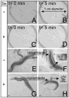
Fig. 1
Fig. 1
Selenium causes paralysis independent of muscle function. Animals paralyzed by high dose Se-exposure responded normally to treatment with levamisole, an anthelmintic which functions as a cholinergic agonist and induces muscle contraction in C. elegans. “+” indicates exposure to 5 mM Na2SeO3 while “−” indicates mock-exposure to water only. Scale bars (E–H, including insets), 50 μm. (A–D) Nearly all mock-exposed adult WT animals leave a 1 cm diameter circle after 5 min (compare A to B) while paralyzed Se-exposed adults do not (compare C to D). (E–H) When treated with the levamisole, both mock- and Se-exposed adults were observed to hyper-contract (compare (E) and (F), and (G) to (H), respectively) and lay eggs (F and H, insets). Late stage embryos were observed in the eggs released by the Se-exposed animals only (H, inset).
Fig. 2
Fig. 2
Neurons, but not muscles appear structurally damaged by selenium. In adult hermaphrodites, exposure to high dose Se damages cholinergic motor neurons in the ventral cord and those required for egg-laying, but does not affect muscles. “*” = vulva (C–H). “Arrowheads” = VC neurons, “arrows” = HSNs, white color indicates normal and black indicates damaged or missing (E–H). “+” and “−”, and scale bars are as in Fig. 1, except inset scale bars = 5 μm. (A and B) Animals expressing an integrated myosin-GFP translational fusion protein (MYO-3::GFP) were grown for 48 h “+” or “−” Se. The regular linear pattern of the MYO-3::GFP in the head (A) was not grossly altered by Se exposure (B). (C and D) Similarly, in phalloidin staining of F-actin no apparent structural differences were observed between mock- (C) or Se-exposed animals (D), as shown in the vulval region. (E–H) Nearly all mock-exposed WT animals expressed the Pida-1::gfp transgene in all six VC neurons and the two HSNs (E) while Se-exposed animals were significantly more likely (Table 1) to have lost GFP expression in one or more VC neurons (F). 400×, mock-exposed animals were observed to exhibit normal cellular morphology in their VC neurons (G) and HSNs (G, inset) while neurons from Se-exposed animals showed evidence of neurodegeneration, including nuclear swelling (H; VC neuron), nuclear fragmentation (H, inset; HSN), and axonal beading (H, double arrowhead lines). (I and J) Presynaptic densities were visualized using an unc-4::snb-1::GFP reporter construct that expresses a fusion of GFP and the C. elegans synaptobrevin protein, SNB-1 in a subset of cholinergic motor neurons in the ventral cord. A regular pattern of expression was observed after 24 h in mock-exposed adult animals (I). This pattern was disrupted in a significant percentage of the Se-exposed population (J).
Fig. 3
Fig. 3
Selenium alters cholinergic signaling. Pharmacological agents that increase cholinergic signaling at the NMJ, either by preventing breakdown of ACh (aldicarb and eserine salicylate) or through activating AChRs on muscles (levamisole), reduced movement deficits caused by high dose Se-exposure and prevented Se-induced loss of a soluble muscle protein, but could not induce a time dependent increase in paralysis suggesting that ACh levels at the NMJ are decreased after Se-exposure. (A) Animals expressing a myosin::lac-z reporter were histochemically stained for β-galactosidase activity. Reporter activity was reduced in muscles of Se-exposed, but not mock-exposed animals at 24 and 48 h (upper panel). After 24 h exposure to Se, levamisole (mid panel) and aldicarb (lower panel) treatment both suppressed protein degradation in muscle cells. Controls for the Se-exposed worms at 0 h were not determined (ND) because they were no different from mock-exposed. “+” and “−” are as in Fig. 1. (B) The percentage of adult WT (N2) animals paralyzed by aldicarb gradually increases over time. A significant reduction was observed for the Se-exposed populations (△) that were backing deficient prior to aldicarb treatment as compared to mock-exposed populations (■) at ≤90 min. Data was reported for each time point as the average percentages of 20 paralyzed animals with six repetitions (n = 120). Error bars = ±SEM. *p = 3.2 × 10⁻¹⁷, by two-way ANOVA, comparing Se-exposed to mock-exposed animals at the same time points after aldicarb treatment; post hoc analysis by Student’s t-test. (C) Populations of animals exposed to a concentration range (△, 0.1–1000 μm) of eserine salicylate and Se show a dose dependent increase in the percentage of animals that are motile (move both forwards and backwards) which is significantly different when compared to the Se-exposed controls (△, 0 μm). Although animals not exposed to Se, but treated with the two highest concentrations of eserine salicylate (■, 100 and 1000 μM) were observed to move slower than the water only controls (■, 0 μM), they were not significantly different (p > 0.05 comparing 100 and 1000 μM eserine salicylate with water). *p = 1.4 × 10⁻² (one-way ANOVA, comparing Se-exposed and drug treated animals to Se-exposed only animals, post hoc analysis by Student’s t-test). Error bars = ±SEM.
Fig. 4
Fig. 4
Presynaptic cholinergic release is altered by selenium. Mutations in some genes which reduce either post-synaptic muscle reception (lev-1, unc-29, unc-38, unc-63) or affect ACh release from motor neurons (unc-17) adversely alter forward movement behavior in response to high dose Se exposure. (A) Reduction- or loss-of-function mutations in some of the C. elegans AChRs (lev-1, unc-29, unc-38, unc-63) and one GABAR (unc-49), but not others (acr-16, gar-2), conferred additional sensitivity to Se (black bars) when compared to WT animals as indicated by a decrease in the percentage of animals within each population that were able to move forward; black bars = mock-exposed controls. Each graph bar represents an average of four plates with twenty animals each repeated three to four times (average n/graph bar = 300); error bars = ±SEM. “non-α” and “α” refer to the subtypes of the L-AChR. *p = 2.8 × 10⁻²⁰, by one-way ANOVA comparing the Se-exposed mutant strains to the Se-exposed WT strain, post hoc analysis was by Student’s t-test. (B) Populations of animals with a reduction-of-function mutation (e245) in the C. elegans VAChT, unc-17 had significantly less normal forward movement in comparison to WT populations of animal after 24 h (black bars) and 48 h (white bars) of Se exposure. A dominant mutation (e1563) in the gene for the vesicle-associated membrane protein, synaptobrevin (snb-1) was able to suppress this unc-17(e245) Se-induced phenotype such that there was no significant difference (NS) between populations of the unc-17;snb-1 double mutant strain when compared to WT populations exposed for the same amount of time (Student’s t-test). Since animals with the unc-17(e245) mutation do not normally move backwards, forward movement only was scored such that direct comparisons of the three strains could be made. Each graph bar represents at least five plates of twenty animals (n ≥ 100) of each genotype (WT, unc-17, unc-17;snb-1) which were scored at 24 and 48 h to determine the percentage of animals within each population expressing each phenotype; error bars = ±SEM. *p = 6.8 × 10⁻⁷ (two-way ANOVA comparing unc-17 to both WT and unc-17;snb-1 across phenotypes and time, post hoc analysis was by Student’s t-test).
Fig. 5
Fig. 5
Decreased egg-laying rates after selenium-exposure. Animals exposed to high dose Se were not only observed to retain late stage embryos, but to reduce their egg-laying rate overtime, a phenomena that appeared to be partially mediated through oxidative stress-induction. (A and B) Adult animals expressing GFP under the control of the promoter for the pan-neuronal expression gene, unc-119 and maintained continuously in the presence of an adequate food supply do not normally retain embryos past the multicellular stages (A) although late stage embryos (two-fold and beyond) were observed after only 24 h exposure to Se under the same conditions (B). “+” and “−”, and scale bars are as in Fig. 1. (C) The egg-laying rate (#eggs laid/animal/hour) of WT animals was reduced after 6 h of exposure to Se as compared to mock-exposed animals (H2O). Each bar graph represents the average progeny from 30 animals (three plates of ten animals each); error bars = ±SEM. *p = 1.9 × 10⁻², comparing Se-exposed to mock-exposed animals at the same time point, Student’s t-test (two-tailed, unequal variance). (D) The Se-induced reduction in the egg-laying rate was mediated by oxidative stress since treatment with the cellular antioxidant glutathione (GSH) was able to partially rescue this phenotype. In addition, the glrx-21(tm2921) null mutant could not be rescued by the GSH pretreatment. NS, not significant (Student’s t-test). *p = 4.1 × 10⁻¹³-WT and p = 3.8 × 10⁻¹⁴-glrx-21 (one-way ANOVA comparing across conditions within strains). **p = 1.6 × 10⁻²-WT and p = 5.2 × 10⁻⁴-glrx-21, comparing “Se-exposed only” to “Se-exposed pretreated with GSH” animals within the same strain (Student’s t-test).
Fig. 6
Fig. 6
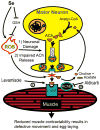
Summary. Motor neuron signaling to muscles is altered by selenium-induced oxidative stress. Reduced glutathione (GSH) normally prevents reactive oxygen species (ROS) from damaging cells. Excess selenium (Se) from the environment can both increase the formation of ROS and decrease the pool of GSH, thus inducing motor neuron (yellow) degeneration and impairing release of acetylcholine (ACh) filled synaptic vesicles (green circles) loaded by the vesicular acetylcholine transporter (VAChT). The reduction in ACh available to muscles reduces contractility, but does not affect the function of muscles since exogenous application of the pharmaceutical aldicarb induces muscle paralysis by artificially increasing ACh in the neuromuscular junction (space between muscle and neuron). Aldicarb inhibits the action of the enzyme acetylcholinesterase (AChE) which normally functions to inactivate ACh by breaking it down into its constituent parts choline (orange circle) and acetate (orange rectangle). In addition, the levamisole-sensitive ACh receptors (L-AChR) still respond by causing muscle hyper-contraction when the AChR agonist levamisole is exogenously applied. ChAT = choline acetyl transferase. (For interpretation of the references to color in this figure legend, the reader is referred to the web version of the article.)

Similar articles

Cited by

References

    1. Alfonso A, Grundahl K, Duerr JS, Han HP, Rand JB. The Caenorhabditis elegans unc-17 gene: a putative vesicular acetylcholine transporter. Science. 1993;261(5121):617–9. - PubMed
    1. Bany IA, Dong M-Q, Koelle MR. Genetic and cellular basis for acetylcholine inhibition of Caenorhabditis elegans egg-laying behavior. J Neurosci. 2003;23(22):8060–9. - PMC - PubMed
    1. Bellinger FP, Raman AV, Reeves MA, Berry MJ. Regulation and function of selenoproteins in human disease. Biochem J. 2009;422(1):11–22. - PMC - PubMed
    1. Bonilla-Ramirez L, Jimenez-Del-Rio M, Velez-Pardo C. Acute and chronic metal exposure impairs locomotion activity in Drosophila melanogaster: a model to study Parkinsonism. Biometals. 2011;24(6):1045–57. - PubMed
    1. Boulin T, Gielen M, Richmond JE, Williams DC, Paoletti P, Bessereau JL. Eight genes are required for functional reconstitution of the Caenorhabditis elegans levamisole-sensitive acetylcholine receptor. Proc Natl Acad Sci U S A. 2008;105(47):18590–5. - PMC - PubMed

Publication types

MeSH terms