. 2012 Jun 22;287(26):21936-49.
doi: 10.1074/jbc.M112.351783. Epub 2012 May 3.

Transcriptional repression of ATF4 gene by CCAAT/enhancer-binding protein β (C/EBPβ) differentially regulates integrated stress response

Affiliations

Transcriptional repression of ATF4 gene by CCAAT/enhancer-binding protein β (C/EBPβ) differentially regulates integrated stress response

Souvik Dey et al. J Biol Chem. .

Abstract

Different environmental stresses induce the phosphorylation of eIF2 (eIF2∼P), repressing global protein synthesis coincident with preferential translation of ATF4. ATF4 is a transcriptional activator of genes involved in metabolism and nutrient uptake, antioxidation, and regulation of apoptosis. Because ATF4 is a common downstream target that integrates signaling from different eIF2 kinases and their respective stress signals, the eIF2∼P/ATF4 pathway is collectively referred to as the integrated stress response. Although eIF2∼P elicits translational control in response to many different stresses, there are selected stresses, such as exposure to UV irradiation, that do not increase ATF4 expression despite robust eIF2∼P. The rationale for this discordant induction of ATF4 expression and eIF2∼P in response to UV irradiation is that transcription of ATF4 is repressed, and therefore ATF4 mRNA is not available for preferential translation. In this study, we show that C/EBPβ is a transcriptional repressor of ATF4 during UV stress. C/EBPβ binds to critical elements in the ATF4 promoter, resulting in its transcriptional repression. Expression of C/EBPβ increases in response to UV stress, and the liver-enriched inhibitory protein (LIP) isoform of C/EBPβ, but not the liver-enriched activating protein (LAP) version, represses ATF4 transcription. Loss of the liver-enriched inhibitory protein isoform results in increased ATF4 mRNA levels in response to UV irradiation and subsequent recovery of ATF4 translation, leading to enhanced expression of its target genes. Together these results illustrate how eIF2∼P and translational control combined with transcription factors regulated by alternative signaling pathways can direct programs of gene expression that are specifically tailored to each environmental stress.


Figures
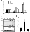
FIGURE 1.
FIGURE 1.
Expression of ATF4 is blocked following UV irradiation despite increased eIF2α∼P. A, wild-type MEF cells were treated with either 40 J/m2 UV-C irradiation (UV) or 1 μm thapsigargin (TG) and cultured for 3 or 6 h as indicated. Alternatively, cells were treated with UV-C irradiation for an hour followed by thapsigargin (UV/TG) for the indicated time. 0 h represents no stress treatment. Total mRNA was then isolated from the cells, and the levels of ATF4 mRNA were measured by qPCR. Values obtained are -fold change compared with the no-treatment control. Each experiment was performed three independent times with error bars representing the S.D. * indicates significance with p < 0.05 compared with non-treated control. # indicates a significant difference between the UV and ER stress treatments after 6 h. B, protein lysates were prepared from wild-type MEF cells treated with the conditions as indicated for A. Levels of ATF4, eIF2α∼P, total eIF2α, CHOP, and β-actin were measured by immunoblot analysis using antibodies specific to the indicated proteins. C, the PATF4-Luc reporter plasmid containing 2.5-kb of the ATF4 promoter was transfected into the wild-type MEF cells, which were then treated with 1 μm thapsigargin or 40 J/m2 UV-C irradiation or subjected to no treatment (NT) as indicated. Firefly luciferase activity was measured as described under “Experimental Procedures,” and the luciferase activity relative to the non-treated sample is presented in the histogram along with the S.D. (error bars).
FIGURE 2.
FIGURE 2.
ATF4 promoter contains critical elements for repression in response to UV irradiation. A, the PATF4-Luc reporter plasmid containing 2.5 kb of the ATF4 promoter was transfected into wild-type MEF cells, and following UV irradiation (UV) or no treatment (NT), luciferase activity was measured. In parallel, 0.5-kb segments were sequentially deleted from the 5′-end of the ATF4 promoter in the PATF4-Luc reporter and analyzed for activity in the wild-type cells treated with UV irradiation or subjected to no treatment. PATF4-Luc activity is presented along with the S.D. (error bars). B, internal 0.5-kb deletions were also constructed in the PATF4-Luc reporter and transfected into wild-type MEF cells followed by exposure to UV irradiation or no treatment. Furthermore, smaller deletions were constructed within the −1 to −0.5-kb region of the ATF4 promoter and assayed in wild-type cells in the presence or absence of UV stress. * designates significance (p < 0.05).
FIGURE 3.
FIGURE 3.
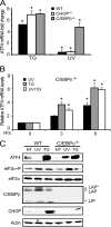
C/EBPβ is required for reduced ATF4 mRNA in response to UV irradiation. Wild-type, CHOP−/−, and C/EBPβ−/− MEF cells were treated with 1 μm thapsigargin (TG) or with 40 J/m2 UV-C irradiation (UV) and cultured for 6 h. A, the levels of ATF4 mRNA were measured by qPCR, and the -fold change in the transcript levels is represented relative to cells not treated with stress with the S.D. indicated by error bars. B, C/EBPβ−/− MEF cells were treated with UV irradiation, thapsigargin, or both (UV/TG) and cultured for 3 or 6 h. Values are relative to the no-treatment control (0), and the S.D. is indicated by error bars. C, protein lysates were prepared from wild-type and C/EBPβ−/− MEF cells subjected to UV irradiation, thapsigargin, or no treatment (NT), and the indicated protein levels were measured by immunoblot analysis. The LAP and LIP isoforms of C/EBPβ are indicated to the right of the panel. Results are representative of three independent experiments. * indicates significance with p < 0.05.
FIGURE 4.
FIGURE 4.
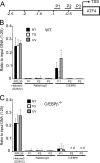
C/EBPβ binds to specific elements in ATF4 promoter. A, schematic representation of the regions of ATF4 promoter analyzed for C/EBPβ binding by ChIP. TSS indicates the transcriptional start site, and the promoter region is illustrated from −2.5 kb to the +1 position. The region designated P1 includes the sequence −978 to −800 bp in the ATF4 promoter, and P2 and P3 represent regions −628 to −470 bp and −334 to −194 bp, respectively. B, 6 h after exposure to 40 J/m2 UV-C irradiation (UV) or 1 μm thapsigargin (TG) or no treatment (NT), wild-type (WT; B) and C/EBPβ−/− (C) cells were analyzed by ChIP analyses for C/EBPβ binding to the P1, P2, and P3 regions of the ATF4 promoter. The immunoprecipitated DNA was analyzed by qPCR using primer sets specific for each promoter region. C/EBPβ indicates that antibody specific to this transcription factor was used in the ChIP assay; rabbit IgG antibody was used as a control. ChIP analyses were also carried out with the control histone H3 Lys-4 (D2B12) antibody and were analyzed by qPCR using primer sets for RPL30. Data are represented as a ratio of the input sample (1:25) and are the mean and S.D. (error bars) of three different experiments. * indicates significance with p < 0.05. n.d. indicates that the C/EBPβ binding to the P2 and P3 was not detected in the C/EBPβ−/− cells.
FIGURE 5.
FIGURE 5.
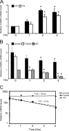
C/EBPβ mRNA is stabilized following UV irradiation. A, wild-type MEF cells were exposed to 1 μm thapsigargin (TG) or 40 J/m2 UV-C irradiation (UV) and cultured for up to 4 h as indicated. RNA was prepared from these cells, and C/EBPβ mRNA levels were measured by qPCR. The amount of C/EBPβ transcript is presented relative to the no-treatment control (0), and the S.D. is indicated by error bars. * indicates significance with p < 0.05. B, measurements of C/EBPβ mRNA half-life were carried out by first treating cells with UV irradiation or thapsigargin. 1 h after the initiation of the stress regimen, transcription was blocked by treating the cells with 20 μm actinomycin D (UV/AD or TG/AD), and then cells were cultured for up to an additional 4 h. Alternatively, cells were treated with actinomycin D (AD) alone. C/EBPβ mRNA levels were measured by qPCR at the indicated time intervals. Values are representative of the mean with the S.D. indicated by error bars. C, the half-life of the C/EBPβ mRNA for each of the stress arrangements was determined by plotting the transcript levels versus the length of time of the actinomycin D treatment in a semilogarithmic graph.
FIGURE 6.
FIGURE 6.
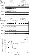
LIP isoform of C/EBPβ is differentially expressed during UV and ER stress. CEBPβ−/− MEF cells and the wild-type counterpart were treated with 40 J/m2 UV-C irradiation (UV) (A) or 1 μm thapsigargin (TG) (B) and were cultured for up to 24 h as indicated. Protein lysates were prepared from the treated cells, and the levels of ATF4, LIP, LAP, and β-actin were measured by immunoblot analyses. Each panel is representative of three independent experiments. C, the levels of the LIP isoform of C/EBPβ were quantified by densitometry and are represented as relative levels of the LIP band as compared with the no-treatment control (0).
FIGURE 7.
FIGURE 7.
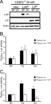
LIP is a potent repressor of ATF4 transcription. A, C/EBPβ−/− MEF cells were cotransfected with the PATF4-Luc reporter and plasmids specifically expressing either the LIP or LAP isoforms of C/EBPβ or the parent vector (Vec). The transfected cells were treated with UV irradiation (UV) or subjected to no treatment (NT), and the levels of LAP, LIP, and β-actin were measured by immunoblot analyses. Levels of PATF4-Luc activity were also measured in the cells expressing LAP (PATF4-Luc + LAP) (B) or LIP (PATF4-Luc + LIP) (C) as compared with the cells containing PATF4-Luc and the expression vector alone (PATF4-Luc). Luciferase activity is presented in the histograms with the luciferase activity in the non-treated wild-type cells being represented as a value of 1. Values were derived from three independent experiments with the S.D. indicated by error bars. * indicates significance with p < 0.05. Tsf, transfection of cells with the indicated expression plasmids.
FIGURE 8.
FIGURE 8.
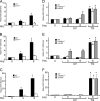
Loss of LIP in C/EBPβ-ΔuORF cells alleviates repression of ATF4 transcription. A, wild-type, C/EBPβ−/−, and C/EBPβ-ΔuORF MEF cells were transfected with the PATF4-Luc plasmid and treated with either with 1 μm thapsigargin (TG) or 40 J/m2 UV-C irradiation (UV) or subjected to no stress treatment (NT). PATF4-Luc expression was measured and is represented in the histogram with the non-treated cells indicated as a value of 1. Values were determined from three independent experiments with the S.D. indicated by error bars. B, the wild-type (WT), C/EBPβ−/−, and C/EBPβ-ΔuORF cells were treated with UV or thapsigargin stress for up to 6 h, and the levels of ATF4 mRNA were determined by qPCR. Mean values are presented in the histograms with the S.D. indicated by error bars. C, alternatively, the levels of the indicated proteins in the stressed wild-type (WT) and C/EBPβ-ΔuORFuORF) cells were measured by immunoblot analyses. The zero time indicates no stress treatment. D, levels of ATF4 translational control were measured in wild-type, C/EBPβ−/−, and C/EBPβ-ΔuORF cells that were transfected with the PTK-ATF4-Luc reporter. Following UV or thapsigargin treatment, luciferase activity was measured and is presented in the histograms relative to no stress treatment (NT) with a value of 1. The luciferase measurements were from three independent experiments with the S.D. indicated by error bars.
FIGURE 9.
FIGURE 9.
Alleviation of ATF4 repression in C/EBPβ-ΔuORF cells causes increased expression of ATF4 target genes in response to UV irradiation. Wild-type and ATF4−/− MEF cells were treated with 1 μm thapsigargin for up to 6 h, and the levels of ASNS (A), CAT-1 (B), and CHOP (C) mRNAs were measured by qPCR. Wild-type, C/EBPβ−/−, and C/EBPβ-ΔuORF cells were treated with 40 J/m2 UV-C irradiation (UV) or thapsigargin (TG) and cultured for up to 6 h as indicated. The levels of ASNS (D), CAT-1 (E), and CHOP (F) mRNAs were measured by qPCR. Values are presented relative to the no-treatment controls (0), and the S.D. for each is indicated by an error bar. * indicates significance with p < 0.05.
FIGURE 10.
FIGURE 10.
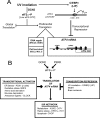
LIP repression of ATF4 transcription reduces levels of ATF4 mRNA available for preferential translation in response to eIF2α∼P during UV stress. A, model for LIP repression of ATF4 expression during UV stress. In response to UV irradiation, GCN2 phosphorylation of eIF2α lowers the levels of eIF2-GTP, resulting in reduced global translation. Additionally, eIF2α∼P leads to preferential translation of genes involved in repair of damaged DNA and those that thwart apoptosis, although the underlying mechanisms have not yet been determined (61). UV irradiation triggers repressed transcription of the ATF4 gene by increased C/EBPβ association at the ATF4 promoter sequences between −1000 and −879 bp. The LIP isoform of C/EBPβ is central for ATF4 repression, and this regulation is suggested to involve LIP engagement with promoter target sequences when LIP is dimerized with other bZIP proteins. The mechanism by which UV irradiation triggers increased LIP regulation of the ATF4 promoter is suggested to involve enhanced stabilization of C/EBPβ mRNA. The resulting lowered ATF4 mRNA levels diminish the amount of transcripts available for preferential translation in response to eIF2α∼P. The resulting loss of ATF4 expression during UV stress impedes the induction of its ISR target genes. B, a combination of transcriptional and translational control of ATF4 directs the gene expression program of the ISR. The eIF2 kinase GCN2 is activated by nutritional deprivation or UV irradiation, whereas PERK is regulated by ER stress. The resulting induced eIF2α∼P can lead to preferential translation of ATF4 by a mechanism involving delayed ribosome reinitiation, which ribosomes to bypass an inhibitory uORF in the ATF4 mRNA. Activation of ATF4 transcription by many different stresses enhances the amount of ATF4 mRNA available for translation in response to eIF2α∼P. Transcription factors that activate the ATF4 promoter include PDX1 in islet β-cells of the pancreas upon ER stress, NRF2 in response to oxidative stress, and CLOCK, which facilitates resistance to anticancer agents cisplatin and etoposide. As a consequence, there will be enhanced levels of ATF4 that directly activate the transcription of ISR target genes involved in metabolism, the redox status of cells, and regulation of apoptosis. Examples of target genes for each ISR category are illustrated. Alternatively, the ATF4 promoter can be repressed by a different set of stress conditions. The LIP isoform of C/EBPβ directly facilitates repression of ATF4 transcription in response to UV irradiation. This would result in low levels of ATF4 mRNA available for preferential translation during UV stress despite high levels of eIF2α∼P, thus lowering the expression of the ATF4 target genes in the ISR. NASH, non-alcoholic steatohepatitis.

Similar articles

Cited by

References

    1. Sonenberg N., Hinnebusch A. G. (2009) Regulation of translation initiation in eukaryotes: mechanisms and biological targets. Cell 136, 731–745 - PMC - PubMed
    1. Jackson R. J., Hellen C. U., Pestova T. V. (2010) The mechanism of eukaryotic translation initiation and principles of its regulation. Nat. Rev. Mol. Cell Biol. 11, 113–127 - PMC - PubMed
    1. Walter P., Ron D. (2011) The unfolded protein response: from stress pathway to homeostatic regulation. Science 334, 1081–1086 - PubMed
    1. Wek R. C., Jiang H. Y., Anthony T. G. (2006) Coping with stress: eIF2 kinases and translational control. Biochem. Soc. Trans. 34, 7–11 - PubMed
    1. Harding H. P., Novoa I., Zhang Y., Zeng H., Wek R., Schapira M., Ron D. (2000) Regulated translation initiation controls stress-induced gene expression in mammalian cells. Mol. Cell 6, 1099–1108 - PubMed

Publication types