. 2012 Jul 6;287(28):23489-501.
doi: 10.1074/jbc.M112.365874. Epub 2012 May 2.

Role of deleted in breast cancer 1 (DBC1) protein in SIRT1 deacetylase activation induced by protein kinase A and AMP-activated protein kinase

Affiliations

Role of deleted in breast cancer 1 (DBC1) protein in SIRT1 deacetylase activation induced by protein kinase A and AMP-activated protein kinase

Veronica Nin et al. J Biol Chem. .

Abstract

The NAD(+)-dependent deacetylase SIRT1 is a key regulator of several aspects of metabolism and aging. SIRT1 activation is beneficial for several human diseases, including metabolic syndrome, diabetes, obesity, liver steatosis, and Alzheimer disease. We have recently shown that the protein deleted in breast cancer 1 (DBC1) is a key regulator of SIRT1 activity in vivo. Furthermore, SIRT1 and DBC1 form a dynamic complex that is regulated by the energetic state of the organism. Understanding how the interaction between SIRT1 and DBC1 is regulated is therefore essential to design strategies aimed to activate SIRT1. Here, we investigated which pathways can lead to the dissociation of SIRT1 and DBC1 and consequently to SIRT1 activation. We observed that PKA activation leads to a fast and transient activation of SIRT1 that is DBC1-dependent. In fact, an increase in cAMP/PKA activity resulted in the dissociation of SIRT1 and DBC1 in an AMP-activated protein kinase (AMPK)-dependent manner. Pharmacological AMPK activation led to SIRT1 activation by a DBC1-dependent mechanism. Indeed, we found that AMPK activators promote SIRT1-DBC1 dissociation in cells, resulting in an increase in SIRT1 activity. In addition, we observed that the SIRT1 activation promoted by PKA and AMPK occurs without changes in the intracellular levels of NAD(+). We propose that PKA and AMPK can acutely activate SIRT1 by inducing dissociation of SIRT1 from its endogenous inhibitor DBC1. Our experiments provide new insight on the in vivo mechanism of SIRT1 regulation and a new avenue for the development of pharmacological SIRT1 activators targeted at the dissociation of the SIRT1-DBC1 complex.


Figures

FIGURE 1.
FIGURE 1.
Characterization of assay used to measure cellular SIRT1 activity. A, basic scheme showing the steps followed to measure cellular SIRT1 activity. Detailed information is provided under “Experimental Procedures.” B, measurement of endogenous cellular SIRT1 activity in 293T cells. Activity was measured in cellular extracts in the absence of exogenous NAD+ (−NAD+), with the addition of 100 μm NAD+ (+NAD), or with NAD+ plus 2 mm nicotinamide (NAD+Nic), with NAD+ plus 100 μm suramin (NAD+Sur), or NAD+ plus 10 μm EX527 (NAD+EX527). C, 293T cells were transfected with different amounts of a FLAG-SIRT1-coding plasmid. Cell lysates were immunoblotted with anti-SIRT1, anti-FLAG, and anti-tubulin antibodies. The graph on the left is the cellular SIRT1 activity measured 24 h after the transfection. The graph on the right shows the relationship between SIRT1 expression levels and cellular SIRT1 activity. D, SIRT1 activity was measured in MEFs obtained from WT and SIRT1 KO mice. E, SIRT1 and DBC1 were knocked down in HepG2 cells with specific siRNAs, and SIRT1 activity was assessed. Activity is shown as -fold change with respect to the control. *, p < 0.05 (ANOVA test, n = 3). Cell lysates were immunoblotted with anti-SIRT1, anti-DBC1, and anti-tubulin antibodies. F, cellular SIRT1 activity was measured in 293T cells transfected with FLAG-SIRT1, FLAG-SIRT1 + Myc-DBC1, or FLAG-SIRT1 + ΔLZ Myc-DBC1, a mutant DBC1 that does not have the leucine zipper domain and does not bind to SIRT1. *, p < 0.05 (ANOVA test, n = 3). G, cellular SIRT1 activity was measured in MEFs from WT, DBC1 KO, and AMPK (α1α2) KO mice. The activity of 1 unit of purified recombinant human SIRT1 was measured in parallel for the same time. *, p < 0.05 (ANOVA test, n = 3). Error bars represent S.D. AFU, arbitrary fluorescence units.
FIGURE 2.
FIGURE 2.
cAMP/PKA increase SIRT1 activity by mechanism that is independent of changes in NAD levels. SIRT1 activity was measured in A549 cells (A), mouse embryonic fibroblasts (B), and HepG2 cells (C) treated with 10 μm forskolin (FSK) or 100 μm cpt-cAMP for the indicated times. *, p < 0.01 (ANOVA, n = 3–9). SIRT1 activity was normalized to time 0. D, -fold change in NAD+ concentration in HepG2 cells after forskolin (10 μm) or cpt-cAMP (100 μm) incubation for 10 min (n = 3). E, SIRT1 was knocked down in HepG2 cells with specific siRNA, and SIRT1 activity was assessed after stimulation with 10 μm forskolin for 10 min. Activity is shown as -fold change with respect to the control. *, p < 0.05 (ANOVA test, n = 3). Cell lysates were immunoblotted with anti-SIRT1 and anti-tubulin antibodies. F, cells were pretreated for 45 min with the PKA inhibitor H89 (30 μm) or (Rp)-cAMP (100 μm) and then stimulated with 10 μm forskolin or 100 μm cpt-cAMP for 10 min. SIRT1 activity was normalized to the control. * shows significant difference with respect to the control (p < 0.01, ANOVA, n = 3). G, human recombinant purified SIRT1 activity in the presence of different compounds was measured in vitro. 0.2 unit of SIRT1 was incubated with the compounds at the indicated concentrations. H, HepG2 cells were pretreated for 45 min with the PKA inhibitor H89 (30 μm) or (Rp)-cAMP (Rp; 100 μm) and then stimulated with 10 μm forskolin for 10 min. Immunofluorescence for Ac-p53 and total p53 was analyzed using specific antibodies. I, cells were incubated with the PKA activator 6-MB-cAMP (100 μm) and the EPAC activator 8-CPT-2′-O-Me-cAMP; (100 μm) for 10 min before harvesting. SIRT1 activity was measured and normalized to the control. *, p < 0.01 (t test, n = 3). Error bars represent S.D. AU, arbitrary units.
FIGURE 3.
FIGURE 3.
SIRT1 activation by PKA is AMPK-dependent. A, activity of human recombinant purified SIRT1 (0.2 unit) was measured using a fluorometric assay after performing a kinase assay with the catalytic subunit of PKA in the presence or absence of 200 μm ATP. B, AMPK activation was measured by immunoblot using anti-Thr(P)-172 antibody in different cell lines after treatment with 10 μm forskolin (FSK) for different times. Compound C (CC; 10 μm) and (Rp)-cAMP (Rp; 100 μm) were added 2 h prior to the addition of forskolin. C, SIRT1 activity in A549 cells was measured after a 2-h incubation with A769662 (100 μm), AICAR (2 mm), and oligomycin (5 μm). Activity was expressed as the percentage of activity with respect to the control. *, p < 0.05 (ANOVA test, n = 3). AMPK activation by the different compounds was confirmed by Western blot (right) with anti-Thr(P)-172 antibody. D, determination of intracellular NAD+ levels in A549 cells treated as described in C. E, SIRT1 activity was measured in MEFs from WT, AMPK KO, and SIRT1 KO mice. Cells were incubated with A769662 (100 μm), AICAR (2 mm), or oligomycin (5 μm) for 2 h before measuring SIRT1 activity. SIRT1 activity was normalized to the respective control for each cell type. SIRT1 KO cells showed no detectable activity. * and **, p < 0.05 (ANOVA test, n = 3). F, SIRT1 activity was determined in MEFs from WT mice. The AMPK inhibitor compound C (10 μm) was added to the cells 2 h before starting the treatments. Cells were incubated with A769662 (100 μm), A769662 + compound C, AICAR (2 mm), AICAR + compound C, oligomycin (5 μm), and oligomycin + compound C for 2 h before measuring SIRT1 activity. SIRT1 activity was normalized to control. * and **, p < 0.05 (ANOVA test, n = 3). G, intracellular NAD+ levels in WT MEFs treated as described in F. H, AMPK was inhibited in HepG2 cells by a pretreatment with compound C (10 μm) for 2 h, and SIRT1 activity was assessed after stimulation with 10 μm forskolin for 10 min. Activity is shown as -fold change with respect to the control. *, p < 0.05 (ANOVA test, n = 3). I, SIRT1 activity was measured in AMPK WT and KO (α1α2) MEFS treated with 10 μm forskolin for the indicated times. *, p < 0.05 (ANOVA test, n = 3). Error bars represent S.D. AFU, arbitrary fluorescence units; P-AMPK, phosphorylated AMPK.
FIGURE 4.
FIGURE 4.
SIRT1 activation by cAMP/PKA/AMPK pathway depends on DBC1. A, SIRT1 activity was measured in WT and DBC1 KO MEFs. Cells were incubated with A769662 (100 μm) or A769662 + compound C (CC), AICAR (2 mm) or AICAR + compound C, and oligomycin (5 μm) or oligomycin + compound C for 2 h before measuring SIRT1 activity. Compound C was used at 10 μm and was preincubated for 2 h. Activity in the WT cells was normalized to the WT control, and activity in the KO cells was normalized to the KO control. SIRT1 activity in the control was always higher in DBC1 KO than in WT MEFs (see Fig. 1). *, p < 0.05 (ANOVA test, n = 3). B, DBC1 was knocked down in HepG2 cells with siRNA, and SIRT1 activity was assessed after stimulation with 10 μm forskolin (FSK) for 10 min. Activity is shown as -fold change with respect to the control. *, p < 0.05 (ANOVA test, n = 3). C, SIRT1 activity in DBC1 WT and KO MEFS treated with 10 μm forskolin for the indicated times. *, p < 0.05 (ANOVA test, n = 3). Error bars represent S.D.
FIGURE 5.
FIGURE 5.
AMPK activation induces dissociation of SIRT1 from DBC1. A and B, SIRT1-DBC1 interaction was evaluated by co-immunoprecipitation in A549 cells. A, cells were treated with resveratrol (100 μm), oligomycin (5 μm), or A769662 (100 μm) for 2 h before performing immunoprecipitation (IP) for DBC1. Proteins were immunoblotted with anti-SIRT1, anti-DBC1, anti-phosphorylated AMPK (P-AMPK) (Thr-172), and anti-AMPK antibodies. B, cells were treated with resveratrol (RSV; 100 μm) or resveratrol + compound C (CC; 10 μm) for 2 h before performing immunoprecipitation. Immunoprecipitates were immunoblotted with anti-SIRT1 and anti-DBC1 antibodies. The graph shows the average of three independent experiments. * Denotes difference to control, and ** denotes difference to RSV+CC. Error bars represent S.D. C, SIRT1-DBC1 interaction was evaluated by co-immunoprecipitation in 293T cells after transfection of FLAG-SIRT1, Myc-DBC1, and CA AMPα or dominant-negative AMPKα (DN). Immunoprecipitates were immunoblotted with anti-FLAG and anti-Myc antibodies.
FIGURE 6.
FIGURE 6.
cAMP-PKA activation promotes dissociation of SIRT1 from DBC1 by AMPK-dependent mechanism. A–C, co-immunoprecipitation (IP) of SIRT1 with DBC1 in A549 cells. DBC1 was immunoprecipitated from cell lysates, and immunoprecipitates were immunoblotted with anti-SIRT1 and anti-DBC1 antibodies. In A, cells were stimulated with forskolin (FSK) + isobutylmethylxanthine (IBMX) (10 and 70 μm, respectively) for the indicated times or the PKA activator 6-MB-cAMP (100 μm) for 10 min (′). B, cells were pretreated with the PKA inhibitor H89 (30 μm for 45 min) and then stimulated with forskolin and isobutylmethylxanthine (10 and 70 μm, respectively) for 10 min. C, cells were pretreated with compound C (CC; 10 μm) for 2 h and then stimulated as in B.
FIGURE 7.
FIGURE 7.
Activation of AMPK and PKA leads to SIRT1 phosphorylation. A, 293T cells were transfected with FLAG-SIRT1 or FLAG-SIRT1 plus a CA form of AMPK. The cell cultures were loaded with H332PO4, FLAG-SIRT1 was immunoprecipitated, and the radioactivity incorporated was visualized by autoradiography. B, 293T cells were transfected with FLAG-SIRT1, loaded with H332PO4, and later treated with 10 μm resveratrol for 2 h. SIRT1 phosphorylation was visualized by autoradiography. C, A549 cells were incubated with resveratrol (10 μm) or resveratrol and compound C (CC; 10 μm) for 2 h. Compound C was added 45 min before resveratrol. Samples were immunoblotted with anti-phospho (P)-SIRT1 (Ser-47). D, HepG2 cells were incubated with A769662 (100 μm) or A769662 and compound C (10 μm) for 2 h. E, Western blot with anti-phospho-SIRT1 (Ser-47) in 293T cells transfected with WT SIRT1 (FLAG (F)-SIRT1) or FLAG-S47RSIRT1. F, WT and the triple mutant (TM; S47R,S605R,S615R) SIRT1 were transfected in 293T cells. After 24 h, cells were incubated with forskolin (FSK; 10 μm) for 20 min. FLAG-SIRT1 was immunoprecipitated (IP), and the interaction was evaluated by immunoblot with anti-DBC1 and anti-FLAG antibodies. The graph shows the average of four experiments (**, p < 0.001; ANOVA test). G, SIRT1 activity was measured in the same experimental conditions described in B. Error bars represent S.D.
FIGURE 8.
FIGURE 8.
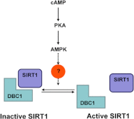
Proposed mechanism of SIRT1 regulation by PKA and AMPK.

Similar articles

Cited by

References

    1. Liu Y., Dentin R., Chen D., Hedrick S., Ravnskjaer K., Schenk S., Milne J., Meyers D. J., Cole P., Yates J., 3rd, Olefsky J., Guarente L., Montminy M. (2008) A fasting inducible switch modulates gluconeogenesis via activator/coactivator exchange. Nature 456, 269–273 - PMC - PubMed
    1. Rodgers J. T., Lerin C., Haas W., Gygi S. P., Spiegelman B. M., Puigserver P. (2005) Nutrient control of glucose homeostasis through a complex of PGC-1α and SIRT1. Nature 434, 113–118 - PubMed
    1. Bordone L., Motta M. C., Picard F., Robinson A., Jhala U. S., Apfeld J., McDonagh T., Lemieux M., McBurney M., Szilvasi A., Easlon E. J., Lin S. J., Guarente L. (2006) Sirt1 regulates insulin secretion by repressing UCP2 in pancreatic β cells. PLoS Biol. 4, e31. - PMC - PubMed
    1. Moynihan K. A., Grimm A. A., Plueger M. M., Bernal-Mizrachi E., Ford E., Cras-Mneur C., Permutt M. A., Imai S. (2005) Increased dosage of mammalian Sir2 in pancreatic β cells enhances glucose-stimulated insulin secretion in mice. Cell Metab. 2, 105–117 - PubMed
    1. Schenk S., McCurdy C. E., Philp A., Chen M. Z., Holliday M. J., Bandyopadhyay G. K., Osborn O., Baar K., Olefsky J. M. (2011) Sirt1 enhances skeletal muscle insulin sensitivity in mice during caloric restriction. J. Clin. Investig. 121, 4281–4288 - PMC - PubMed

Publication types

MeSH terms