. 2012 May 15;109(20):7865-70.
doi: 10.1073/pnas.1200081109. Epub 2012 May 1.

MicroRNAs miR-125a and miR-125b constitutively activate the NF-κB pathway by targeting the tumor necrosis factor alpha-induced protein 3 (TNFAIP3, A20)

Affiliations

MicroRNAs miR-125a and miR-125b constitutively activate the NF-κB pathway by targeting the tumor necrosis factor alpha-induced protein 3 (TNFAIP3, A20)

Sang-Woo Kim et al. Proc Natl Acad Sci U S A. .

Abstract

Constitutive activation of the NF-κB pathway is associated with diffuse large B-cell lymphoma (DLBCL) pathogenesis, but whether microRNA dysfunction can contribute to these events remains unclear. Starting from an integrative screening strategy, we uncovered that the negative NF-κB regulator TNFAIP3 is a direct target of miR-125a and miR-125b, which are commonly gained and/or overexpressed in DLBCL. Ectopic expression of these microRNAs in multiple cell models enhanced K63-linked ubiquitination of proximal signaling complexes and elevated NF-κB activity, leading to aberrant expression of its transcriptional targets and the development of a proproliferative and antiapoptotic phenotype in malignant B cells. Concordantly, genetic inhibition of miR-125a/miR-125b blunted NF-κB signals, whereas rescue assays and genetic modulation of a TNFAIP3-null model defined the essential role of the TNFAIP3 targeting on miR-125a/miR-125b-mediated lymphomagenesis. Importantly, miR-125a/mir-125b effects on TNFAIP3 expression and NF-κB activity were confirmed in a well-characterized cohort of primary DLBCLs. Our data delineate a unique epigenetic model for aberrant activation of the NF-κB pathway in cancer and provide a coherent mechanism for the role of these miRNAs in immune cell activation and hematopoiesis. Further, as miR-125b is a direct NF-κB transcriptional target, our results suggest the presence of a positive self-regulatory loop whereby termination of TNFAIP3 function by miR-125 could strengthen and prolong NF-κB activity.


Conflict of interest statement

The authors declare no conflict of interest.

Figures
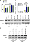
Fig. 1.
Fig. 1.
MiR-125a and miR-125b directly target TNFAIP3. (A) Luciferase constructs containing miR-125a/b binding sites in wild-type (WT) or mutant (Mut) configuration were cotransfected with miR-125a, miR-125b, or control oligonucleotides. MiR-125a and miR-125b inhibited luciferase activity in the TNFAIP3 WT but had no effect in the mutant construct (P < 0.05, Student’s t test). Data shown are mean ± SD of the ratio of luciferase activity (miR-125a or miR-125b vs. control oligo); experiments were performed in triplicate and repeated three times. (B) Lysates from Ly8 cells expressing MSCV, MSCV–miR-125a, or MSCV–miR-125b were immunoprecipitated with anti-Ago2 antibody or nonspecific IgG; RNA was isolated from the IP fractions and real-time RT-PCR used to demonstrate the significant enrichment for TNFAIP3 in the immunoprecipitates of miR-125a and miR-125b loaded RISC. Data shown are mean ± SD of TNFAIP3 relative abundance in the various Ago2-IPs. RISC-associated GAPDH was used for normalization. (C) Western blots depict TNFAIP3 inhibition in DLBCL cell lines stably expressing miR-125a and miR-125b. (D) TNFAIP3 expression is elevated in DLBCL cell lines transiently transfected with miR-125a– or miR-125b–specific antagomiRs. Densitometric quantification of TNFAIP3 expression, normalized by actin, is shown in C and D.
Fig. 2.
Fig. 2.
MiR-125a and miR-125b expression modulates NF-κB activity in DLBCL. (A) Immunoblot analyses of phospho-IκBα and IκBα after TNFα stimulation. Phosphorylation and degradation of IκBα are elevated in DLBCL cell lines expressing miR-125a or miR-125b. Ratios of p-IκBα/IκBα are listed in blue (control) and red fonts (miR-125a or miR-125b) and were normalized to time 0 of MSCV-only cells. (B) Ectopic expression of miR-125a and miR-125b increase accumulation of p65 in the nuclear fraction of DLBCL cell lines. Data shown in A and B were independently confirmed in two to three biological replicates. Densitometric quantification of p65 nuclear expression, normalized by H3, is shown. (C) Heat-map display of quantitative real-time RT-PCR measurements of six independent NF-κB transcriptional targets show significantly higher expression in miR-125a and miR-125b expressing cells (P < 0.05, Student’s t test). Data displayed are mean ± SD of the mRNA fold induction following exposure to TNFα (200 ng/mL for 120 min), normalized by MSCV-only cells. All assays were performed in triplicate and confirmed in three biological replicates. (D) AntagomiR-mediated inhibition of miR-125a or miR-125b expression in DLBCL cell lines with high basal NF-κB activity significantly inhibits this pathway, as determined by an ELISA-based measurement of p65 levels (*P < 0.05, Student’s t test). Data shown are mean ± SD of a representative assay performed in triplicate and independently confirmed in three biological replicates.
Fig. 3.
Fig. 3.
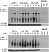
miR-125a and miR-125b expression modulate K63-linkage ubiquitination. Immunoprecipitation of TRAF2 (Upper) or RIP1 (Lower) followed by immunoblotting for K63-linkage ubiquitin, in isogenic Ly8 cells stably expressing miR-125a, miR-125b, or empty vector show that these miRNAs enhance the K63 ubuiquitination that follows engagement of the TNFR1 by TNFα (100 ng/mL). Data were confirmed in four biological replicates. Immunoblotting with TRAF2 and RIP1 antibodies confirm equal amount of these proteins in the pulldowns, and IgG controls confirm the specificity of the IPs.
Fig. 4.
Fig. 4.
Targeting of TNFAIP3 is essential for the miR125a/b–mediated NF-κB activation in DLBCL. (A) Immunoblot analyses of p-IκBα and IκBα show that stable reconstitution of TNFAIP3 expression (Upper) rescued the effects of miR-125a and miR-125b in DLBCL cells; note the markedly lower ratio IκBα phosphorylation/degradation in TNFAIP3–FLAG-expressing cells in comparison with isogenic MSCV–puromycin-expressing cells. MiR-125a/miR-125b–expressing DLBCL cell lines reconstituted with TNFAIP3–FLAG also displayed a limited nuclear accumulation of p65 following TNFα stimulation (Lower). (B) ELISA-based measurement of p65 activity shows that stable expression of miR-125a and miR-125b in the TNFAIP3-null DHL2 cell line has minimal effect on NF-κB function, in statistically significant contrast to the effects of these miRNAs in a TNFAIP3-competent DLBCL cell line (Ly7) (*P < 0.05, Student’s t test). Data shown are mean ± SD (four independent data points) of NF-κB induction following exposure to TNFα (100 ng/mL), normalized by MSCV-only cells.
Fig. 5.
Fig. 5.
MiR-125 targeting of TNFAIP3 enhances DLBCL aggressiveness. (A) DLBCL cell lines stably expressing miR-125a or miR-125b grow at significantly faster pace than their empty-vector isogenic counterparts. (B) Ectopic expression of miR-125a or miR-125b enhanced colony-formation capability in soft agar in all DLBCL models examined. (C) DLBCL cells expressing miR-125a or miR-125b became resistant to apoptosis induced by H202 and serum deprivation. Ctrl (control) and “10%” data indicate the basal apoptosis rate in cells exposed to vehicle or grown in media supplemented with 10% FBS, respectively. (D) Reconstitution of TNFAIP3 expression rescued DLBCLs from the miR-125a and miR-125b–induced antiapoptotic effects. All assays were performed in triplicate and repeated from two to four times; *P < 0.05, Student’s t test.
Fig. 6.
Fig. 6.
MiR-125a/b expression in primary DLBCL modulates TNFAIP3 expression and NF-κB activity. (A) Western blot analysis showed a significant suppression in TNFAIP3 expression in tumors with high (H1–H10) vs. low miR-125a and/or miR-125b levels (L1–L8) (P < 0.01 Mann–Whitney, normalized densitometric values (also Fig. S7). *, samples with copy number gain at the miR-125b1 locus; #, samples with hemizygous loss of miR-125; ^, samples monoallelic mutation or loss of TNFAIP3. (B) ELISA-based quantification of p65 in protein lysates from primary DLBCLs showed significantly higher NF-κB activity (P < 0.01, Student’s t test) in tumors expressing high miR-125a and/or miR-125b levels and consequently low TNFAIP3; p65 measurements were performed in duplicate and all 36 values are included in the display. Data points labeled in red correspond to the samples with copy number gain at the miR-125b1 locus; those with loss of miR-125b are shown in gold; and the samples with monoallelic loss or mutation in TNFAIP3 are shown in black.

Similar articles

Cited by

References

    1. Baltimore D. NF-κB is 25. Nat Immunol. 2011;12:683–685. - PubMed
    1. Hymowitz SG, Wertz IE. A20: From ubiquitin editing to tumour suppression. Nat Rev Cancer. 2010;10:332–341. - PubMed
    1. Skaug B, et al. Direct, noncatalytic mechanism of IKK inhibition by A20. Mol Cell. 2011;44:559–571. - PMC - PubMed
    1. Shembade N, Ma A, Harhaj EW. Inhibition of NF-kappaB signaling by A20 through disruption of ubiquitin enzyme complexes. Science. 2010;327:1135–1139. - PMC - PubMed
    1. Ben-Neriah Y, Karin M. Inflammation meets cancer, with NF-κB as the matchmaker. Nat Immunol. 2011;12:715–723. - PubMed

Publication types

MeSH terms

LinkOut - more resources