

Epigenetic resensitization to platinum in ovarian cancer
- PMID: 22549947
- PMCID: PMC3700422
- DOI: 10.1158/0008-5472.CAN-11-3909
Epigenetic resensitization to platinum in ovarian cancer
Abstract
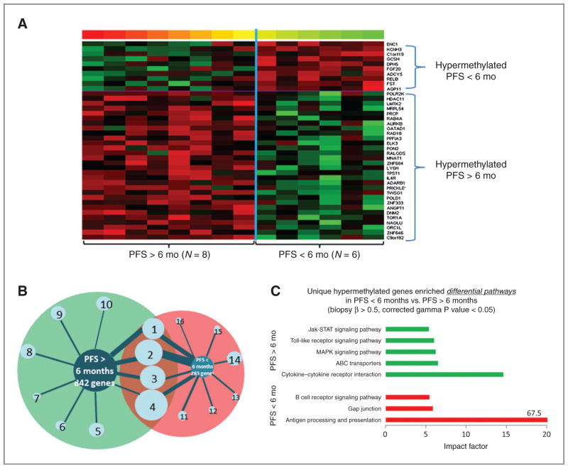
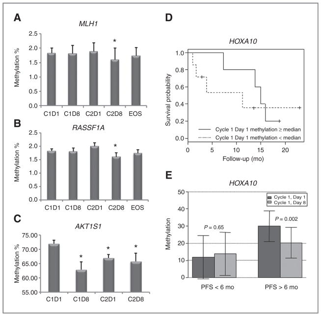
Preclinical studies have shown that hypomethylating agents reverse platinum resistance in ovarian cancer. In this phase II clinical trial, based upon the results of our phase I dose defining study, we tested the clinical and biologic activity of low-dose decitabine administered before carboplatin in platinum-resistant ovarian cancer patients. Among 17 patients with heavily pretreated and platinum-resistant ovarian cancer, the regimen induced a 35% objective response rate (RR) and progression-free survival (PFS) of 10.2 months, with nine patients (53%) free of progression at 6 months. Global and gene-specific DNA demethylation was achieved in peripheral blood mononuclear cells and tumors. The number of demethylated genes was greater (P < 0.05) in tumor biopsies from patients with PFS more than 6 versus less than 6 months (311 vs. 244 genes). Pathways enriched at baseline in tumors from patients with PFS more than 6 months included cytokine-cytokine receptor interactions, drug transporters, and mitogen-activated protein kinase, toll-like receptor and Jak-STAT signaling pathways, whereas those enriched in demethylated genes after decitabine treatment included pathways involved in cancer, Wnt signaling, and apoptosis (P < 0.01). Demethylation of MLH1, RASSF1A, HOXA10, and HOXA11 in tumors positively correlated with PFS (P < 0.05). Together, the results of this study suggest that low-dose decitabine altered DNA methylation of genes and cancer pathways, restoring sensitivity to carboplatin in patients with heavily pretreated ovarian cancer and resulting in a high RR and prolonged PFS.
©2012 AACR
Conflict of interest statement
D. Matei and K.P. Nephew are consultants and on the advisory board of Supergen (now Astex Pharmaceuticals, Inc.).
Figures


Similar articles
-
A randomised, phase II trial of the DNA-hypomethylating agent 5-aza-2'-deoxycytidine (decitabine) in combination with carboplatin vs carboplatin alone in patients with recurrent, partially platinum-sensitive ovarian cancer.Br J Cancer. 2014 Apr 15;110(8):1923-9. doi: 10.1038/bjc.2014.116. Epub 2014 Mar 18. Br J Cancer. 2014. PMID: 24642620 Free PMC article. Clinical Trial.
-
A phase 1 and pharmacodynamic study of decitabine in combination with carboplatin in patients with recurrent, platinum-resistant, epithelial ovarian cancer.Cancer. 2010 Sep 1;116(17):4043-53. doi: 10.1002/cncr.25204. Cancer. 2010. PMID: 20564122 Free PMC article. Clinical Trial.
-
Carboplatin with Decitabine Therapy, in Recurrent Platinum Resistant Ovarian Cancer, Alters Circulating miRNAs Concentrations: A Pilot Study.PLoS One. 2015 Oct 20;10(10):e0141279. doi: 10.1371/journal.pone.0141279. eCollection 2015. PLoS One. 2015. PMID: 26485143 Free PMC article. Clinical Trial.
-
Restoring platinum sensitivity in recurrent ovarian cancer by extending the platinum-free interval: Myth or reality?Cancer. 2017 Sep 15;123(18):3450-3459. doi: 10.1002/cncr.30830. Epub 2017 Jul 5. Cancer. 2017. PMID: 28678350 Review.
-
Platinum versus platinum-combination chemotherapy in platinum-sensitive recurrent ovarian cancer: a meta-analysis using individual patient data.Ann Oncol. 2013 Dec;24(12):3028-34. doi: 10.1093/annonc/mdt406. Epub 2013 Nov 4. Ann Oncol. 2013. PMID: 24190964 Review.
Cited by
-
Targeting the cancer epigenome for therapy.Nat Rev Genet. 2016 Sep 15;17(10):630-41. doi: 10.1038/nrg.2016.93. Nat Rev Genet. 2016. PMID: 27629931 Review.
-
Expression profiling of DNA methyl transferase I (DNMT1) and efficacy of a DNA-hypomethylating agent (decitabine) in combination with chemotherapy in osteosarcoma.J Bone Oncol. 2020 Sep 22;25:100321. doi: 10.1016/j.jbo.2020.100321. eCollection 2020 Dec. J Bone Oncol. 2020. PMID: 33072501 Free PMC article.
-
3D culture adds an extra dimension to targeted epigenetic therapies.Cell Cycle. 2013 Jul 15;12(14):2173-4. doi: 10.4161/cc.25551. Cell Cycle. 2013. PMID: 23803725 Free PMC article. No abstract available.
-
Epigenomic heterogeneity as a source of tumour evolution.Nat Rev Cancer. 2025 Jan;25(1):7-26. doi: 10.1038/s41568-024-00757-9. Epub 2024 Oct 16. Nat Rev Cancer. 2025. PMID: 39414948 Review.
-
Understanding and Targeting Apoptotic Pathways in Ovarian Cancer.Cancers (Basel). 2019 Oct 24;11(11):1631. doi: 10.3390/cancers11111631. Cancers (Basel). 2019. PMID: 31652965 Free PMC article. Review.
References
-
- Bukowski RM, Ozols RF, Markman M. The management of recurrent ovarian cancer. Semin Oncol. 2007;34:S1–15. - PubMed
-
- Wei SH, Chen CM, Strathdee G, Harnsomburana J, Shyu CR, Rahmatpanah F, et al. Methylation microarray analysis of late-stage ovarian carcinomas distinguishes progression-free survival in patients and identifies candidate epigenetic markers. Clin Cancer Res. 2002;8:2246–52. - PubMed
-
- Barton CA, Hacker NF, Clark SJ, O’Brien PM. DNA methylation changes in ovarian cancer: implications for early diagnosis, prognosis and treatment. Gynecol Oncol. 2008;109:129–39. - PubMed
Publication types
MeSH terms
Substances
Grants and funding
LinkOut - more resources
Full Text Sources
Other Literature Sources
Medical
