. 2012 Jul;32(13):2608-17.
doi: 10.1128/MCB.00182-12. Epub 2012 Apr 30.

The super elongation complex family of RNA polymerase II elongation factors: gene target specificity and transcriptional output

Affiliations

The super elongation complex family of RNA polymerase II elongation factors: gene target specificity and transcriptional output

Zhuojuan Luo et al. Mol Cell Biol. 2012 Jul.

Abstract

The elongation stage of transcription is highly regulated in metazoans. We previously purified the AFF1- and AFF4-containing super elongation complex (SEC) as a major regulator of development and cancer pathogenesis. Here, we report the biochemical isolation of SEC-like 2 (SEC-L2) and SEC-like 3 (SEC-L3) containing AFF2 and AFF3 in association with P-TEFb, ENL/MLLT1, and AF9/MLLT3. The SEC family members demonstrate high levels of polymerase II (Pol II) C-terminal domain kinase activity; however, only SEC is required for the proper induction of the HSP70 gene upon stress. Genome-wide mRNA-Seq analyses demonstrated that SEC-L2 and SEC-L3 control the expression of different subsets of genes, while AFF4/SEC plays a more dominant role in rapid transcriptional induction in cells. MYC is one of the direct targets of AFF4/SEC, and SEC recruitment to the MYC gene regulates its expression in different cancer cells, including those in acute myeloid or lymphoid leukemia. These findings suggest that AFF4/SEC could be a potential therapeutic target for the treatment of leukemia or other cancers associated with MYC overexpression.


Figures

Fig 1
Fig 1
Characterization of AFF2 and AFF3-containing complexes. (A) Purification of AFF2 and AFF3 complexes. Flag affinity purifications were performed to purify the AFF2- or AFF3-containing complex. The purified complexes were then subjected to silver staining and MudPIT analyses. (B) Comparison among AFF2-, AFF3-, and AFF4-containing complexes. P-TEFb, ENL, and AF9 were identified in all of the Flag-AFF2, -AFF3, and -AFF4 purifications. A low level of AFF4 was also detected in the AFF2 purification. However, ELL1, ELL2, and ELL3 could not be reproducibly detected by MudPIT analyses in the purified AFF2 and AFF3 complexes. The CDK9 purification contains AFF2, AFF3, and all of the subunits of previously identified P-TEFb-containing complexes, including SEC, BRD4/P-TEFb, and HEXIM1/P-TEFb. Numbers are distributed normalized spectral abundance factors (dNSAF) (59) averaged across 5, 7, 7, and 4 replicate analyses for AFF2, AFF3, AFF4, and CDK9, respectively. (C) Interaction between AFF family members and P-TEFb, ENL, ELL2, and ELL3 assessed by endogenous coimmunoprecipitation. 293T cell lysates were immunoprecipitated using antibodies to AFF1, AFF2, AFF3, AFF4, CDK9, or normal rabbit IgG; Western blotting was carried out to determine the levels of CDK9, cyclin T1 (CCNT1), ENL, ELL2, and ELL3 in the input and immunoprecipitated samples. CDK9, cyclin T1, and ENL were found to associate with all of the AFF proteins, while ELL2 and ELL3 were detected only in the AFF1-, AFF4-, and CDK9-immunoprecipitated samples. (D and E) Size exclusion chromatography of the Flag-AFF2 (D) and Flag-AFF3 (E) complexes were analyzed by silver staining and Western blotting. Western blotting using a Flag antibody reveals that the AFF2 complex is eluted from fractions 11 to 14 (D) and that the AFF3 complex is eluted from fractions 10 to 14 (E). The presence of ENL, AF9, CDK9, and cyclin T1 in these two complexes, as detected by Western blotting, is shown. (F) Schematic models of different SECs. The P-TEFb-containing complex SEC also contains a dynamic combination of ELL1 to ELL3, AFF1 and AFF4, and AF9/ENL. EAF1/2 can exist in SEC through binding to ELL1 to ELL3. The AFF2 and AFF3 complexes, named SEC-L2 and SEC-L3, respectively, contain P-TEFb and AF9/ENL. The presence of ELL- and EAF-related proteins has not been experimentally confirmed but is expected based on sequence conservation among AFF family members.
Fig 2
Fig 2
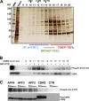
In vitro Pol II CTD kinase activities of P-TEFb-containing complexes. (A) Purification and size exclusion chromatography of P-TEFb complexes. P-TEFb complexes were isolated from Flag-CDK9-expressing HEK293T cells by Flag purification. Size exclusion chromatography was used to separate different P-TEFb complexes, including SEC, SEC-L2, and SEC-L3 (peak from fractions 10 to 14), the BRD4/P-TEFb complex (peak in fractions 14 and 15), and the HEXIM1/7SK/P-TEFb complex (peak from fraction 15 to 19) (28, 43). The fractions were analyzed by silver staining. (B) Pol II CTD kinase activity analyses of the fractionated P-TEFb complexes. The amount of each fraction used was adjusted to ensure that similar amounts of CDK9 were present in each assay with [γ-32P]ATP and recombinant Pol II CTD. The reaction products were then subjected to SDS-PAGE and autoradiography to assess the phosphorylated Pol II levels in each reaction. (C) Pol II CTD kinase activity analyses of SEC, SEC-L2, and SEC-L3. Flag-AFF4 (SEC), Flag-AFF2 (SEC-L2), Flag-AFF3 (SEC-L3), and Flag-CDK9 were assayed for Pol II CTD kinase activity as for panel B. Flag purification from wild-type HEK293T cells was used as a negative control (CTR). Triangles indicate decreasing titrations of CDK9-containing complexes.
Fig 3
Fig 3
AFF4, but not AFF2 or AFF3, is required for proper HSP70 induction upon heat shock. (A) Schematic models indicating the positions of qPCR primer sets along the HSP70 gene. (B) AFF4, but not AFF2 or AFF3, is recruited to the HSP70 gene upon heat shock. HEK293T cells were left untreated or heat shocked by incubating at 42°C for 2 h. Non-heat-shocked and heat-shocked cells were used in ChIP assays with AFF2, AFF3, AFF4, and Pol II antibodies. The nonexpressed beta-globin gene (Hemo) served as a negative control. Error bars represent standard deviations. (C and D) RT-qPCR (C) and Northern blot (D) analyses of HSP70 mRNA levels upon AFF2, AFF3, and AFF4 knockdown. HEK293T cells were transfected with control (CTR), AFF2, AFF3, and AFF4 siRNA; 72 h after transfection, the cells were left untreated or heat shocked by incubating at 42°C for 2 h. Total RNA was extracted. (C) HSP70 mRNA levels in each sample were measured by RT-qPCR. GAPDH served as an internal control. Error bars represent standard deviations. (D) Total RNA (5 μg) from each sample was assessed by Northern blot analysis with a riboprobe complementary to HSP70. Hybridization with a histone H1B (HIST1H1B) riboprobe was used as a loading control.
Fig 4
Fig 4
SEC, SEC-L2, and SEC-L3 control the expression of different subsets of genes. (A and B) RT-qPCR showing the specificity and efficiency of shRNA-mediated AFF2, AFF3, and AFF4 knockdown in HEK293T cells. (C) MA plots showing differentially expressed genes from RNAi of AFF2, AFF3, and AFF4. The y axis (M) of each of the nine plots shows the log2 change (fold) of gene expression levels of RNAi over wild-type values; the x axis (A) of each plot shows the log2 average fragment per million reads per kb of exon as reported by Cufflinks. Columns show MA plots; rows depict differentially expressed genes. For example, the plot in the top row and middle column shows how the differentially expressed genes identified in the AFF2 RNAi are expressed in the AFF3 RNAi condition. This analysis indicates that AFF4-regulated genes are very differently regulated in the AFF2 and AFF3 RNAi conditions, but AFF2- and AFF3-regulated genes are more similar to each other. (D) Venn diagram of the differentially expressed genes from RNAi of AFF2, AFF3, and AFF4.
Fig 5
Fig 5
SEC regulates MYC gene expression. (A and B) AFF4 and Pol II occupancy at the MYC (A) and ADAMTS1 loci (B). Genome-wide analyses of AFF4 and Pol II occupancy in HEK293T cells by ChIP-Seq demonstrate that AFF4 is recruited to the MYC (A) and ADAMTS1 genes (B) and travels with Pol II throughout the transcript unit. (C and D) mRNA levels of MYC (C) and ADAMTS1 (D) are reduced by the depletion of AFF4 but not AFF2 or AFF3. Genome browser tracks of the MYC (C) and ADAMTS1 (D) loci are shown for shRNA-mediated knockdown of AFF2, AFF3, or AFF4 and for a nontargeting (NonT) shRNA. (E and F) RT-qPCR analyses of MYC and ADAMTS1 mRNA levels upon siRNA-mediated AFF2, AFF3, and AFF4 knockdown. HEK293T cells were transfected with control (CTR), AFF2, AFF3, and AFF4 siRNA; 72 h after transfection, the cells were harvested and total RNA was extracted. MYC (E) and ADAMTS1 (F) mRNA levels in each sample were measured by RT-qPCR. GAPDH served as an internal control. Error bars represent standard deviations.
Fig 6
Fig 6
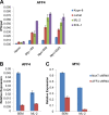
MYC expression in leukemia cells is regulated by AFF4. (A) AFF4 is recruited to the MYC gene in different leukemia cell lines. ChIP of AFF4 in ALL cell lines (Jurkat and Kopn-8) and AML cell lines (EOL-1 and ML-2) demonstrates the recruitment of AFF4 to the MYC gene in these cell lines. (B) RT-qPCR analysis showing the efficiency of AFF4 knockdown in leukemia cell lines SEM (human ALL with MLL-AFF1 translocation) and ML-2 (human AML cells). (C) RT-qPCR analysis of MYC mRNA levels upon AFF4 knockdown in leukemic cell lines. SEM and ML-2 were transfected with nontargeting or AFF4 shRNA. At 72 h after transfection and puromycin selection, total RNA was extracted and MYC mRNA levels were assessed by RT-qPCR. Expression is relative to GAPDH. Error bars represent standard deviations.

Similar articles

Cited by

References

    1. Bitoun E, Davies KE. 2005. The robotic mouse: unravelling the function of AF4 in the cerebellum. Cerebellum 4:250–260 - PubMed
    1. Bitoun E, Oliver PL, Davies KE. 2007. The mixed-lineage leukemia fusion partner AF4 stimulates RNA polymerase II transcriptional elongation and mediates coordinated chromatin remodeling. Hum. Mol. Genet. 16:92–106 - PubMed
    1. Boettiger AN, Levine M. 2009. Synchronous and stochastic patterns of gene activation in the Drosophila embryo. Science 325:471–473 - PMC - PubMed
    1. Bres V, Yoh SM, Jones KA. 2008. The multi-tasking P-TEFb complex. Curr. Opin. Cell Biol. 20:334–340 - PMC - PubMed
    1. Bursen A, Moritz S, Gaussmann A, Dingermann T, Marschalek R. 2004. Interaction of AF4 wild-type and AF4.MLL fusion protein with SIAH proteins: indication for t(4;11) pathobiology? Oncogene 23:6237–6249 - PubMed

Publication types

MeSH terms