. 2012;7(4):e34914.
doi: 10.1371/journal.pone.0034914. Epub 2012 Apr 23.

Dasatinib as a bone-modifying agent: anabolic and anti-resorptive effects

Affiliations

Dasatinib as a bone-modifying agent: anabolic and anti-resorptive effects

Antonio Garcia-Gomez et al. PLoS One. 2012.

Abstract

Background: Bone loss, in malignant or non-malignant diseases, is caused by increased osteoclast resorption and/or reduced osteoblast bone formation, and is commonly associated with skeletal complications. Thus, there is a need to identify new agents capable of influencing bone remodeling. We aimed to further pre-clinically evaluate the effects of dasatinib (BMS-354825), a multitargeted tyrosine kinase inhibitor, on osteoblast and osteoclast differentiation and function.

Methods: For studies on osteoblasts, primary human bone marrow mensenchymal stem cells (hMSCs) together with the hMSC-TERT and the MG-63 cell lines were employed. Osteoclasts were generated from peripheral blood mononuclear cells (PBMC) of healthy volunteers. Skeletally-immature CD1 mice were used in the in vivo model.

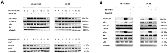
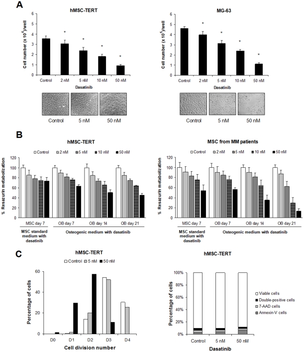
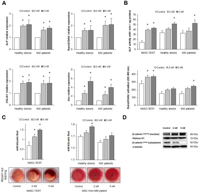
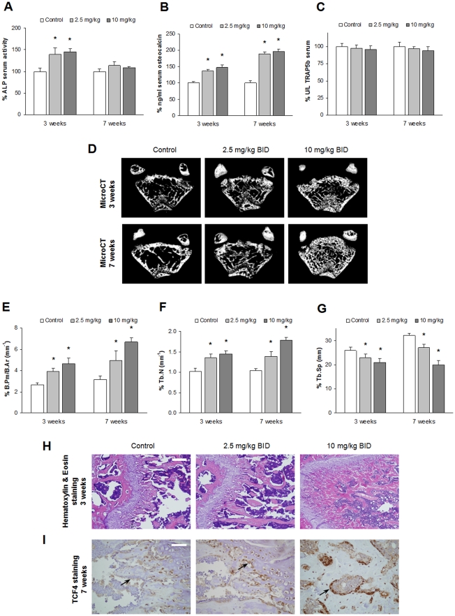
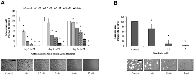
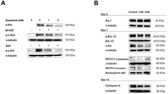
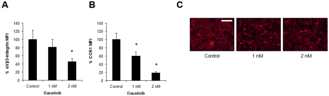
Results: Dasatinib inhibited the platelet derived growth factor receptor-β (PDGFR-β), c-Src and c-Kit phosphorylation in hMSC-TERT and MG-63 cell lines, which was associated with decreased cell proliferation and activation of canonical Wnt signaling. Treatment of MSCs from healthy donors, but also from multiple myeloma patients with low doses of dasatinib (2-5 nM), promoted its osteogenic differentiation and matrix mineralization. The bone anabolic effect of dasatinib was also observed in vivo by targeting endogenous osteoprogenitors, as assessed by elevated serum levels of bone formation markers, and increased trabecular microarchitecture and number of osteoblast-like cells. By in vitro exposure of hemopoietic progenitors to a similar range of dasatinib concentrations (1-2 nM), novel biological sequelae relative to inhibition of osteoclast formation and resorptive function were identified, including F-actin ring disruption, reduced levels of c-Fos and of nuclear factor of activated T cells 1 (NFATc1) in the nucleus, together with lowered cathepsin K, αVβ3 integrin and CCR1 expression.

Conclusions: Low dasatinib concentrations show convergent bone anabolic and reduced bone resorption effects, which suggests its potential use for the treatment of bone diseases such as osteoporosis, osteolytic bone metastasis and myeloma bone disease.


Conflict of interest statement

Competing Interests: FYL is an employee of Bristol-Myers Squibb. This does not alter the authors' adherence to all the PLoS ONE policies on sharing data and materials. There are no patents or products in development to declare.

Figures

Figure 1
Figure 1. Dasatinib inhibits PDGFR-β, c-Kit and c-Src phosphorylation in mesenchymal and osteoblast-like cell lines.
(A) Mesenchymal (hMSC-TERT) and osteoblast-like (MG-63) cell lines were pretreated with different concentrations of dasatinib for 6 hours and then exposed to PDGF-BB or SCF for 20 minutes before protein lysates were generated. Immunoblotting with specific antibodies against total and phosphorylated PDGFR-β, c-Kit and c-Src were performed. (B) Modulation of downstream signaling after dasatinib treatment. Similarly to experimental conditions in (A), the hMSC-TERT and the MG-63 cell lines were pretreated with 50 nM dasatinib for 6 hours, stimulated with PDGF-BB for 20 minutes and then cell harvested for protein isolation. Immunoblotting is shown for total and phosphorylated forms of PDGFR-β, c-Src, Erk 1/2, Akt and p38 mitogen activated protein kinase (MAPK).
Figure 2
Figure 2. Dasatinib reduces the number of viable cells by inhibition of mesenchymal and OB cell proliferation.
(A) Dasatinib decreases the number of MSC and OB cells in culture. The hMSC-TERT and the MG-63 cell lines were cultured for 7 days in maintenance medium in the absence or presence of increasing dasatinib concentrations, and then the number of cells at each condition was counted with a haemocytometer and a Trypan Blue solution. Representative micrographs are shown. (B) Dasatinib reduced the number of viable cells in osteogenic cultures in a time and concentration-dependent manner. MSCs were maintained in osteogenic medium for 7, 14 or 21 days in the presence of different dasatinib concentrations, and percentage of viable cells was evaluated with the alamarBlue assay on OBs derived from the hMSC-TERT (left) and from primary MSCs from MM patients (right). Data are expressed as the mean ± SD from three experiments. Statistically significant differences from control are indicated as *P<0.05. (C) Dasatinib (5–50 nM) reduces the number of cell divisions in the hMSC-TERT cell line (left) but does not induce apoptosis (right). MSCs were stained with PKH67 and cultured in osteogenic medium for 7 days in the absence or presence of dasatinib; at the time of collection, cells were also stained with Annexin-V-PE and 7-AAD and analyzed by flow cytometry.
Figure 3
Figure 3. Dasatinib promotes osteogenic differentiation of MSCs from MM patients and healthy donors and of the hMSC-TERT cell line.
(A) Dasatinib upregulates the expression of bone-formation markers in the osteogenic differentiation process. Primary MSCs from MM patients and healthy donors were cultured in osteogenic medium in the presence (2–5 nM) or absence of dasatinib, and total RNA was isolated on days 7 and 14. Real-time qRT-PCR was used to determine the expression of several OB related markers: ALP was determined at day 7, whereas the transcription factors Runx2/Cbfa1 and Osterix (Osx), and collagen I type A 1 (COLIA1) were measured at day 14. Expression levels for each gene were normalized to GAPDH expression and referred to those in the absence of dasatinib. Graphs illustrate mean values from samples from 5 healthy donors and 5 MM patients ± SEM (bars) *P<0.05. (B) Dasatinib increases ALP and Runx2/Cbfa1 activities in osteoprogenitor cells. In the hMSC-TERT cell line and in primary hMSCs derived from three myeloma patients and three healthy donors, ALP activity was measured at day 7 (upper graph) and Runx2/Cbfa1 activity was measured at day 14 (lower graph) after the addition of dasatinib to the osteogenic differentiation medium. Data are represented as the mean ± SD from three experiments. (C) Dasatinib (2–5 nM) augments bone matrix mineralization in OBs derived from the hTERT-MSC cell line (left) or MSCs from healthy donors and myeloma patients (right), as assessed by alizarin red staining quantification. Data are represented as the mean ± SD from three experiments with the hMSC-TERT cell line, and as the mean (5 MM patients and 5 healthy donors) ± SEM in experiments with primary MSCs. Statistically significant differences from controls are indicated as *, where P < 0.05. Micrographs show matrix mineralization after alizarin red staining of correspondent differentiated OBs. (D) Both dephospho- and phospho-β-catenin levels were determined by immunoblotting in cytosolic or nuclear lysates of pre-OBs differentiated from the hMSC-TERT cell line in the absence or presence of dasatinib. Histone H1 and α-tubulin were used as loading controls for nuclear or cytosolic protein fractions.
Figure 4
Figure 4. Dasatinib promotes trabecular bone formation in vivo.
(A, B, C) Five-week-old CD1 mice were treated with vehicle (control) or with dasatinib in a 2.5 mg/kg BID or a 10 mg/kg BID regimen for 3 or 7 weeks, and serum levels were determined for ALP (A), osteocalcin (B) or TRAP5b (C) before initiation of the experiment and at each time point. Graphs are plotted as mean values of fold change from baseline levels for the mentioned factors in sera ± SEM (bars). *, P<0.05 indicates significant differences between levels for each time and dose of dasatinib and untreated mice at the same conditions (control). (D) Representative micro-CT analyses of equivalent cross-sections of distal femurs are shown for each dasatinib concentration and time of treatment. (E, F, G) Trabecular bone morphometric parameters from micro-CT images were quantitated by CT-Analyser software for bone perimeter per area (E), trabecular number (F) and trabecular separation (G). *, P<0.05 relative to vehicle control at each time-point; n = 3 femurs per group. (H) Representative femur sections treated with both dasatinib doses for 3 weeks and stained with hematoxylin and eosin. Bar = 50 µm. (I) Representative images of Tcf4 immunohistochemistry in dasatinib-treated femurs for 7 weeks. OB-like cells immunostained for Tcf4 can be observed lining the trabeculae (arrows). Bar = 12.5 µm.
Figure 5
Figure 5. Dasatinib treatment inhibits OC formation and resorption activity.
(A) PBMCs from healthy donors were cultured in medium containing M-CSF/RANKL for 21 days in the absence or presence of dasatinib for the indicated times, and OCs were counted (as assessed by TRAP+ staining and the presence of more than three nuclei). Representative micrographs of TRAP staining for OCs treated with dasatinib for 3 weeks are shown. Bar =  0 μm. (B) OCs were generated on calcium-coated slides, and the effect of different dasatinib concentrations on OC resorption was evaluated by calculation of the total area of resorbed lacunae. Graphs represent mean values of samples from OCs derived from three healthy donors ± SEM (bars). *, P<0.05 indicates significant differences between dasatinib-treated cultures and untreated control at the same conditions. Representative micrographs of resorbed lacunae on the calcium-coated wells are shown. Bar = 30 μm.
Figure 6
Figure 6. Dasatinib regulates the expression of important molecules/factors for OC formation, differentiation and activity.
(A) Dasatinib inhibits c-Fms, c-Src, and c-Kit tyrosine kinase phosphorylation in committed OC precursors. PBMCs were differentiated in osteoclastogenic medium for 7 days, pretreated with 1 nM or 2 nM dasatinib or vehicle, and exposed to 50 ng/mL M-CSF or 50 nM SCF for 20 minutes prior to protein isolation. Immunoblotting with specific antibodies was performed as indicated. (B) PBMCs were maintained in osteoclastogenic medium for indicated times in absence or presence of 1 nM or 2 nM dasatinib. Immunoblots are shown for PU.1, Erk1/2, p-Erk1/2, c-Fos, NFATc1 (both in nuclear and cytoplasmic protein fractions) and cathepsin K.
Figure 7
Figure 7. Further consequences of dasatinib treatment on OC function.
Expression of αVβ3-integrin (CD51/61) (A) and CCR1 (CD191) (B) was evaluated by flow cytometry in pre-OCs after culture in an osteoclastogenic medium in the absence or presence of dasatinib for 2 weeks. Graphs represent the mean values of the median fluorescence intensity (MFI) percentage from OCs derived from three healthy donors ± SD (bars). *, P<0.05 indicates significant differences between dasatinib-treated cultures and untreated control. (C) The integrity of the F-actin ring in multinucleated OC precursors (obtained like in A and B) was evaluated by phalloidin-rhodamine staining, whereas nuclei were visualized with DAPI. Representative micrographs for each condition are reported. Bar = 50 µm.

Similar articles

Cited by

References

    1. Rachner TD, Khosla S, Hofbauer LC. Osteoporosis: now and the future. Lancet. 2011;377:1276–1287. - PMC - PubMed
    1. Vallet S, Smith MR, Raje N. Novel bone-targeted strategies in oncology. Clin Cancer Res. 2010;16:4084–4093. - PubMed
    1. Weilbaecher KN, Guise TA, McCauley LK. Cancer to bone: a fatal attraction. Nat Rev Cancer. 2011;11:411–425. - PMC - PubMed
    1. Basak GW, Srivastava AS, Malhotra R, Carrier E. Multiple myeloma bone marrow niche. Curr Pharm Biotechnol. 2009;10:345–346. - PubMed
    1. Yaccoby S. Advances in the understanding of myeloma bone disease and tumour growth. Br J Haematol. 2010;149:311–321. - PMC - PubMed

Publication types

MeSH terms