. 2012 Jun 15;287(25):21303-15.
doi: 10.1074/jbc.M111.326694. Epub 2012 Apr 26.

Expression of mitochondrial non-coding RNAs (ncRNAs) is modulated by high risk human papillomavirus (HPV) oncogenes

Affiliations

Expression of mitochondrial non-coding RNAs (ncRNAs) is modulated by high risk human papillomavirus (HPV) oncogenes

Claudio Villota et al. J Biol Chem. .

Abstract

The study of RNA and DNA oncogenic viruses has proved invaluable in the discovery of key cellular pathways that are rendered dysfunctional during cancer progression. An example is high risk human papillomavirus (HPV), the etiological agent of cervical cancer. The role of HPV oncogenes in cellular immortalization and transformation has been extensively investigated. We reported the differential expression of a family of human mitochondrial non-coding RNAs (ncRNAs) between normal and cancer cells. Normal cells express a sense mitochondrial ncRNA (SncmtRNA) that seems to be required for cell proliferation and two antisense transcripts (ASncmtRNAs). In contrast, the ASncmtRNAs are down-regulated in cancer cells. To shed some light on the mechanisms that trigger down-regulation of the ASncmtRNAs, we studied human keratinocytes (HFK) immortalized with HPV. Here we show that immortalization of HFK with HPV-16 or 18 causes down-regulation of the ASncmtRNAs and induces the expression of a new sense transcript named SncmtRNA-2. Transduction of HFK with both E6 and E7 is sufficient to induce expression of SncmtRNA-2. Moreover, E2 oncogene is involved in down-regulation of the ASncmtRNAs. Knockdown of E2 in immortalized cells reestablishes in a reversible manner the expression of the ASncmtRNAs, suggesting that endogenous cellular factors(s) could play functions analogous to E2 during non-HPV-induced oncogenesis.


Figures
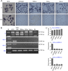
FIGURE 1.
FIGURE 1.
Immortalization of HFK with HPV induces down-regulation of the ASncmtRNAs. A, expression of the SncmtRNA and the ASncmtRNAs in normal HFK or HFK immortalized with HPV-16 (HFK698 cells) or 18 (18Nco cells) was analyzed by ISH. The ASncmtRNAs are down-regulated in the immortalized cells as well as in the corresponding tumorigenic cell lines SiHa (HPV-16-transformed) and HeLa (HPV-18-transformed). Magnification was ×40. B, expression of SncmtRNA-1 (S), ASncmtRNA-1 (AS-1), and ASncmtRNA-2 (AS-2) in the different cell lines was evaluated by RT-PCR amplification. The internal control used was 18S rRNA. M, 100-bp ladder. C, the relative intensity of the bands corresponding to the different non-coding mitochondrial RNAs was determined by densitometry and normalized against the intensity of the 150-bp amplicon from 18S rRNA.
FIGURE 2.
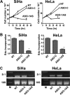
FIGURE 2.
Knockdown of SncmtRNA-1 induces inhibition of proliferation in SiHa and HeLa cells. A, cell proliferation. SiHa and HeLa cells (2.5 × 10⁵ cells/well) were plated onto 12-well plates and the next day were transfected with 100 nm ASO-1 AS or ASO-Control (ASO-C) using Lipofectamine2000, or left untreated (NT). At the indicated time periods, cells were harvested and counted. The experiments were carried out in triplicate, and the vertical bars show the mean ± S.D. The asterisk at 48 h post-transfection indicates p < 0.05. B, DNA synthesis rate was determined with the Click-iT® EdU Alexa Fluor® 488 kit (Molecular Probes), according to the manufacturer's directions. After 48 h of transfection as described in A, cells were pulsed with 10 μm EdU for 2 h at 37 °C, harvested, fixed in 3.7% p-formaldehyde in PBS, and saponin-permeabilized. EdU was detected with and co-stained with DAPI. The cells were analyzed by fluorescence microscopy in an Olympus BX-51 microscope. For each experiment, the number of EdU-positive cells was counted in at least quadruplicate versus the total amount of cells (DAPI staining). The results are presented as the mean ± S.D. The asterisk on the ASO-1AS bars indicates p < 0.01 (SiHa) and p < 0.05 (HeLa), respectively. C, knockdown of the SncmtRNA-1 was confirmed by RT-PCR on total RNA extracted from SiHa and HeLa cells treated as in A. A representative gel from the experiment run in triplicate is shown. Transfection with ASO-1AS induces a marked reduction of the SncmtRNA-1 (S-1) as compared with the controls. The amplification of 18S rRNA (18S) used as loading control was not affected by the same treatment. M, 100-bp ladder.
FIGURE 3.
FIGURE 3.
HPV-16- or HPV-18-immortalized cells express a novel sense transcript or SncmtRNA-2. A, schematic structure of the SncmtRNA-1 and strategy to amplify the transcript between the loop and the IR. Red line, 16S mitochondrial rRNA; blue line, IR. The primers used for amplification are indicated. B, PCR amplification of cDNA from the indicated cells, using primers 1 and 2. The 210-bp fragment corresponds to the SncmtRNA-1, whereas the 150-bp fragment corresponds to the SncmtRNA-2. Lanes labeled 698 correspond to HFK698 cells. C, PCR amplification of HFK698 cDNA between primer 1 and primers 2, 3, 4, 5, 6, 7, and 8 (lanes 1–7, respectively). No amplification was observed when using primers 1 and 8 (lane 7). The inset shows the amplification fragments obtained with HFK698 cDNA and corresponding to lanes 5 and 6. M, 100-bp ladder. D and E, SncmtRNA-1 and SncmtRNA-2 contain a double-stranded region. D, RNA from SiHa cells in 2× SCC was incubated in the presence (even lanes) or absence (odd lanes) of 50 μg/ml RNase A for 15 s at room temperature. RNA was recovered and amplified by RT-PCR using primers 1 and 6 (lanes 1 and 2), primers 11 and 6 covering the double-stranded region (lanes 3 and 4), and primers 9 and 10 (lanes 5 and 6) targeted to the 3′ single-stranded region (see A). Only the double-stranded region was resistant to RNase digestion (lanes 3 and 4). E, similar results were obtained with total RNA from HFK698 cells. Amplification of the double amplicons with primers 1 and 6 (lanes 1 and 2) was abolished after digestion, and a similar result was obtained with primers 9 and 10 (lanes 5 and 6). These results indicate that the single-stranded regions (the loop and the 3′ region) of the SncmtRNA-2 were digested by RNase A. Amplification between primers 11 and 6, covering the double-stranded region of the SncmtRNA-2, was not affected by the enzyme (lanes 3 and 4). M, 100-bp ladder. F, schematic structure of the SncmtRNA-1 and SncmtRNA-2, indicating the primers used for amplification. Oligonucleotides 12 and 13 were used for specific RT-PCR amplification or for ISH. Primers 14 and 15 were used as controls.
FIGURE 4.
FIGURE 4.
Specific detection of SncmtRNA-1 and SncmtRNA-2. A, sequence of probes 12 (specific for SncmtRNA-1) and 13 (specific for SncmtRNA-2) targeted to the linker region between the IR and the 5′-end of the 16S mtrRNA (see Fig. 3F, showing the position of these primers and control primers 14 and 15). B, PCR amplification of cDNA obtained from the indicated cells using primers 12 and 2 (SncmtRNA-1; Fig. 3F). A single fragment of 190 bp was obtained in all samples. C, same as B but using primers 13 and 2 (SncmtRNA-2; Fig. 3F). A 130-bp fragment corresponding to SncmtRNA-2 was obtained only with cDNA from HFK698 and 18Nco cells. M, 100-bp ladder. D, PCR amplification of SncmtRNA-1 and -2 from HFK698 cDNA. The expected fragments of 190 and 130 bp were obtained using primer 2 in combination with primers 12 or 13 (lanes 1–4). No amplification was observed when primer 2 was used in combination with primer 14 (9 nt complementary to the 5′-end of the 16S rRNA) or primer 15 (9 nt complementary to the 3′-end of the IR) of the SncmtRNA-1. Lane M, 100-bp ladder. E, differential expression of the SncmtRNA-1 and SncmtRNA-2. The expression of SncmtRNA-1 (S-1) and SncmtRNA-2 (S-2) in the indicated cells was determined by ISH using probe 12 and probe 13, respectively. The SncmtRNA-2 was expressed only in the immortalized cells HFK698 and 18Nco. Notice that only HFK expresses the ASncmtRNAs (AS). Magnification was ×40.
FIGURE 5.
FIGURE 5.
The SncmtRNA-2 is not present in isolated mitochondria. Mitochondria were isolated from ∼5 × 10⁸ HFK698 or 18Nco cells as described under “Experimental Procedures.” The final mitochondrial fraction was treated with RNase A to eliminate contamination from cytoplasmic RNA, followed by extraction of mitochondrial RNA with TRIzol. In parallel, total RNA was also extracted from HFK698 and 18Nco cells. Amplification of fragments of 190 bp (SncmtRNA-1), 130 bp (SncmtRNA-2), 320 bp (COX I mRNA), and 150 bp corresponding to 18S rRNA was observed in total RNA from both immortalized cell lines (T lanes). Only the amplicons of 190 bp of the SncmtRNA-1 and 320 bp of the COX I mRNA were amplified from mitochondrial RNA (Mt lanes) of HFK698 and 18Nco cells, whereas no amplification was obtained of the SncmtRNA-2 or 18S rRNA. M, 100-bp ladder.
FIGURE 6.
FIGURE 6.
Expression of SncmtRNA-2 is induced by HPV-16 E6 and E7. A, expression of the SncmtRNA-1, SncmtRNA-2, and the ASncmtRNAs was determined in HFK transduced with E6, E7, or E6/E7. The SncmtRNA-1 was expressed in all cells, including HFK698. The SncmtRNA-2 was expressed only in HFK transduced with E6 and E7 and in HFK698. Expression of the ASncmtRNAs was maintained in all cells except HFK698. Magnification was ×20. B, PCR amplification confirmed that the SncmtRNA-2 was expressed only in HFK698 and HFK transduced with E6 and E7. C, total RNA from HFK698 cells, HFK, and HFK transduced with E6, E7, and E6/E7 was used for RT-PCR amplification of HPV-16 E6 mRNA. E6 is expressed in HFK transduced with E6 or E6/E7 and HFK698 cells. D, same as C, but amplification was carried out with primers corresponding to HPV-16 E7 mRNA. Lane M, 100-bp ladder.
FIGURE 7.
FIGURE 7.
Down-regulation of the ASncmtRNA and HPV oncogenes. A, HFK698 cells express E1^E4 and E5 mRNA. B, HFK698 cells were transduced with a GFP-expressing lentiviral vector encoding an shRNA complementary to E4. The GFP-expressing cells were sorted, and expression of E1^E4 mRNA was determined by RT-PCR amplification. E4 was knocked down in HFK698 cells transduced with the E4shRNA. C, expression of the SncmtRNA-1 and the ASncmtRNAs in HFK698 cells transduced with E4shRNA or empty vector (ConshRNA). Expression of the ASncmtRNAs was reestablished only in HFK698 cells transduced with the E4shRNA. Magnification was ×20. D, HFK cells were transformed with a plasmid containing HPV-18 E1^E4 or with empty vector. E1^E4 was expressed only in HFK transformed with E1^E4-containing vector. E, HFK cells transformed with HPV-18 E1^E4 plasmid or with the empty vector were subjected to fluorescent in situ hybridization and compared with untreated HFK. The expression of the SncmtRNA-1 and the ASncmtRNAs was not affected by the expression of E1^E4. Magnification was ×20.
FIGURE 8.
FIGURE 8.
HPV E2 is involved in down-regulation of the ASncmtRNAs. A, schematic illustration of the HPV-16 genome, indicating the position of E6, E7, E1, E2, E4, E5, and L2 genes. The absence of L1 is for simplicity. The transplicing reaction between a fragment of E1 with a fragment of E2 to generate E1^E4 is shown. B, expression of E2 mRNA in 18Nco cells was determined by RT-PCR amplification. A fragment of 330 bp was obtained with cDNA of 18Nco cells (lanes 1 and 2). E2 was knocked down by an ASO complementary to the NH2-terminal coding region (18 E2-ASO; lanes 3 and 4) but not by a control oligonucleotide (C-ASO; lanes 5 and 6). 24 h after transfection with the 18E2-ASO, the cells were cultured in fresh medium for another 24 h. The expression of E2 was reestablished (Recovery; lanes 7 and 8). C, analysis of the expression of SncmtRNA-1 and -2 (S) and ASncmtRNAs (AS) by fluorescent in situ hybridization. Hybridization revealed that the expression of the ASncmtRNAs was recovered in 18Nco cells transfected with the 18E2-ASO. Once the expression of E2 was reestablished, expression of the ASncmtRNAs was down-regulated (Recovery). D, the same results were obtained with HFK698 cells transfected with an E2-ASO complementary to HPV-16 E2 (16 E2-ASO). The expression of the ASncmtRNAs was reestablished after knocking down HPV-16 E2. Recovery of E2 expression resulted in down-regulation of the ASncmtRNAs (Recovery). Magnification was ×20.

Similar articles

Cited by

References

    1. Hanahan D., Weinberg R. A. (2011) Hallmarks of cancer. The next generation. Cell 144, 646–674 - PubMed
    1. D'Agostino D. M., Bernardi P., Chieco-Bianchi L., Ciminale V. (2005) Mitochondria as functional targets of proteins coded by human tumor viruses. Adv. Cancer Res. 94, 87–142 - PubMed
    1. McLaughlin-Drubin M. E., Munger K. (2008) Viruses associated with human cancer. Biochim. Biophys. Acta 1782, 127–150 - PMC - PubMed
    1. McLaughlin-Drubin M. E., Münger K. (2009) Oncogenic activities of human papillomaviruses. Virus Res. 143, 195–208 - PMC - PubMed
    1. zur Hausen H. (2009) Papillomaviruses in the causation of human cancers. A brief historical account. Virology 384, 260–265 - PubMed

Publication types

MeSH terms

LinkOut - more resources