. 2012 Jul;86(13):7256-67.
doi: 10.1128/JVI.07222-11. Epub 2012 Apr 24.

Hepatitis C virus attachment mediated by apolipoprotein E binding to cell surface heparan sulfate

Affiliations

Hepatitis C virus attachment mediated by apolipoprotein E binding to cell surface heparan sulfate

Jieyun Jiang et al. J Virol. 2012 Jul.

Abstract

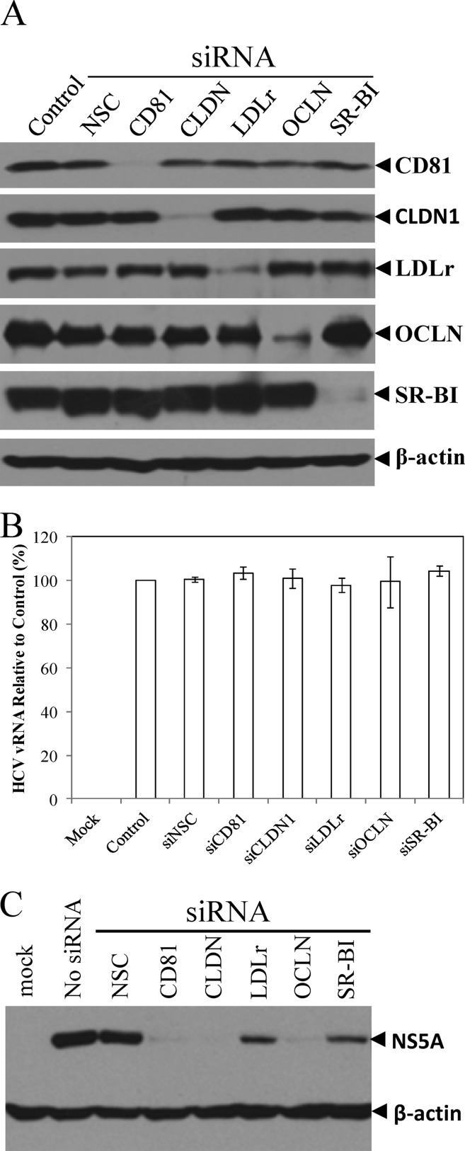
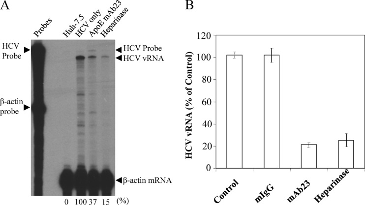
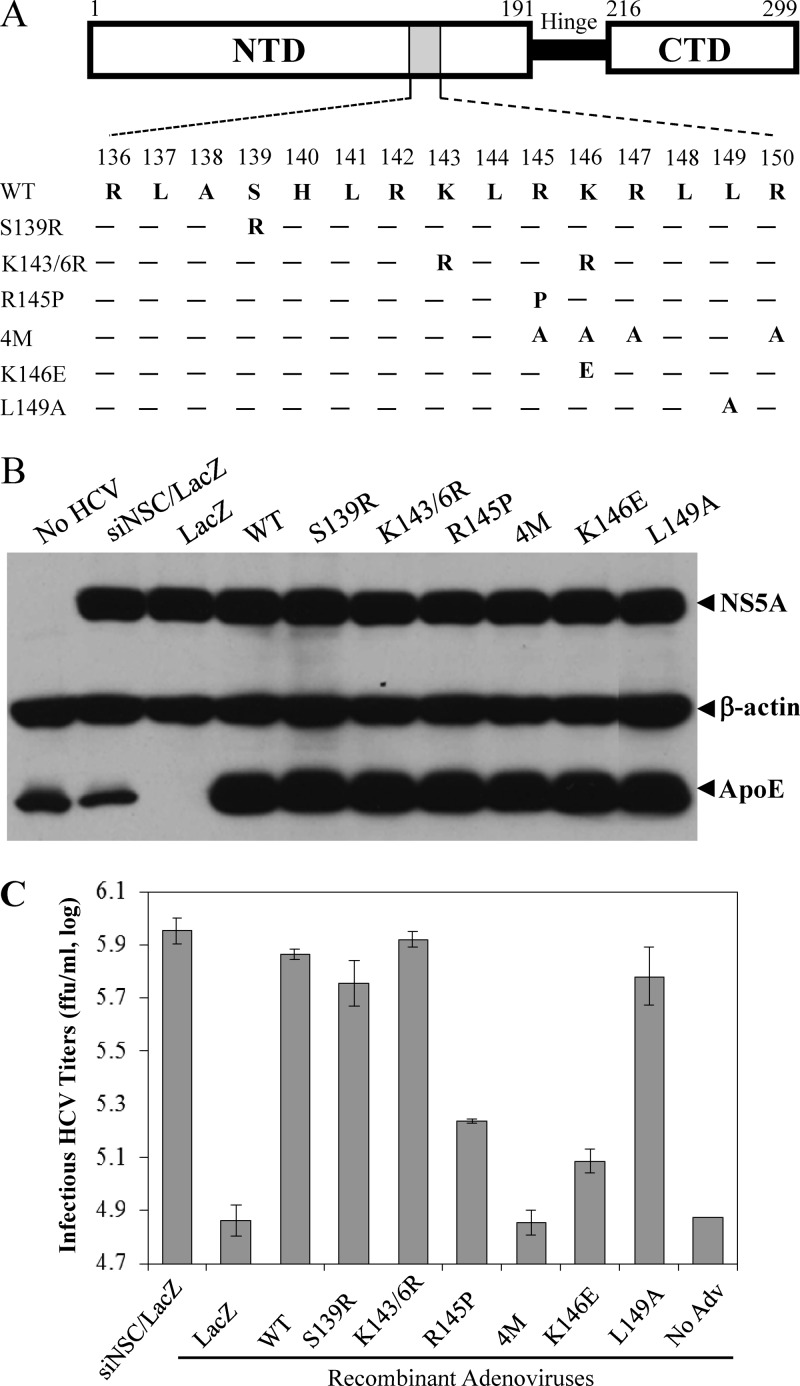
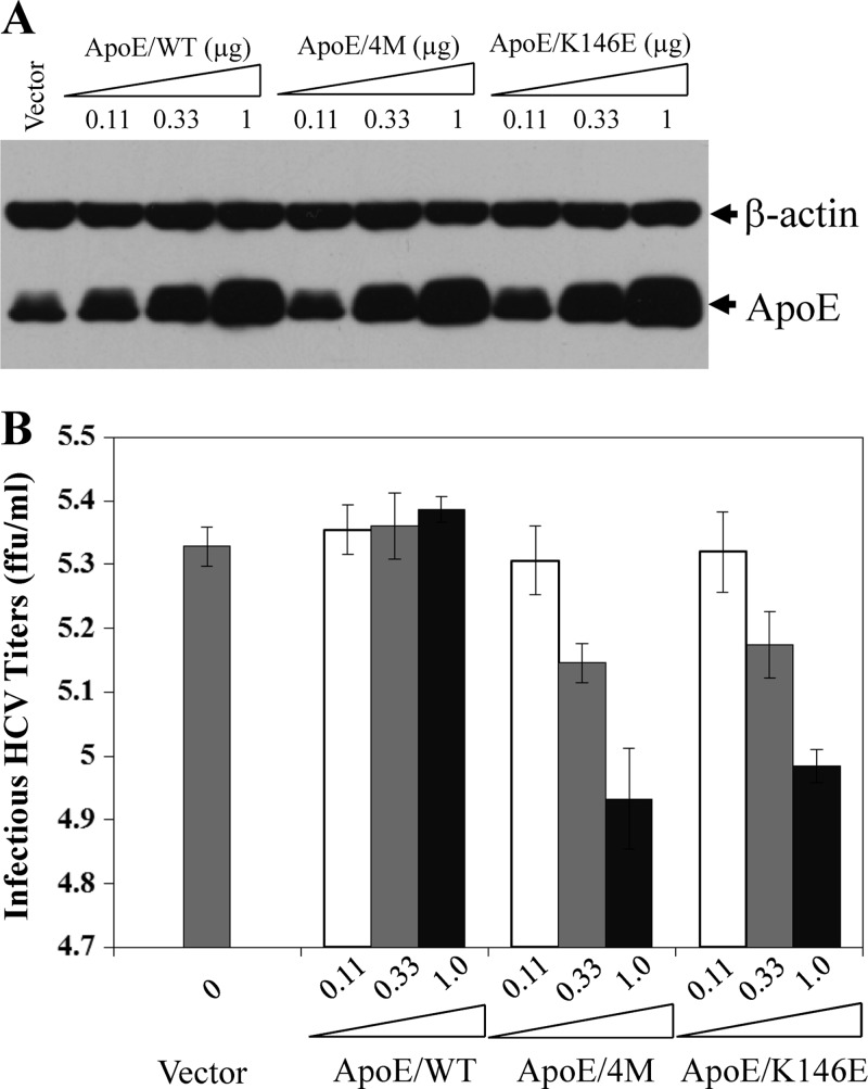
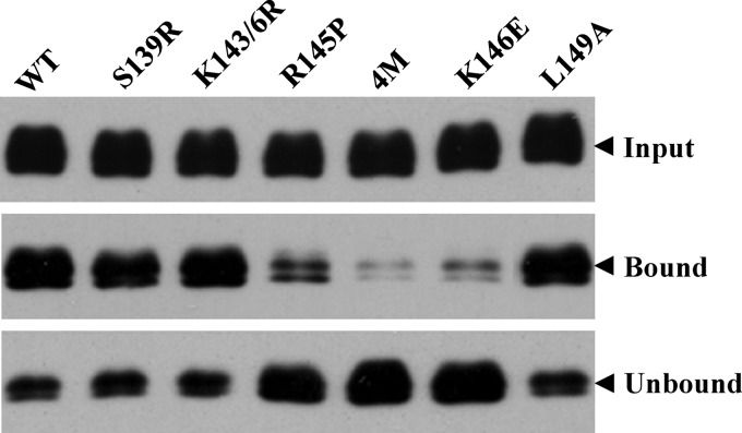
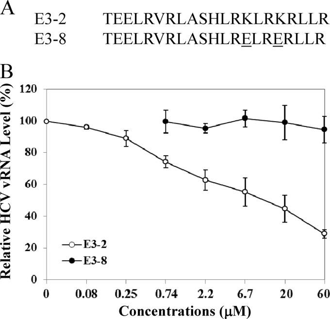
Viruses are known to use virally encoded envelope proteins for cell attachment, which is the very first step of virus infection. In the present study, we have obtained substantial evidence demonstrating that hepatitis C virus (HCV) uses the cellular protein apolipoprotein E (apoE) for its attachment to cells. An apoE-specific monoclonal antibody was able to efficiently block HCV attachment to the hepatoma cell line Huh-7.5 as well as primary human hepatocytes. After HCV bound to cells, however, anti-apoE antibody was unable to inhibit virus infection. Conversely, the HCV E2-specific monoclonal antibody CBH5 did not affect HCV attachment but potently inhibited HCV entry. Similarly, small interfering RNA-mediated knockdown of the key HCV receptor/coreceptor molecules CD81, claudin-1, low-density lipoprotein receptor (LDLr), occludin, and SR-BI did not affect HCV attachment but efficiently suppressed HCV infection, suggesting their important roles in HCV infection at postattachment steps. Strikingly, removal of heparan sulfate from the cell surface by treatment with heparinase blocked HCV attachment. Likewise, substitutions of the positively charged amino acids with neutral or negatively charged residues in the receptor-binding region of apoE resulted in a reduction of apoE-mediating HCV infection. More importantly, mutations of the arginine and lysine to alanine or glutamic acid in the receptor-binding region ablated the heparin-binding activity of apoE, as determined by an in vitro heparin pulldown assay. HCV attachment could also be inhibited by a synthetic peptide derived from the apoE receptor-binding region. Collectively, these findings demonstrate that apoE mediates HCV attachment through specific interactions with cell surface heparan sulfate.


Figures

Fig 1
Fig 1
Inhibition of HCV infection by the Fab fragments of MAb23. (A) Analysis of MAb23 and Fab and Fc fragments. The MAb23 whole IgG (IgG) and Fab and Fc fragments (1 μg each) were analyzed by 10% SDS-PAGE and stained with Coomassie blue. The sizes of protein markers are indicated on the right. (B) Inhibition of HCV infection by the MAb23 Fab fragments. Huh-7.5 cells were incubated with HCV on ice for 2 h in the presence of various amounts (0.04 to 1 μg/ml) of the Fab fragments (Fab), 1 μg/ml of the Fc fragment (Fc), or 1 μg/ml of normal mouse IgG (mIgG). Upon removal of the unbound HCV and washing of cells with PBS, the HCV-infected cells were incubated with medium at 37°C. At 24 h p.i., total cellular RNAs were extracted with TRI Reagent/RNAzol (Molecular Research Center, Inc.) and the levels of positive-stranded HCV RNA were determined by RPA as previously described (16). The β-actin mRNA was used as an internal control. The protected HCV RNA products migrated about 40 nucleotides faster due to a mismatched sequence derived from the vector DNA.
Fig 2
Fig 2
Inhibition of HCV attachment by MAb23. The effects of MAb23 on HCV attachment were determined by incubating Huh-7.5 cells with HCV on ice for 2 h in the absence (control) or presence of various concentrations (0.04, 0.2, and 1 μg/ml) of MAb23 or a normal mouse IgG (mIgG). The unbound HCV was removed, and the HCV-bound cells were washed with PBS and then incubated at 37°C for 24 h. To determine the effects of MAb23 on HCV entry, HCV was allowed to bind to Huh-7.5 cells on ice for 2 h prior to the addition of MAb23 or mIgG. After HCV attachment on ice for 2 h, the unbound HCV was aspirated and the cells were washed with PBS. Subsequently, the HCV-bound cells were incubated with cell culture medium containing 0.04, 0.2, and 1 μg/ml of MAb23 or mIgG at 37°C for 6 h. Cell culture medium was replaced with fresh medium without antibody. At 24 h p.i. at 37°C, the levels of NS5A in the HCV-infected cells were detected by Western blotting (A), while the infectious HCV titers in the supernatants were determined by limiting dilution and IFA staining for HCV NS3-positive cells (B). Mean values and standard deviations of three experiments are shown in panel B. Attach, HCV attachment assay; Entry, HCV entry assay.
Fig 3
Fig 3
Inhibition of HCV infection of primary human hepatocytes by apoE MAb23. Freshly isolated primary human hepatocytes (PHHs) in 12-well plates were purchased from Celsis In Vitro Technologies (Baltimore, MD) and maintained according to the manufacturer's instructions. PHHs were infected with HCV in the absence (control) or presence of mIgG (50 μg/ml) or apoE MAb23 (10 and 50 μg/ml) at 37°C for 8 h. At 48 h p.i., HCV RNAs in the infected PHHs were isolated with TRI reagent (Invitrogen). The levels of positive-strand HCV RNA were determined by qRT-PCR as described in Materials and Methods. The average levels of relative HCV RNA to the control (100%), from triplicate experiments, are shown.
Fig 4
Fig 4
Effects of MAb23 and HCV E2-specific MAb (CBH5) on HCV attachment and infection. (A) Inhibition of HCV attachment by apoE but not E2 MAb. Huh-7.5 cells were incubated with HCV on ice in the presence of various amounts of MAb23 or CBH5. Normal mouse IgG (mIgG) and human IgG (hIgG) were used as negative controls. After 2 h incubation on ice, the unbound HCV was removed and cells were washed three times with PBS. The vRNA of the cell-bound HCV was extracted with RNeasy minikit (Qiagen) and quantified by RPA as previously described (24, 36). (B) Inhibition of HCV infection by MAb23 and CBH5. HCV infection was the same as in panel A. After washing with PBS, the HCV-infected cells were incubated at 37°C for 24 h. Total RNAs were extracted from cells, and the levels of positive-stranded HCV RNA were determined by RPA. The β-actin mRNA was used as an internal control. Numbers on the top indicate the amounts of MAb (μg/ml). The levels of HCV RNA relative to the control (%) are shown at the bottom with 100% representing the RNA level in the absence of antibodies.
Fig 5
Fig 5
Effects of siRNA-mediated knockdown of key HCV receptors on HCV attachment and infection. Huh-7.5 cells in 12-well plates were transfected with 0.2 nmol of each siRNA specific to CD81, claudin-1, LDLr, occludin, and SR-BI using RNAiMax reagent (Invitrogen) as described in Materials and Methods. (A) At 48 h p.t., the levels of CD81, claudin-1, LDLr, occludin, and SR-BI expression were determined by Western blotting using specific antibodies to each protein as indicated on the right. (B) Effects of silencing CD81, claudin-1, LDLr, occludin, and SR-BI expression on HCV attachment. At 48 h after siRNA transfection, Huh-7.5 cells were incubated with HCV at 37°C for 2 h. Upon extensive washing, total cellular RNA was extracted with RNAzol reagent. The levels of HCV vRNA were quantified by qRT-PCR and were converted to a percentage of the control with 100% representing the level of HCV vRNA without antibody treatment. Average levels of HCV vRNA and deviations from three independent experiments are shown. (C) Effects of the siRNA-mediated knockdown of the above receptors on HCV infection. At 48 h after siRNA transfection, Huh-7.5 cells were incubated with HCV on ice for 2 h, followed by extensive washing. The HCV-attached Huh-7.5 cells were incubated at 37°C for 24 h. HCV NS5A in the cell lysates was detected by Western blotting using β-actin as a control.
Fig 6
Fig 6
(A) Effects of heparinase treatment on HCV attachment. Heparan sulfate on the cell surface was removed by treatment of Huh-7.5 cells with 1 unit/ml of heparinase I (Sigma) at 37°C for 1 h as described previously (38). The heparinase-treated Huh-7.5 cells were subsequently incubated with HCV on ice for 2 h. The unattached HCV was aspirated and the HCV-attached Huh-7.5 cells were washed three times with PBS. Total cellular RNA was prepared using an RNeasy mini purification kit (Qiagen). The apoE-specific MAb23 was used as a positive control. The HCV vRNA in 50 μg of total RNA was quantified by RPA as previously described (16, 34). The β-actin mRNA was used as an internal control to normalize the amounts of total RNA used between different samples. (B) Quantification of HCV vRNA by qRT-PCR. HCV attachment inhibition experiments were carried out the same way as in panel A except for the use of a 12-well plate of Huh-7.5 cells. Upon HCV attachment and extensive washing of the heparinase-treated Huh-7.5 cells, total cellular RNA was extracted with RNAzol reagent (Molecular Research Center, Inc.). The HCV vRNA was quantified by qRT-PCR using the StepOnePlus PCR system (Applied Biosystems). Percentage of average HCV vRNA levels and deviations derived from three independent experiments are shown. The HCV vRNA level relative to that of the control (100%) is shown.
Fig 7
Fig 7
Site-directed mutagenesis analysis of the apoE receptor-binding region. (A) Mutations of the N-terminal receptor-binding region. Three domains of apoE are schematically shown: the N-terminal domain (NTD) containing the receptor-binding region (gray box), the hinge region (Hinge), and the C-terminal domain (CTD) involved in interactions with phospholipids. Numbers indicate amino acid positions from the N terminus of mature apoE. Amino acid residues of the receptor-binding region (136 to 150) are shown underneath their positions. Each amino acid mutation is highlighted at its corresponding position. Wild-type and mutant apoE were ectopically expressed by recombinant adenoviruses (see Materials and Methods). (B) Effects of ectopic expression of wild-type and mutant apoE on HCV replication. Huh-7.5 cells were transfected with 50 nM apoE-specific siRNA. At 24 h p.t., Huh-7.5 cells were superinfected with HCV at an MOI of 5 at 37°C for 3 h, followed by adenovirus at an MOI of 6 at 37°C for 6 h. At 24 h p.i., cell lysates were harvested and used for the detection of apoE and NS5A by Western blotting with β-actin as a control. (C) Determination of infectious HCV titers in the supernatants by IHC. The supernatants of the experiments described in panel A were collected at 24 h p.i. The infectious HCV titers in the supernatants were determined by serial dilution.
Fig 8
Fig 8
Dominant negative effects of the 4M and K146E apoE mutants. Huh-7.5 cells in 12-well cell culture plates were transfected with increasing amounts (0.11, 0.33, and 1 μg) of pCMV6-XL5 vectors expressing wild-type apoE, the apoE mutant with arginine-to-alanine mutations at residues 145, 146, 147 and 149 (4M), and the apoE mutant with a lysine to glutamic acid mutation at amino acid residue 146 (K146E), respectively. The total amount of DNA was kept constant at 1 μg using vector DNA. DNA was transfected into Huh-7.5 cell using DMRIE-C reagent (Invitrogen) by following the manufacturer's instructions. At 6 h p.t., the medium was replaced with DMEM containing 10% FBS. At 24 h p.t., Huh-7.5 cells were infected with HCV at an MOI of 5. At 24 h p.i., cell lysates and the culture media were collected for detection of apoE by Western blotting (A) and for determination of infectious HCV titers by IHC (B). Mean values and standard deviations of three experiments are shown in panel B.
Fig 9
Fig 9
Effects of mutations in the receptor-binding region on the heparin binding of apoE. The endogenous apoE expression was silenced to undetectable levels by a synthetic apoE-specific siRNA as described previously (24). The wild type and individual apoE mutants (indicated on the top) were ectopically expressed by recombinant adenoviruses. The supernatants were collected and subjected to a pulldown assay with heparin-conjugated beads. The heparin-bound (pulldown) and unbound apoE proteins were determined by Western blotting using apoE-specific WuE4 monoclonal antibody and an ECL kit (Pierce).
Fig 10
Fig 10
Inhibition of HCV attachment by a synthetic peptide derived from the apoE receptor-binding region. (A) Huh-7.5 cells in 12-well cell culture plates were incubated with HCV on ice for 2 h in the absence or presence of various concentrations of wild-type (E3-2) and mutant (E3-8) peptides. The unbound HCV was removed by extensive washing with PBS. The vRNA of the cell-bound HCV was isolated with RNAzol reagent (Molecular Research Center). (B) The average levels of HCV vRNA, from triplicate experiments, were determined by qRT-PCR as described in Materials and Methods.

Similar articles

Cited by

References

    1. Acharya P, et al. 2002. Comparison of the stabilities and unfolding pathways of human apolipoprotein E isoforms by differential scanning calorimetry and circular dichroism. Biochim. Biophys. Acta 1584:9–19 - PubMed
    1. Agnello V, Abel G, Elfahal M, Knight GB, Zhang QX. 1999. Hepatitis C virus and other Flaviviridae viruses enter cells via low density lipoprotein receptor. Proc. Natl. Acad. Sci. U. S. A. 96:12766–12771 - PMC - PubMed
    1. Alter MJ. 2006. Epidemiology of viral hepatitis and HIV co-infection. J. Hepatol. 44:S6–S9 - PubMed
    1. André P, et al. 2002. Characterization of low- and very-low-density hepatitis C virus RNA-containing particles. J. Virol. 76:6919–6928 - PMC - PubMed
    1. André P, Perlemuter G, Budkowska A, Brechot C, Lotteau V. 2005. Hepatitis C virus particles and lipoprotein metabolism. Semin. Liver Dis. 25:93–104 - PubMed

Publication types