Review
. 2012 Apr 25;12(5):367-82.
doi: 10.1038/nri3210.

Interferon-inducible effector mechanisms in cell-autonomous immunity

Affiliations
Review

Interferon-inducible effector mechanisms in cell-autonomous immunity

John D MacMicking. Nat Rev Immunol. .

Abstract

Interferons (IFNs) induce the expression of hundreds of genes as part of an elaborate antimicrobial programme designed to combat infection in all nucleated cells - a process termed cell-autonomous immunity. As described in this Review, recent genomic and subgenomic analyses have begun to assign functional properties to novel IFN-inducible effector proteins that restrict bacteria, protozoa and viruses in different subcellular compartments and at different stages of the pathogen life cycle. Several newly described host defence factors also participate in canonical oxidative and autophagic pathways by spatially coordinating their activities to enhance microbial killing. Together, these IFN-induced effector networks help to confer vertebrate host resistance to a vast and complex microbial world.


Conflict of interest statement

The author declares no competing financial interests.

Figures
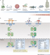
Figure 1
Figure 1. Evolution of IFN-induced cell-autonomous host defence.
a | The evolution of cell-autonomous immunity and the emergence of interferon (IFN)-induced effector mechanisms around the protochordate–vertebrate split (∼530 million years ago). b | Cell-autonomous host defence proteins are canonically induced by IFNs via three receptor complexes with high affinities for their ligands (Ka < 10 nM−1). The first receptor complex is a tetramer — composed of two chains of IFNγ receptor 1 (IFNGR1) and two chains of IFNGR2 — that engages type II IFN (that is, IFNγ) dimers. The second is a heterodimer of IFNα/β receptor 1 (IFNAR1) and IFNAR2 that binds to the type I IFNs: a family consisting of 13 different IFNα subtypes and one IFNβ subtype in humans. In the third receptor complex, interleukin-10 receptor 2 (IL-10R2) associates with IFNλ receptor 1 (IFNLR1; also known as IL-28Rα) to bind to three different type III IFN (that is, IFNλ) ligands (see Ref. 8). Following receptor–ligand engagement, signals are transduced through signal transducer and activator of transcription 1 (STAT1) homodimers in response to IFNγ or through STAT1–STAT2 heterodimers in response to type I IFNs or IFNλ. Following their recruitment to the receptor complexes, these STAT molecules are phosphorylated by receptor-bound tyrosine kinases (namely, Janus kinases (JAKs) and tyrosine kinase 2 (TYK2)). Phosphorylated STAT1 homodimers (also known as GAF) translocate to the nucleus to bind to IFNγ-activated site (GAS) promoter elements to promote the IFN-induced expression of antimicrobial effector genes, some of which also require transactivation by IFN-regulatory factor 1 (IRF1) and IRF8. In the case of type I and III IFN signalling, phosphorylated STAT1–STAT2 dimers form a complex with IRF9 to yield IFN-stimulated gene factor 3 (ISGF3); this complex also translocates to the nucleus, where it binds to IFN-stimulated response elements (ISREs) in the promoters of different or overlapping IFN-stimulated effector genes.
Figure 2
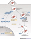
Figure 2. Cell-autonomous mechanisms used by IFN-induced proteins against intracellular bacteria.
Interferon (IFN)-inducible proteins are required for host resistance to intracellular bacteria. a | Specific immunity-related GTPases (IRGs), guanylate-binding proteins (GBPs) and other GTPases translocate to compartmentalized bacteria in phagosomes or inclusion bodies. Here, different membrane regulatory complexes — IRGM1–snapin, GBP1–sequestosome 1 (SQSTM1) and GBP7–ATG4B — are assembled. These complexes initiate autophagic capture and SNARE-mediated fusion of the bacterial compartments with lysosomes,,,. In addition, IRGM3–IRGA6 (or IRGB10) mediate vacuole disruption,,, and GBP7 (and possibly leucine-rich repeat kinase 2 (LRRK2)) help to assemble NADPH oxidase 2 (NOX2) on bacterial phagosomes, which mediates bacterial killing. Using this pathway, these GTPases can also deliver antimicrobial peptides (AMPs) to the autophagolysosome and, in the case of human IRGM, may instigate mitochondrial fission before autophagy. Other IFN-inducible components, such as natural resistance-associated macrophage protein 1 (NRAMP1), help to exclude Mn2+ and Fe2+ from the bacterial phagosome, while importing protons (H+) into this compartment. Nitric oxide synthase 2 (NOS2), which synthesizes NO, works in concert with NOX2, which produces reactive oxygen species such as superoxide (O2) and hydrogen peroxide (H2O2), to produce compound intermediates like peroxynitrite (not shown) that are highly bactericidal. b | An emerging signature for the recognition of some escaped bacteria in the cytosol is ubiquitylation (either single or multiple modifications with monoubiquitin and/or polyubiquitin chains) (see Ref. 47). SQSTM1, NDP52 and optineurin bind to ubiquitylated bacteria to initiate innate immune signalling and to recruit the autophagic machinery via LC3 family members. In addition, GBP1 and GBP2 polymerize around cytosolic bacteria in a ubiquitin-independent process that may recruit specific antimicrobial partners, while galectin 3 and galectin 8 bind to exposed glycans on the bacteria and, in the case of galectin 8, recruit NDP52 and downstream autophagic effectors. SQSTM1 also activates a second antibacterial pathway involving diacylglycerol (DAG) and protein kinase Cδ (PKCδ) to induce NOX2 complex assembly. Dashed lines indicate possible routes and consequences. PtdIns(4,5)P2, phosphatidylinositol-4,5-bisphosphate; PtdIns(3,4,5)P3, phosphatidylinositol-3,4,5-trisphosphate; TBK1, TANK-binding kinase 1.
Figure 3
Figure 3. Cell-autonomous mechanisms used by IFN-induced proteins against intracellular protozoa.
Different intracellular strategies are used by interferon (IFN)-inducible proteins against protozoa. Nitric oxide synthase 2 (NOS2) exerts potent parasiticidal activity, while GKS-containing immunity-related GTPases (IRGs) appear to be directly involved in parasite vacuole disruption once they reach the parisitophorous compartment. This proceeds via autophagy-independent trafficking after release from IRGM1–IRGM3 or ATG5 and is mediated by cooperative IRG loading,,. Guanylate-binding proteins (GBPs) — specifically GBP1–GBP2 and GBP1–GBP5 complexes — also traffic to the parasitophorous vacuole, with uncharacterized effects on parasite control. Natural resistance-associated macrophage protein 1 (NRAMP1) is important for restricting the uptake of Mn2+ and Fe2+ by this compartment, whereas indoleamine 2,3-dioxygenase 1 (IDO1) and/or IDO2 limit amino acid acquisition via the depletion of L-tryptophan. Dashed lines indicate possible routes or consequences.
Figure 4
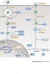
Figure 4. Cell-autonomous mechanisms used by IFN-induced proteins against viruses.
Multiple strategies are used by interferon (IFN)-inducible proteins to combat viruses. IFN-inducible effectors function at nearly every stage of the pathogen life cycle. For example, interferon-inducible transmembrane proteins (IFITMs) and tripartite motif proteins (TRIMs) act during viral entry and uncoating, and myxoma resistance proteins (MXs) block nucleocapsid transport. Inhibition of RNA reverse transcription, protein translation and stability is mediated by APOBEC3 (apolipoprotein B mRNA-editing enzyme, catalytic polypeptide 3), SAMHD1 (SAM-domain- and HD-domain-containing protein 1), ADAR1 (adenosine deaminase, RNA-specific 1), NOS2 (nitric oxide synthase 2), OASs (2′-5′ oligoadenylate synthases), RNase L, ISG20 (IFN-stimulated gene 20 kDa protein), PKR (IFN-induced, RNA-activated protein kinase) and ISG15. Finally, viperin and tetherin help to prevent viral assembly and release, respectively. Some of the effectors (such as MX proteins) appear to operate in both the nucleus and the cytosol (not shown).

Similar articles

Cited by

References

    1. Jones JD, Dangl JL. The plant immune system. Nature. 2006;444:323–329. doi: 10.1038/nature05286. - DOI - PubMed
    1. Marathe R, Guan Z, Anandalakshmi R, Zhao H, Dinesh-Kumar SP. Study of Arabidopsis thaliana resistome in response to cucumber mosaic virus infection using whole genome microarray. Plant Mol. Biol. 2004;55:501–520. doi: 10.1007/s11103-004-0439-0. - DOI - PubMed
    1. Beutler B, et al. Genetic analysis of host resistance: Toll-like receptor signaling and immunity at large. Annu. Rev. Immunol. 2006;24:353–389. doi: 10.1146/annurev.immunol.24.021605.090552. - DOI - PubMed
    1. Mascie-Taylor CG, Karim E. The burden of chronic disease. Science. 2003;302:1921–1922. doi: 10.1126/science.1092488. - DOI - PubMed
    1. Bezbradica JJ, Medzhitov R. Integration of cytokine and heterologous receptor signaling pathways. Nature Immunol. 2009;10:333–339. doi: 10.1038/ni.1713. - DOI - PubMed

Publication types