. 2012 May;33(1):17-30.
doi: 10.1007/s10974-012-9292-y. Epub 2012 Apr 17.

Pathogenic properties of the N-terminal region of cardiac myosin binding protein-C in vitro

Affiliations

Pathogenic properties of the N-terminal region of cardiac myosin binding protein-C in vitro

Suresh Govindan et al. J Muscle Res Cell Motil. 2012 May.

Abstract

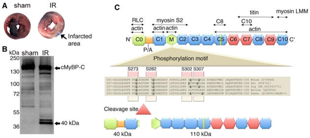
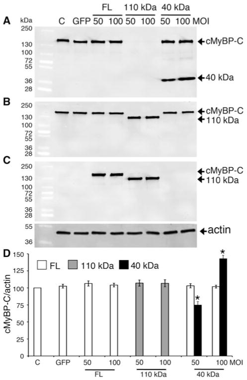
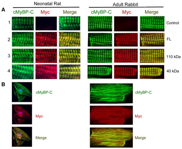
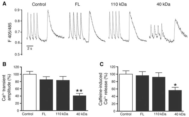
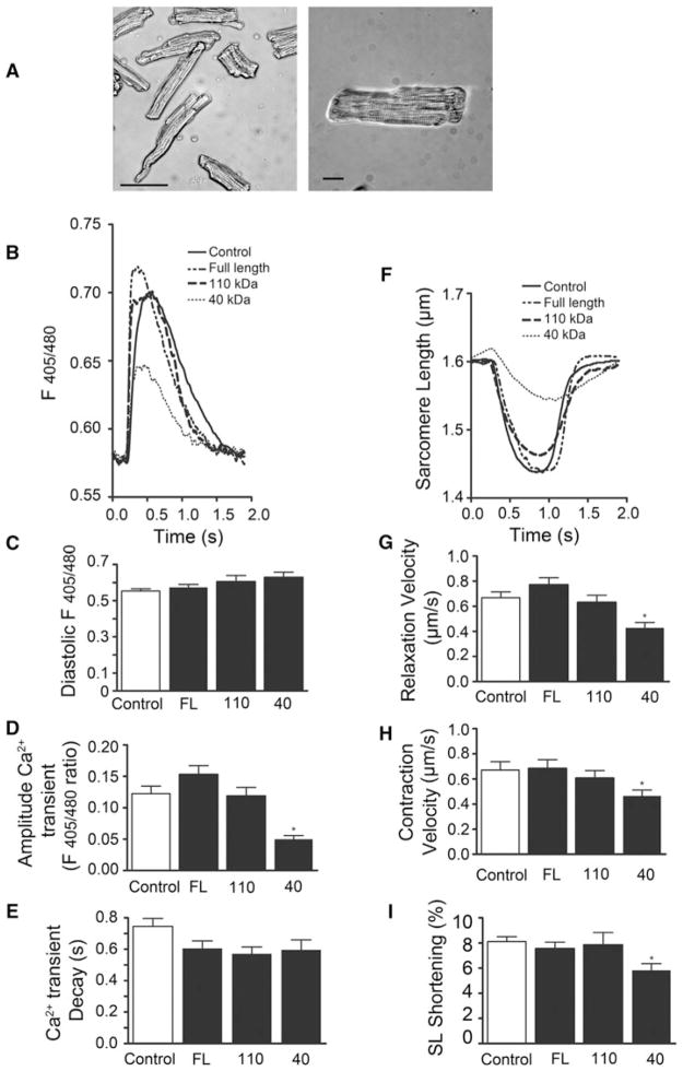
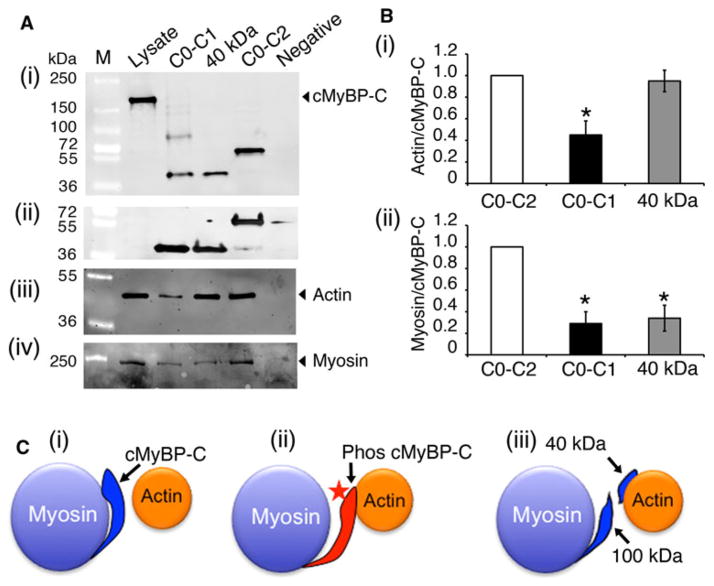
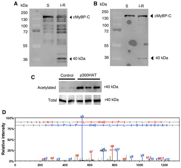
Cardiac myosin binding protein-C (cMyBP-C) plays a role in sarcomeric structure and stability, as well as modulating heart muscle contraction. The 150 kDa full-length (FL) cMyBP-C has been shown to undergo proteolytic cleavage during ischemia-reperfusion injury, producing an N-terminal 40 kDa fragment (mass 29 kDa) that is predominantly associated with post-ischemic contractile dysfunction. Thus far, the pathogenic properties of such truncated cMyBP-C proteins have not been elucidated. In the present study, we hypothesized that the presence of these 40 kDa fragments is toxic to cardiomyocytes, compared to the 110 kDa C-terminal fragment and FL cMyBP-C. To test this hypothesis, we infected neonatal rat ventricular cardiomyocytes and adult rabbit ventricular cardiomyocytes with adenoviruses expressing the FL, 110 and 40 kDa fragments of cMyBP-C, and measured cytotoxicity, Ca(2+) transients, contractility, and protein-protein interactions. Here we show that expression of 40 kDa fragments in neonatal rat ventricular cardiomyocytes significantly increases LDH release and caspase 3 activity, significantly reduces cell viability, and impairs Ca(2+) handling. Adult cardiomyocytes expressing 40 kDa fragments exhibited similar impairment of Ca(2+) handling along with a significant reduction of sarcomere length shortening, relaxation velocity, and contraction velocity. Pull-down assays using recombinant proteins showed that the 40 kDa fragment binds significantly to sarcomeric actin, comparable to C0-C2 domains. In addition, we discovered several acetylation sites within the 40 kDa fragment that could potentially affect actomyosin function. Altogether, our data demonstrate that the 40 kDa cleavage fragments of cMyBP-C are toxic to cardiomyocytes and significantly impair contractility and Ca(2+) handling via inhibition of actomyosin function. By elucidating the deleterious effects of endogenously expressed cMyBP-C N-terminal fragments on sarcomere function, these data contribute to the understanding of contractile dysfunction following myocardial injury.


Figures

Fig. 1
Fig. 1
cMyBP-C interacting partners and generation of the 40 kDa cleavage fragment. Ischemia–reperfusion injury induces myocardial infarction, resulting in tissue damage and necrosis. Three month-old mice were induced with 60-min ischemia and 24-h reperfusion, as described previously (Sadayappan et al. 2009). Following I–R injury, hearts were fixed with 2 % Evan’s Blue in PBS, sectioned and staining with 2 % triphenyl tetrazolium chloride. Compared to sham operated mouse heart, I–R injured heart shows severe infarct region (a). The 40 kDa fragment is the predominant N′ fragment of cMyBP-C that is released in the sarcomere during I–R injury (b). Ten micrograms of total myofilament proteins were used to perform western blot analysis with anti-cMyBP-C2–14 antibodies. The antibody detects all the N′ fragments of cMyBP-C in which the 40 kDa is predominant. Although the 40 kDa fragment runs at the 40 kDa position in the gel, MS/MS sequence analysis identified the site of cleavage and molecular weight as 29 kDa (Sadayappan et al. 2008; Govindan et al. 2012). (c) cMyBP-C consists of 11 modules labeled C0–C10 from the N- to the C-terminus and belongs to the intracellular Ig super-family which is composed of repeating domains of Ig (Plaque) and fibronectin type-3 (Hexagon). The interacting regions of cMyBP-C with titin, myosin, actin and regulatory light chain are shown. The C8 and C10 domains that interact with C5 and C7 of cMyBP-C are marked. The cardiac-specific regions (C0 and M domain, and an insert in C5 domain) are marked in light green. A proline-alanine (P/A)-rich linker sequence located between the C0 and C1 domains is shown. The four-phosphorylation sites, Ser-273, Ser-282, Ser-302 and Ser-307, are highlighted across mouse to human species. The calpain cleavage occurs at 272–280 residues, resulting in both 40 (residues 1–271) and 110 kDa (residues 272–1270) fragments
Fig. 2
Fig. 2
Overexpression of the 40 kDa fragments in NRVCMs does not affect expression of endogenous cMyBP-C. Representative western blot analyses show the expression of the transgenic FL, 110 and 40 kDa fragments. Fifteen micrograms of total lysates from infected NRVCMs for 48 h were used for SDS-PAGE, followed by western blot analyses with respective antibodies. FL of both endogenous and transgenic cMyBP-C and 40 kDa fragments were recognized with anti-cMyBP-C2–14 antibodies (N′-specific, (a)). FL of both endogenous and transgenic cMyBP-C and 110 kDa proteins were recognized with anti-cMyBP-CC10 antibodies (C-terminal-specific, (b)). FL and 110 kDa cMyBP-C were transgenically tagged with Myc and recognized with anti-Myc antibodies (c). Data are summarized for respective antibodies (n = 4, (d)). Sarcomeric α-actin was used as a loading control
Fig. 3
Fig. 3
Overexpression of 40 kDa fragments is cytotoxic to neonatal cardiomyocytes. Cardiomyocytes were infected with 50 and 100 MOI of respective adenoviruses for 48 h. LDH release in medium (a), MTT conversion to formazan for cell viability (b), and caspase-3 activity (c) were assessed as described in “Materials and methods” section. *P < 0.001 * versus control (n = 4). Data for LDH activity were shown as activities, units/liter, and as absorption units at 540 nm for MTT assay. For caspase-3 activity, absorption values were expressed as % of control, which was set as 100 %
Fig. 4
Fig. 4
Exogenously expressed 40 kDa peptides incorporate into the sarcomere and co-localize with endogenous cMyBP-C. Immunofluorescent staining of cMyBP-C with anti-cMyBP-C2–14 (1, 2 and 4) and anti-cMyBP-CC10 polyclonal antibodies (3) with anti-Myc monoclonal antibodies (a, 60×). Green and red identify cMyBP-C and Myc tag, respectively. Immunofluorescent staining of cMyBP-C and 40 kDa proteins in neonatal cardiomyocytes and adult rabbit cardiomyocytes are shown, respectively (b, 20×). Data demonstrate that exogenously expressed 40 kDa fragments properly localize to the sarcomere and co-localized with cMyBP-C
Fig. 5
Fig. 5
N-terminal 40 kDa fragments of cMyBP-C induce abnormal Ca2+ handling in neonatal cardiomyocytes. Representative Ca2+ transients and caffeine (10 mM)-releasable SR Ca2+ content (a). Averaged Ca2+ transient amplitudes (b) and caffeine-releasable SR Ca2+ content (c) normalized to control. *P < 0.05; **P <0.001 (n = 3; 10 NRVCMs/time). One-way ANOVA with Tukey’s post hoc analysis
Fig. 6
Fig. 6
N-terminal 40 kDa fragments of cMyBP-C induce abnormal Ca2+ handling and contractile dysfunction. Isolated adult rabbit cardiomyocytes cultured for 24 h. Scale bar 100 μM (panel 1); scale bar 20 μM (panel 2) (a). Representative averaged Ca2+ transients in adult rabbit cardiomyocytes infected with empty control vector, FL cMyBP-C, 110 kDa fragment cMyBP-C, or 40 kDa fragment cMyBP-C (b). Averaged diastolic calcium levels (c), Ca2+ transient amplitudes (d), and transient decay (tau) (e). Representative SL shortening (f) and averaged relaxation velocities (g), contraction velocities (h), and SL shortening (i). *P < 0.001; **P <0.01; ***P < 0.05 (n = 3 experiments; ≥10 cells/experiment). One-way ANOVA with Tukey’s post hoc analysis
Fig. 7
Fig. 7
The 40 kDa fragment interacts with actin. Pull-down assay was performed to determine the direct interaction between N′ region of cMyBP-C with either actin or myosin (a). Ten micrograms of recombinant His-tagged C0–C1, 40 kDa and C0–C2 peptides were mixed with 200 μg of total mouse heart lysates (lysate). Following this, the protein complex was pulled down with Ni–NTA beads (see “Materials and methods” section), and the proteins were separated by SDS-PAGE and western blotted with anti-cMyBP-C2–14 (i), anti-His antibodies (ii), anti-sarcomeric actin (iii) and anti-α-myosin heavy chain (iv) antibodies. Two micrograms of total lysates were used for positive control, and Ni–NTA beads-alone (without peptides) was used as a negative control. Quantitation data show (b) that 40 kDa interacts with actin in the same ratio as C0–C2 (i), but has significantly reduced binding with myosin in the same ratio as C0–C1 fragments (ii). *P <0.001 versus C0–C2, n = 4. A schematic diagram illustrates the 40 kDa-actin interaction (c). In the absence of cMyBP-C phosphorylation (i), the position of the actin-binding site of cMyBP-C would lie about 3 nm from the thin filament and tightly interact with myosin S2. Phosphorylation (star) of cMyBP-C (ii) would extend the cross-bridge to the surface of the thin filament and lose the packing of the rod portion of the myosin molecule. The 40 kDa fragment may possibly constantly remain with actin (ii) to inhibit its movements during cross-bridge cycling
Fig. 8
Fig. 8
cMyBP-C is acetylated in vivo and in vitro. cMyBP-C and its fragments were immunoprecipitated using ventricular lysates from sham (S) and I–R injured mouse hearts with goat anti-cMyBP-C2–14 antibodies and western blotted with rabbit anti-cMyBP-C antibodies (a). A representative blot of the respective antibodies was shown in (a) and (b). To determine whether cMyBP-C and its fragments are acetylated in vivo, the immunoprecipitated samples were western blotted with rabbit anti-acetylated lysine antibodies (b). The His-tagged recombinant 40 kDa (r40) was acetylated in vitro (c) and western blotted with anti-acetylated lysine antibodies (top) and rabbit anti-cMyBP-C2–14 antibodies (bottom). A sample LC–MS/MS spectrum of “VAGASLLKacPPVVK” that identified K185 as one of the acetylation sites in the 40 kDa fragment (d). To determine the acetylation site, the immunoprecipitated FL cMyBP-C and 40 kDa fragments from the I–R injured hearts were subjected to mass spectrometry according to the method described previously (Kim et al. 2006). Annotation of representative tandem mass spectra of trypsin-digested 40 kDa fragments showing the K185 acetylation

Similar articles

Cited by

References

    1. Barefield D, Sadayappan S. Phosphorylation and function of cardiac myosin binding protein-C in health and disease. J Mol Cell Cardiol. 2010;48:866–875. - PMC - PubMed
    1. Cohen S, Brault JJ, Gygi SP, Glass DJ, Valenzuela DM, Gartner C, Latres E, Goldberg AL. During muscle atrophy, thick, but not thin, filament components are degraded by MuRF1-dependent ubiquitylation. J Cell Biol. 2009;185:1083–1095. - PMC - PubMed
    1. Communal C, Sumandea M, de Tombe P, Narula J, Solaro RJ, Hajjar RJ. Functional consequences of caspase activation in cardiac myocytes. Proc Natl Acad Sci USA. 2002;99:6252–6256. - PMC - PubMed
    1. Copeland O, Sadayappan S, Messer AE, Steinen GJ, van der Velden J, Marston SB. Analysis of cardiac myosin binding protein-C phosphorylation in human heart muscle. J Mol Cell Cardiol. 2010;49:1003–1011. - PubMed
    1. Decker RS, Decker ML, Kulikovskaya I, Nakamura S, Lee DC, Harris K, Klocke FJ, Winegrad S. Myosin-binding protein C phosphorylation, myofibril structure, and contractile function during low-flow ischemia. Circulation. 2005;111:906–912. - PubMed

Publication types

MeSH terms