

Osteoclast fusion and fission
- PMID: 22527205
- PMCID: PMC3349023
- DOI: 10.1007/s00223-012-9600-y
Osteoclast fusion and fission
Abstract
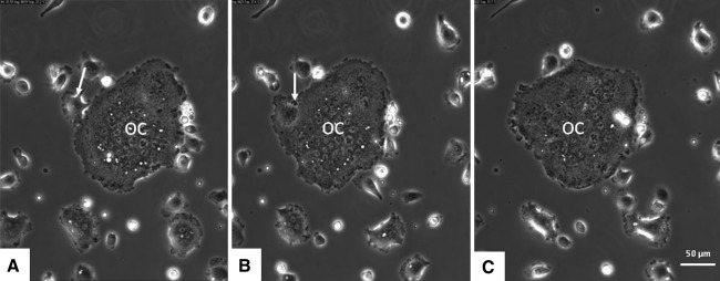
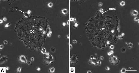
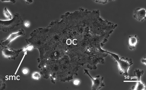
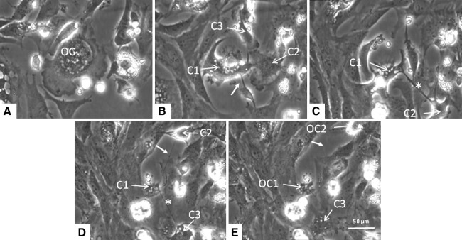
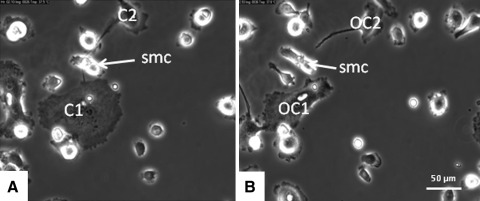
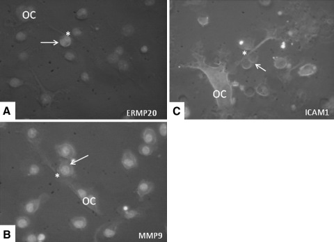
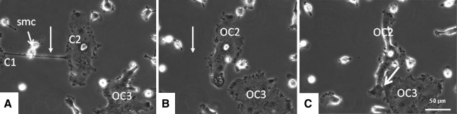
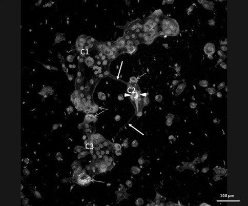
Osteoclasts are specialized multinucleated cells with the unique capacity to resorb bone. Despite insight into the various steps of the interaction of osteoclast precursors leading to osteoclast formation, surprisingly little is known about what happens with the multinucleated cell itself after it has been formed. Is fusion limited to the short period of its formation, or do osteoclasts have the capacity to change their size and number of nuclei at a later stage? To visualize these processes we analyzed osteoclasts generated in vitro with M-CSF and RANKL from mouse bone marrow and native osteoclasts isolated from rabbit bones by live cell microscopy. We show that osteoclasts fuse not only with mononuclear cells but also with other multinucleated cells. The most intriguing finding was fission of the osteoclasts. Osteoclasts were shown to have the capacity to generate functional multinucleated compartments as well as compartments that contained apoptotic nuclei. These compartments were separated from each other, each giving rise to a novel functional osteoclast or to a compartment that contained apoptotic nuclei. Our findings suggest that osteoclasts have the capacity to regulate their own population in number and function, probably to adapt quickly to changing situations.
Figures
Comment in
-
"Fusion and fission" unveils remarkable insights into osteoclast plasticity.Calcif Tissue Int. 2012 Aug;91(2):157-8; author reply 159. doi: 10.1007/s00223-012-9620-7. Epub 2012 Jul 1. Calcif Tissue Int. 2012. PMID: 22752618 Free PMC article. No abstract available.
Similar articles
-
The generation of highly enriched osteoclast-lineage cell populations.Bone. 2002 Jan;30(1):164-70. doi: 10.1016/s8756-3282(01)00654-8. Bone. 2002. PMID: 11792580
-
PDK1 is important lipid kinase for RANKL-induced osteoclast formation and function via the regulation of the Akt-GSK3β-NFATc1 signaling cascade.J Cell Biochem. 2020 Nov;121(11):4542-4557. doi: 10.1002/jcb.29677. Epub 2020 Feb 12. J Cell Biochem. 2020. PMID: 32048762
-
Jaw and long bone marrow derived osteoclasts differ in shape and their response to bone and dentin.Biochem Biophys Res Commun. 2011 Jun 3;409(2):205-10. doi: 10.1016/j.bbrc.2011.04.120. Epub 2011 May 3. Biochem Biophys Res Commun. 2011. PMID: 21565168
-
Osteoclast differentiation by RANKL and OPG signaling pathways.J Bone Miner Metab. 2021 Jan;39(1):19-26. doi: 10.1007/s00774-020-01162-6. Epub 2020 Oct 20. J Bone Miner Metab. 2021. PMID: 33079279 Review.
-
Osteoclast formation at the bone marrow/bone surface interface: Importance of structural elements, matrix, and intercellular communication.Semin Cell Dev Biol. 2021 Apr;112:8-15. doi: 10.1016/j.semcdb.2020.05.016. Epub 2020 Jun 17. Semin Cell Dev Biol. 2021. PMID: 32563679 Review.
Cited by
-
Osteoclast Fusion: Physiological Regulation of Multinucleation through Heterogeneity-Potential Implications for Drug Sensitivity.Int J Mol Sci. 2020 Oct 19;21(20):7717. doi: 10.3390/ijms21207717. Int J Mol Sci. 2020. PMID: 33086479 Free PMC article. Review.
-
Osteoclasts recycle via osteomorphs during RANKL-stimulated bone resorption.Cell. 2021 Mar 4;184(5):1330-1347.e13. doi: 10.1016/j.cell.2021.02.002. Epub 2021 Feb 25. Cell. 2021. PMID: 33636130 Free PMC article.
-
Macrophages and Bone Remodeling.J Bone Miner Res. 2023 Mar;38(3):359-369. doi: 10.1002/jbmr.4773. Epub 2023 Feb 3. J Bone Miner Res. 2023. PMID: 36651575 Free PMC article. Review.
-
Osteoclastogenesis Requires Primary Cilia Disassembly and Can Be Inhibited by Promoting Primary Cilia Formation Pharmacologically.Cells Tissues Organs. 2024;213(3):235-244. doi: 10.1159/000531098. Epub 2023 May 22. Cells Tissues Organs. 2024. PMID: 37231815 Free PMC article.
-
Osteoclast Multinucleation: Review of Current Literature.Int J Mol Sci. 2020 Aug 8;21(16):5685. doi: 10.3390/ijms21165685. Int J Mol Sci. 2020. PMID: 32784443 Free PMC article. Review.
References
-
- Zambonin ZA, Teti A, Primavera MV. Monocytes from circulating blood fuse in vitro with purified osteoclasts in primary culture. J Cell Sci. 1984;66:335–342. - PubMed
MeSH terms
Substances
LinkOut - more resources
Full Text Sources
Research Materials
