. 2012;8(4):e1002630.
doi: 10.1371/journal.ppat.1002630. Epub 2012 Apr 5.

Virion assembly factories in the nucleus of polyomavirus-infected cells

Affiliations

Virion assembly factories in the nucleus of polyomavirus-infected cells

Kimberly D Erickson et al. PLoS Pathog. 2012.

Abstract

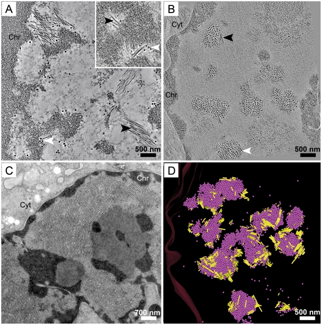
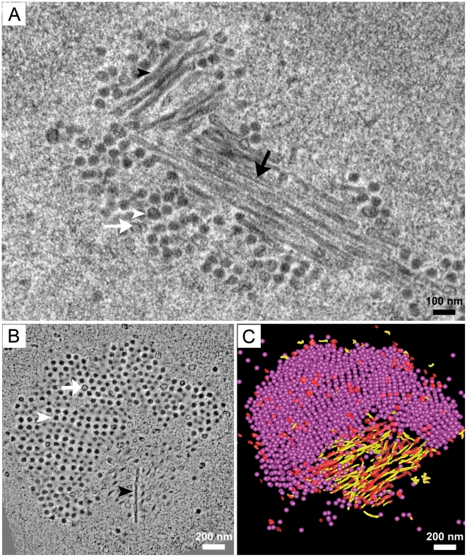
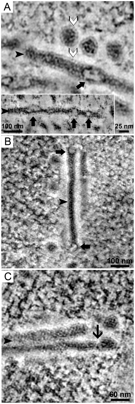
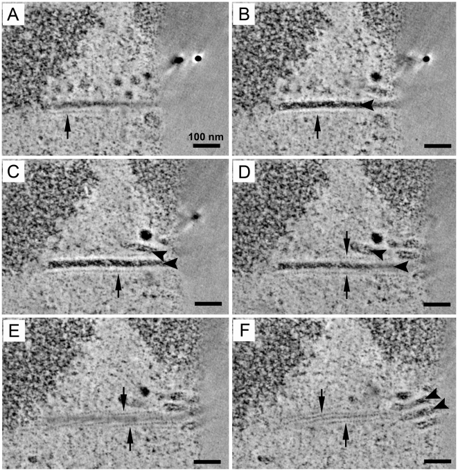
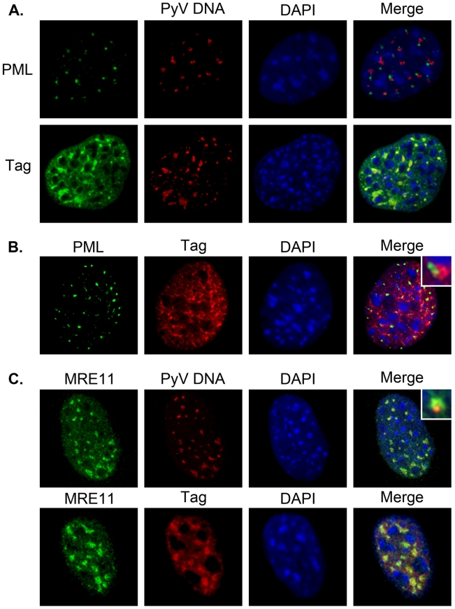
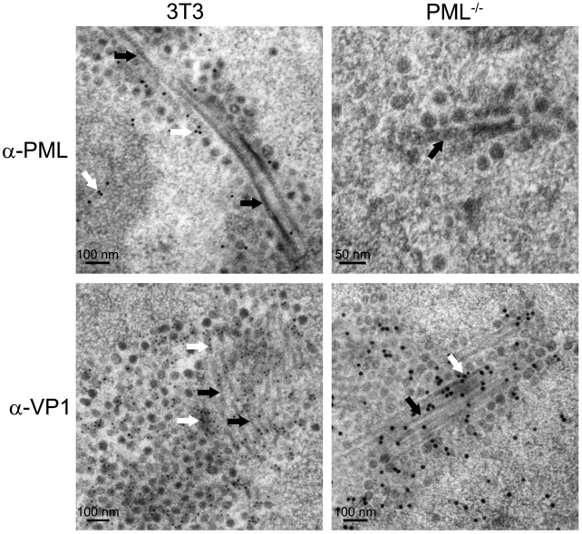
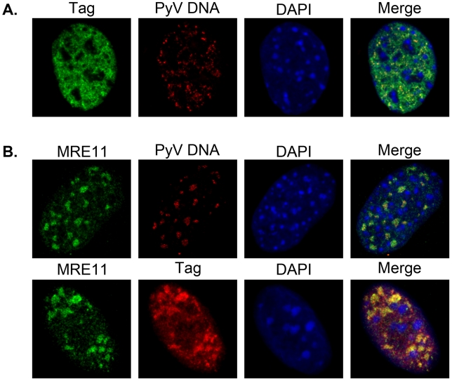
Most DNA viruses replicate in the cell nucleus, although the specific sites of virion assembly are as yet poorly defined. Electron microscopy on freeze-substituted, plastic-embedded sections of murine polyomavirus (PyV)-infected 3T3 mouse fibroblasts or mouse embryonic fibroblasts (MEFs) revealed tubular structures in the nucleus adjacent to clusters of assembled virions, with virions apparently "shed" or "budding" from their ends. Promyelocytic leukemia nuclear bodies (PML-NBs) have been suggested as possible sites for viral replication of polyomaviruses (BKV and SV40), herpes simplex virus (HSV), and adenovirus (Ad). Immunohistochemistry and FISH demonstrated co-localization of the viral T-antigen (Tag), PyV DNA, and the host DNA repair protein MRE11, adjacent to the PML-NBs. In PML⁻/⁻ MEFs the co-localization of MRE11, Tag, and PyV DNA remained unchanged, suggesting that the PML protein itself was not responsible for their association. Furthermore, PyV-infected PML⁻/⁻ MEFs and PML⁻/⁻ mice replicated wild-type levels of infectious virus. Therefore, although the PML protein may identify sites of PyV replication, neither the observed "virus factories" nor virus assembly were dependent on PML. The ultrastructure of the tubes suggests a new model for the encapsidation of small DNA viruses.


Conflict of interest statement

The authors have declared that no competing interests exist.

Figures

Figure 1
Figure 1. Temporal progression of PyV nuclear assembly.
Images from dual-axis tomograms of high pressure frozen, Epon-embedded, 300 nm thick sections of 3T3 cells infected with PyV (MOI of 10–20 pfu/cell) and harvested at 32 hpi. A) Tubular structures (black arrowhead) are present in the periphery of the condensed chromatin (Chr) adjacent to occasional virions (white arrowhead). B) A 1 nm section extracted from a 2×2 montage over six serial sections (1.8 µm thick) of a 3T3 nucleus in which the interchromatin space is partially filled with virion clusters and each cluster is associated with tubular structures. As infection proceeds, the number of virus clusters (white arrowhead) increases while the tubular structures (black arrowhead) are less prominent. C) Late in infection virions fill the entire interchromatin space and tubular structures are not seen. D) 3-D model of the 2×2 montage over six serial sections (each 300 nm thick) of a PyV-infected 3T3 nucleus. The model represents 1.8 µm thick section of the nucleus showing the connections between virion clusters and tubular structures. An image extracted from the tomogram is shown in (B); a video of the tomograms of each section and the model can be found as supporting information Video S1. Pink spheres, full virions; yellow cylinders, tubular structures. Chr, host condensed chromatin; Cyt, cytoplasm.
Figure 2
Figure 2. Tomographic reconstruction of a virus factory.
3T3 cells were infected with PyV as described for Figure 1. A) 70 nm thin section of Epon-embedded cells showing tubular structures adjacent to a virus cluster. (Black arrowhead, “full” tubular structure; white arrowhead, full virion; black arrow, “empty” tubular structure; white arrow, empty virion) B) A cluster of virions packed in a highly ordered array at the periphery of a bundle of tubular structures. One of the tubular structures appears in the plane (black arrowhead). Empty (white arrow) and full (white arrowhead) virions are identified in the virus clusters. C) 3-D model of (B) showing ≈2000 full assembled virions (pink spheres) and ≈2% empty virions (red spheres) in a 300 nm thick section. The tubular structures are either filled with electron-dense material (yellow cylinders) or appear empty (red cylinders). (See also Supporting information Video S2).
Figure 3
Figure 3. Tubule ultrastructure and association with virions.
PyV-infected 3T3 cells (see Figure 1) were processed by high pressure freezing and cryo-substitution for electron microscopy and a series of images was extracted from dual-axis tomograms of Epon-embedded, 300 nm thick sections. A) Two distinct virions show a lighter and well-arranged density at their periphery (white arrowhead) corresponding to capsid protein density as well as a dense core suggesting DNA. The tubular structure exhibited a similar organization with a lighter and well-arranged density at its periphery (white arrowhead) as well as a patched and electron-dense core (black arrowhead). Occasionally, “bulges” or opaque regions (thick black arrows, also see inset) are seen along the length of the tube. B) A tubular structure (black arrowhead) exhibiting bulges forming at both ends (thick black arrows). The bulges are defined by an inner layer of dense material surrounded by well-arranged lighter densities. C) A mature virion is partially attached to the tubular structure (black arrowhead). A constriction point is seen where the virion may be shed from the tubular structure (thin black arrow). (See also Supporting information Figure S1).
Figure 4
Figure 4. Periodic densities are associated with the tubes.
Images from a dual-axis, tomographic reconstruction of PyV-infected 3T3 cells (described in Figure 1) extracted in silico every 5 nm. A–F) Sections (5 nm) were extracted from the tomogram every 6 nm in the z-plane allowing a longitudinal view of a tubular structure (black arrowhead) and its associated periodical filamentous structures (black arrow). The electron-dense material within the tubular structure has a diameter of 33 nm. The fine, periodic structures were 10 nm from the core with a periodicity of 8–12 nm.
Figure 5
Figure 5. PyV DNA and T-antigen are localized near PML-NBs.
C57 MEFs were infected at an MOI of 30–40 pfu/cell. At 24 or 28 hpi cells were fixed, permeabilized, and co-stained with either anti-PML, anti-Tag and/or anti-MRE11 antibodies followed by AlexaFluor-conjugated secondary antibodies, a fluorescently-labeled PyV DNA FISH probe, and DAPI staining of nuclei. A) PyV-infected C57 MEFs at 24 hpi stained for either PML (top) or Tag (bottom) followed by fluorescent in situ hybridization (FISH) for PyV DNA. B) PyV-infected C57 MEFs at 28 hpi co-stained for PML and Tag. C) C57 MEFs at 24 hpi co-stained for either MRE11 and PyV DNA (by FISH) or MRE11 and Tag. All images represent a 0.1 mm z-stack slice. Insets show enlarged regions from image to illustrate the localization of proteins in relation to each other (B) or PyV DNA (C).
Figure 6
Figure 6. Tubular structures contain VP1.
PyV-infected 3T3 cells or PML−/− MEFs (MOI of 10–20 pfu/cell) at 32 hpi were frozen by high pressure and processed by cryo-substitution for immunoelectron microscopy. Thin sections (45 nm or 70 nm) of Lowicryl-embedded samples were stained with either anti-PML or anti-VP1 antibodies followed by a secondary antibody conjugated to 10 or 15 nm colloidal gold. Top panels: 45 nm sections stained for the PML protein; white arrows, anti-PML staining, black arrow, tubular structures. Bottom panels: 70 nm sections stained for VP1; white arrows, anti-VP1 staining, black arrows, tubular structures.
Figure 7
Figure 7. PyV DNA and T-antigen localization in PyV-infected PML−/− MEFs.
PML−/− MEFs were infected with PyV at an MOI of 30–40 pfu/cell. At 22 or 24 hpi cells were fixed, permeabilized, and co-stained with either anti-Tag and/or anti-MRE11a antibodies followed by AlexaFluor-conjugated secondary antibodies, a fluorescently-labeled PyV DNA FISH probe, and DAPI staining of nuclei. A) FISH for PyV DNA at 24 hpi followed by antibody staining for Tag. B) Infected cells were stained by FISH for PyV DNA at 22 hpi followed by antibody staining for MRE11 or co-stained for MRE11 and Tag. All images represent a 0.1 mm z-stack slice.

Similar articles

Cited by

References

    1. Wileman T. Aggresomes and Pericentriolar Sites of Virus Assembly: Cellular Defense or Viral Design? Annu Rev Microbiol. 2007;61:149–167. - PubMed
    1. Novoa RR, Calderita G, Arranz R, Fontana J, Granzow H, et al. Virus factories: associations of cell organelles for viral replication and morphogenesis. Biol Cell. 2005;97:147–172. - PMC - PubMed
    1. den Boon JA, Diaz A, Ahlquist P. Cytoplasmic Viral Replication Complexes. Cell Host Micro. 2010;8:77–85. - PMC - PubMed
    1. Suhy DA, Giddings TH, Jr, Kirkegaard K. Remodeling the endoplasmic reticulum by poliovirus infection and by individual viral proteins: an autophagy-like origin for virus-induced vesicles. J Virol. 2000;74:8953–8965. - PMC - PubMed
    1. Gillespie LK, Hoenen A, Morgan G, Mackenzie JM. The endoplasmic reticulum provides the membrane platform for biogenesis of the flavivirus replication complex. J Virol. 2010;84:10438–10447. - PMC - PubMed

Publication types

MeSH terms