. 2012 Jun 1;287(23):18937-52.
doi: 10.1074/jbc.M111.313262. Epub 2012 Apr 11.

Roles of Kruppel-associated Box (KRAB)-associated Co-repressor KAP1 Ser-473 Phosphorylation in DNA Damage Response

Affiliations

Roles of Kruppel-associated Box (KRAB)-associated Co-repressor KAP1 Ser-473 Phosphorylation in DNA Damage Response

Chen Hu et al. J Biol Chem. .

Abstract

The Kruppel-associated box (KRAB)-associated co-repressor KAP1 is an essential nuclear co-repressor for the KRAB zinc finger protein superfamily of transcriptional factors. Ataxia telangiectasia mutated (ATM)-Chk2 and ATM- and Rad3-related (ATR)-Chk1 are two primary kinase signaling cascades activated in response to DNA damage. A growing body of evidence suggests that ATM and ATR phosphorylate KAP1 at Ser-824 in response to DNA damage and regulate KAP1-dependent chromatin condensation, DNA repair, and gene expression. Here, we show that, depending on the type of DNA damage that occurs, KAP1 Ser-473 can be phosphorylated by ATM-Chk2 or ATR-Chk1 kinases. Phosphorylation of KAP1 at Ser-473 attenuated its binding to the heterochromatin protein 1 family proteins and inhibited its transcriptional repression of KRAB-zinc finger protein (KRAB-ZFP) target genes. Moreover, KAP1 Ser-473 phosphorylation induced by DNA damage stimulated KAP1-E2F1 binding. Overexpression of heterochromatin protein 1 significantly inhibited E2F1-KAP1 binding. Elimination of KAP1 Ser-473 phosphorylation increased E2F1-targeted proapoptotic gene expression and E2F1-induced apoptosis in response to DNA damage. Furthermore, loss of phosphorylation of KAP1 Ser-473 led to less BRCA1 focus formation and slower kinetics of loss of γH2AX foci after DNA damage. KAP1 Ser-473 phosphorylation was required for efficient DNA repair and cell survival in response to DNA damage. Our studies reveal novel functions of KAP1 Ser-473 phosphorylation under stress.


Figures
FIGURE 1.
FIGURE 1.
Characterization of anti-KAP1 Ser-473 phosphorylation antibody. A, comparison of sequences of Chk2 peptide substrates of Strap, E2F1, and Cdc25C with KAP1 466–479 and KAP1 817–830. The consensus sequence is shown. B, HeLa cells stably infected with lentiviral vectors expressing control (Mock-Si) or KAP1 (KAP1-Si) shRNA were treated with 10 Gy of IR. Cell lysates were collected 5 h after radiation, and KAP1 phosphorylation at Ser-473 (KAP1S473p) and Ser-824 (KAP1S824p) and phosphorylation of H2AX (γH2AX) were analyzed by Western blotting (WB). C, FLAG M2-agarose-purified FLAG-KAP1 from 293T cells was treated with or without calf intestine alkaline phosphatase (CIP) (30 units) at 37 °C for 30 min, and KAP1 S473p and total KAP1 levels were analyzed by Western blotting. D, 293T cells were transfected with FLAG-tagged KAP1 WT or S473A, S473D, or S473E mutant plasmid. Cell lysates were immunoprecipitated with anti-FLAG M2-agarose beads and probed with anti-KAP1 S473p and anti-KAP1 antibodies. E, GST-KAP1 was expressed in E. coli BL21 and purified with GST beads, and FLAG-Chk2 was purified from FLAG-Chk2-transiently transfected 293T cells using anti-FLAG M2-agarose. In the presence or absence of ATP, GST-KAP1 was incubated with or without FLAG-Chk2. The reaction product was separated by SDS-PAGE and analyzed by Western blot analysis. Ctrl, control.
FIGURE 2.
FIGURE 2.
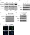
DNA damage induces KAP1 Ser-473 phosphorylation. A, U2OS, HeLa, and A549 cells were treated with 0, 15, or 25 J/m2 UV irradiation and harvested 12 h later. KAP1 S473p, total KAP1, and actin were analyzed by Western blot assay. B, HeLa cells were treated with different doses of IR as indicated and harvested 6 h later, and the total cell lysates were analyzed by immunoblotting with the indicated antibodies. C, HeLa cells were treated with different concentrations of etoposide for 8 h. KAP1 S473p, KAP1, and actin were analyzed by Western blot assay. D, HeLa cells were treated with different doses of doxorubicin as indicated for 12 h. KAP1 S473p, KAP1, and actin were analyzed by Western blot assay. E, HeLa cells were treated with 0 (Ctrl) or 30 μm etoposide for 8 h, and KAP1 S473p (green) was detected by immunofluorescence staining.
FIGURE 3.
FIGURE 3.
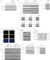
KAP1 Ser-473 is phosphorylated by ATM-Chk2 and ATR-Chk1 pathways in different cells under different types of DNA damage. A, HeLa cells were pretreated with different doses of wortmannin (Wort) or caffeine as indicated for 1 h and then treated with 10 Gy of γ-radiation. Whole cell lysates were collected 8 h after radiation, KAP1 S473p, KAP1 S824p, and total KAP1 were analyzed by Western blot assay. B, 293T cells were transfected with the indicated short interfering RNA (-Si) for ATM, ATR, Chk1, Chk2, PKCδ, or MK2 or irrelevant siRNA (Ctrl-Si) for 66 h and then treated with etoposide (30 μm) for another 6 h before harvesting. Cell lysates were subjected to Western blot analysis using the indicated antibodies. Relative KAP1 S473p levels were quantitated with NIH ImageJ software and are listed. C, 293T cells were transfected with vector (Ctrl), FLAG-Chk2 WT, or Chk2 kinase-dead mutant FLAG-Chk2 D347A (D347A) for 36 h. Cell lysates were analyzed by immunoblotting with the indicated antibodies. D, HeLa cells were transfected with FLAG-Chk2 WT or FLAG-Chk2 D347A for 24 h. FLAG-Chk2 WT (red) or D347A (red) and KAP1 S473p (green) were detected by immunofluorescence staining. E, HCT116 cells were treated with 0 (−), 5 (+), or 10 mm (++) Chk2 inhibitor II (Sigma-Aldrich, C3742) for 1 h followed by treatment of 30 μm etoposide for 6 h. Cell lysates were subjected to Western blotting as indicated. F, 293T cells were co-transfected with GFP-KAP1 and FLAG-Chk2 for 24 h, and then cells were either left untreated (−) or treated with 10 mm (+) Chk2 inhibitor II for another 2 h. KAP1-Chk2 binding was detected by FLAG M2-agarose bead IP and Western blot assay. G, HCT116 (Chk2 wild type) and HCT15 (Chk2-deficient) cells were treated with different doses of etoposide or UV irradiation as indicated for 6 h, and cell lysates were subjected to Western blot analysis using the indicated antibodies. H, HCT15 cells were treated with different doses of IR or UV irradiation as indicated, cells were harvested 6 h after treatment, and lysates were subjected to Western blotting as indicated. I, HCT15 cells were pretreated with 2 μm Chk1 inhibitor (PD407824, Sigma) for 1 h followed by 25 J/m2 UV treatment. Cells were harvested about 6 h after UV treatment, and cell lysates were then subjected to Western blot analysis as indicated.
FIGURE 4.
FIGURE 4.
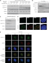
Comparison of KAP1 S824p and S473p in response to DNA damage. A, KAP1 knockdown stable HeLa cells were transfected with vector (−) or siRNA-resistant FLAG-KAP1 WT or Ser-473 or Ser-824 site mutants for 24 h, and then treated with etoposide (30 μm) for 1.5 and 7 h. Cell lysates were subjected to Western blot analysis using the indicated antibodies. B, HeLa cells were treated with IR and harvested at different time points. Cell lysates were subjected to Western blot analysis with antibodies as indicated. C, HeLa cells were pretreated with 30 μm etoposide for 1 h and then transferred to drug-free medium. Cells were harvested at different time point as indicated, and cell lysates were subjected to Western blotting. D, HeLa cells were treated with 0 (control) or 30 μm etoposide for 8 h, fixed, and stained for KAP1 S473p (green) and γH2AX (red). DNA (blue) is stained with DAPI. The images were analyzed by confocal microscopy. E, HeLa cells were irradiated with 10 Gy of IR, harvested at different time points, fixed, and stained for KAP1 S473p (green) and γH2AX (red). DNA (blue) is stained with DAPI. Ctrl, control.
FIGURE 5.
FIGURE 5.
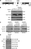
DNA damage-induced KAP1 S473p attenuates KAP1 binding affinity to HP1 family proteins by changing structure at HP1 binding domain. A, schematic representation of the domain organization of KAP1. Bromo, bromodomain. Phosphorylated residues Ser-473 and Ser-824 are indicated. B, HeLa cells were transfected with vector (ctrl) or FLAG-HP1α, β, or γ for 24 h followed by 30 μm etoposide treatment for 12 h. KAP1-HP1 binding was detected by anti-FLAG M2-agarose bead immunoprecipitation and Western blot assay. C, FLAG-KAP1 WT or Ser-473 site mutant plasmids were co-transfected with GFP-HP1 α-, β-, or γ-overexpressing plasmids as indicated into 293T cells for 24 h. KAP1-HP1 binding was detected by anti-FLAG M2-agarose bead IP and Western blot assay. D, HeLa cells were treated with etoposide for 6 h, endogenous KAP1 was precipitated using KAP1 antibody (ab10483), and co-precipitation of endogenous HP1 was detected by Western blot.
FIGURE 6.
FIGURE 6.
Phosphorylation of KAP1 Ser-473 regulates its transcription co-repression activity. A, schematic illustration of GAL4-TK-luciferase reporter (GTK-luciferase reporter) and GAL4-KRAB repressor protein. B, KAP1 knockdown stable HeLa cells were co-transfected with GAL4-TK-luciferase reporter and Renilla luciferase plasmid or different amounts of RNAi-resistant FLAG-tagged KAP1 WT, S473A, S473D, or S473E plasmid in the presence or absence of GAL4-KRAB plasmid. Relative luciferase repression (upper panel) was calculated by comparing firefly luciferase activity with Renilla luciferase activity. Error bars represent mean ± S.D. (n = 4) in each case. Phosphorylation-mimicking mutants (S473D and S473E) increased (p < 0.01) KAP1 transcriptional repression activity compared with WT KAP1, and there is no significant difference between wild-type KAP1 and S473A mutant. KAP1 levels were confirmed by Western blot (lower panel). C, HeLa cells were infected with pll3.7-DsRed (shN) or pll3.7-DsRed-shKAP1 (shKAP1) lentiviral vectors, and siRNA-resistant lenti-ZsGreen-FLAG-KAP1 (WT, S473A, S473D, or S473E) plasmids for 60 h, green and red positive cells were collected by FACS, a quantitative RT-PCR assay was performed to analyze KAP1-ZNF target gene expression as indicated, and KAP1 expression was detected by Western blots.
FIGURE 7.
FIGURE 7.
KAP1 Ser-473 phosphorylation regulates E2F1 and etoposide-induced cell apoptosis. A, HA-tagged E2F1 was co-transfected with FLAG-KAP1 WT, S473A, or S824A expression plasmid into 293T cells for 24 h followed by treatment with etoposide (30 μm) for another 10 h, and KAP1-E2F1 binding was detected by anti-FLAG M2-agarose bead IP and Western blot assay. B, 293T cells were co-transfected with HA-E2F1, Myc-KAP1, FLAG-Chk2 WT, or FLAG-Chk2 D347A plasmid as indicated for 36 h. KAP1-E2F1 binding was detected by anti-Myc-agarose IP and Western blot assay. C, 293T cells were transfected with FLAG-tagged E2F1, GFP-tagged KAP1, and HP1 expression plasmids as indicated. Cells were harvested 36 h after transfection, and E2F1-KAP1 binding was detected by anti-FLAG M2-agarose bead IP and Western blot assay. D, an H1299 cell line stably expressing the E2F1-ER construct was transfected with KAP1 wild type or Ser-473 mutant plasmid as indicated for 24 h, and then cells were either left untreated (−) or treated with 100 nm 4-hydroxytamoxifen (4-OHT) (+) for another 12 h. The mRNA levels of E2F1-mediated apoptosis genes (Apaf-1, ASK1, Bim, p73, and caspase 7) were determined by semiquantitative RT (Semi-qRT)-PCR, and KAP1 protein expression levels were analyzed by Western blot assay. E, SaoS2 cells were co-transfected with either vector (Ctrl) or HA-E2F1 and KAP1 WT or KAP1 S473A expression plasmids for 20 h, and then cells were treated with etoposide (20 μm) for another 12 h. Cells were stained with Annexin V-FITC and propidium iodide (PI), and analyzed by FACS (upper panel). KAP1, E2F1, and actin protein levels were examined by Western blot (WB) (lower panel). F, Mock control (shN) or KAP1 knockdown (shKAP1) HeLa cells were infected with siRNA-resistant lenti-ZsGreen-FLAG-KAP1 (WT, S473A, S473D, S824A, or S824D) for 36 h followed by treatment with different doses of etoposide for another 24 h. Cell survival was analyzed by WST-1 assay. Error bars represent mean ± S.D. (n = 4) in each case.
FIGURE 8.
FIGURE 8.
A, HeLa cells were treated with 20 μm etoposide for 1 h and washed with PBS three times. Cells were collected at different time points after etoposide withdrawal, and KAP1 S473p (green), BRCA1 (red), and DAPI (blue) were detected by immunofluorescence staining. B, Mock control or KAP1 knockdown (shKAP1) stable HeLa cells were co-transfected with GFP and KAP1 siRNA-resistant FLAG-tagged KAP1 wild type, S473A, or S824A plasmid for 24 h, and then cells were treated the same as in A. Cells were stained for BRCA1 (red) (lower panels) and analyzed by confocal microscopy. The average number of BRCA1 foci per nucleus of GFP-positive cells was quantified (upper panel). A minimum of 30 cells per sample were counted in triplicate. C, the average number of γH2AX foci per nucleus was determined using the same assay as in B.

Similar articles

Cited by

References

    1. Friedman J. R., Fredericks W. J., Jensen D. E., Speicher D. W., Huang X. P., Neilson E. G., Rauscher F. J., 3rd (1996) KAP-1, a novel corepressor for the highly conserved KRAB repression domain. Genes Dev. 10, 2067–2078 - PubMed
    1. Cammas F., Mark M., Dollé P., Dierich A., Chambon P., Losson R. (2000) Mice lacking the transcriptional corepressor TIF1β are defective in early postimplantation development. Development 127, 2955–2963 - PubMed
    1. Weber P., Cammas F., Gerard C., Metzger D., Chambon P., Losson R., Mark M. (2002) Germ cell expression of the transcriptional co-repressor TIF1β is required for the maintenance of spermatogenesis in the mouse. Development 129, 2329–2337 - PubMed
    1. Peng H., Begg G. E., Schultz D. C., Friedman J. R., Jensen D. E., Speicher D. W., Rauscher F. J., 3rd (2000) Reconstitution of the KRAB-KAP-1 repressor complex: a model system for defining the molecular anatomy of RING-B box-coiled-coil domain-mediated protein-protein interactions. J. Mol. Biol. 295, 1139–1162 - PubMed
    1. Lechner M. S., Begg G. E., Speicher D. W., Rauscher F. J., 3rd (2000) Molecular determinants for targeting heterochromatin protein 1-mediated gene silencing: direct chromoshadow domain-KAP-1 corepressor interaction is essential. Mol. Cell. Biol. 20, 6449–6465 - PMC - PubMed

Publication types

MeSH terms