Epithelial cell responses to infection with human papillomavirus
- PMID: 22491770
- PMCID: PMC3346303
- DOI: 10.1128/CMR.05028-11
Epithelial cell responses to infection with human papillomavirus
Abstract
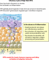
Human papillomavirus (HPV) infection of the genital tract is common in young sexually active individuals, the majority of whom clear the infection without overt clinical disease. Most of those who do develop benign lesions eventually mount an effective cell-mediated immune (CMI) response, and the lesions regress. Regression of anogenital warts is accompanied histologically by a CD4(+) T cell-dominated Th1 response; animal models support this and provide evidence that the response is modulated by antigen-specific CD4(+) T cell-dependent mechanisms. Failure to develop an effective CMI response to clear or control infection results in persistent infection and, in the case of the oncogenic HPVs, an increased probability of progression to high-grade intraepithelial neoplasia and invasive carcinoma. Effective evasion of innate immune recognition seems to be the hallmark of HPV infections. The viral infectious cycle is exclusively intraepithelial: there is no viremia and no virus-induced cytolysis or cell death, and viral replication and release are not associated with inflammation. HPV globally downregulates the innate immune signaling pathways in the infected keratinocyte. Proinflammatory cytokines, particularly the type I interferons, are not released, and the signals for Langerhans cell (LC) activation and migration, together with recruitment of stromal dendritic cells and macrophages, are either not present or inadequate. This immune ignorance results in chronic infections that persist over weeks and months. Progression to high-grade intraepithelial neoplasia with concomitant upregulation of the E6 and E7 oncoproteins is associated with further deregulation of immunologically relevant molecules, particularly chemotactic chemokines and their receptors, on keratinocytes and endothelial cells of the underlying microvasculature, limiting or preventing the ingress of cytotoxic effectors into the lesions. Recent evidence suggests that HPV infection of basal keratinocytes requires epithelial wounding followed by the reepithelization of wound healing. The wound exudate that results provides a mechanistic explanation for the protection offered by serum neutralizing antibody generated by HPV L1 virus-like particle (VLP) vaccines.
Figures


Similar articles
-
Immune responses to human papilloma viruses.Indian J Med Res. 2009 Sep;130(3):266-76. Indian J Med Res. 2009. PMID: 19901436 Review.
-
Host responses to infection with human papillomavirus.Curr Probl Dermatol. 2014;45:58-74. doi: 10.1159/000355964. Epub 2014 Mar 13. Curr Probl Dermatol. 2014. PMID: 24643178 Review.
-
HPV - immune response to infection and vaccination.Infect Agent Cancer. 2010 Oct 20;5:19. doi: 10.1186/1750-9378-5-19. Infect Agent Cancer. 2010. PMID: 20961432 Free PMC article.
-
High-risk HPV oncoproteins E6 and E7 and their interplay with the innate immune response: Uncovering mechanisms of immune evasion and therapeutic prospects.J Med Virol. 2024 Jun;96(6):e29685. doi: 10.1002/jmv.29685. J Med Virol. 2024. PMID: 38783790 Review.
-
Immunobiology of HPV and HPV vaccines.Gynecol Oncol. 2008 May;109(2 Suppl):S15-21. doi: 10.1016/j.ygyno.2008.02.003. Gynecol Oncol. 2008. PMID: 18474288 Review.
Cited by
-
Werner Syndrome Protein (WRN) Regulates Cell Proliferation and the Human Papillomavirus 16 Life Cycle during Epithelial Differentiation.mSphere. 2020 Sep 16;5(5):e00858-20. doi: 10.1128/mSphere.00858-20. mSphere. 2020. PMID: 32938703 Free PMC article.
-
Cervical-Vaginal Microbiome and Associated Cytokine Profiles in a Prospective Study of HPV 16 Acquisition, Persistence, and Clearance.Front Cell Infect Microbiol. 2020 Sep 25;10:569022. doi: 10.3389/fcimb.2020.569022. eCollection 2020. Front Cell Infect Microbiol. 2020. PMID: 33102255 Free PMC article.
-
Human papillomavirus in the setting of immunodeficiency: Pathogenesis and the emergence of next-generation therapies to reduce the high associated cancer risk.Front Immunol. 2023 Mar 7;14:1112513. doi: 10.3389/fimmu.2023.1112513. eCollection 2023. Front Immunol. 2023. PMID: 36960048 Free PMC article. Review.
-
Comparison of Expectant and Excisional/Ablative Management of Cervical Intraepithelial Neoplasia Grade 2 (CIN2) in the Era of HPV Testing.Obstet Gynecol Int. 2022 Mar 24;2022:7955290. doi: 10.1155/2022/7955290. eCollection 2022. Obstet Gynecol Int. 2022. PMID: 35371262 Free PMC article.
-
TIME Is Ticking for Cervical Cancer.Biology (Basel). 2023 Jun 30;12(7):941. doi: 10.3390/biology12070941. Biology (Basel). 2023. PMID: 37508372 Free PMC article. Review.
References
-
- Akira S, Hemmi H. 2003. Recognition of pathogen-associated molecular patterns by TLR family. Immunol. Lett. 85:85–95 - PubMed
-
- Alazawi W, et al. 2002. Changes in cervical keratinocyte gene expression associated with integration of human papillomavirus 16. Cancer Res. 62:6959–6965 - PubMed
-
- Arany I, Goel A, Tyring SK. 1995. Interferon response depends on viral transcription in human papillomavirus-containing lesions. Anticancer Res. 15:2865–2869 - PubMed
-
- Arend WP, Palmer G, Gabay C. 2008. IL-1, IL-18, and IL-33 families of cytokines. Immunol. Rev. 223:20–38 - PubMed
-
- Bedoui S, et al. 2009. Cross-presentation of viral and self antigens by skin-derived CD103+ dendritic cells. Nat. Immunol. 10:488–495 - PubMed
Publication types
MeSH terms
Substances
LinkOut - more resources
Full Text Sources
Other Literature Sources
Research Materials

