. 2012 Jun;86(11):6146-58.
doi: 10.1128/JVI.00013-12. Epub 2012 Apr 4.

Functions of the Epstein-Barr virus EBNA1 protein in viral reactivation and lytic infection

Affiliations

Functions of the Epstein-Barr virus EBNA1 protein in viral reactivation and lytic infection

Nirojini Sivachandran et al. J Virol. 2012 Jun.

Abstract

EBNA1 is the only nuclear Epstein-Barr virus (EBV) protein expressed in both latent and lytic modes of infection. While EBNA1 is known to play several important roles in latent infection, the reason for its continued expression in lytic infection is unknown. Here we identified two roles for EBNA1 in the reactivation of latent EBV to the lytic cycle in epithelial cells. First, EBNA1 depletion in latently infected cells was shown to positively contribute to spontaneous EBV reactivation, showing that EBNA1 has a role in suppressing reactivation. Second, when the lytic cycle was induced, EBNA1 depletion decreased lytic gene expression and DNA amplification, showing that it positively contributed to lytic infection. Since we have previously shown that EBNA1 disrupts promyelocytic leukemia (PML) nuclear bodies, we investigated whether this function could account for the effects of EBNA1 on lytic infection by repeating the experiments with cells lacking PML proteins. In the absence of PML, EBNA1 did not promote lytic infection, indicating that the EBNA1-mediated PML disruption is responsible for promoting lytic infection. In keeping with this conclusion, PML silencing was found to be sufficient to induce the EBV lytic cycle. Finally, by generating cells with single PML isoforms, we showed that individual PML isoforms were sufficient to suppress EBV lytic reactivation, although PML isoform IV (PML IV) was ineffective because it was most efficiently degraded by EBNA1. Our results provide the first function for EBNA1 in lytic infection and show that EBNA1 interactions with PML IV lead to a loss of PML nuclear bodies (NBs) that promotes lytic infection.


Figures

Fig 1
Fig 1
EBNA1 expression inhibits spontaneous EBV reactivation. AGS-EBV cells were treated with siRNA against GFP (siGFP) or EBNA1 (siEBNA1) or left untreated (AGS-EBV). (A) Cells were fixed and stained with antibodies against EBNA1 (middle panels) and BZLF1 (right panels), and the percentage of BZLF1-positive cells was determined, where more than 100 cells were counted under each condition. Images with the same antibody treatment were captured using the same exposure times. The bar graph shows average values from three separate experiments with standard deviations. The bar graph on the right shows results using an alternative siRNA against EBNA1 (siEBNA1a) and AllStars negative-control siRNA (siAS). **, P value < 0.01. (B) Equal amounts of cell lysates from untreated AGS-EBV (lane 3) or AGS-EBV treated with siGFP, siEBNA1, siEBNA1a, or AllStars siRNA (siAS) were compared by Western blotting using antibodies for EBNA1, BMRF1, BZLF1, and actin (loading control). (C) Steady-state transcript levels of BZLF1 were monitored by isolating total RNA, synthesizing cDNA, and quantifying transcript levels using primers specific to BZLF1. Values were normalized to the GAPDH housekeeping gene. Average levels of the BZLF1 transcript from three separate experiments are shown for siEBNA1 treatment (with standard deviations) relative to those for the siGFP treatment (set to 1). (D) EBV genomic DNA was quantified by isolating total DNA, amplifying the EBV DS sequence, and normalizing to GAPDH. Average levels (with standard deviations) for siEBNA1 treatment were compared to those for siGFP treatment (set to 1; left graph), and results for siEBNA1a and siAllStars treatment was compared to results for untreated AGS-EBV (set to 1; left panel). ***, P < 0.001.
Fig 2
Fig 2
EBNA1 positively contributes to the EBV lytic cycle after NaB-TPA induction. AGS-EBV cells were treated with siRNA against GFP (siGFP) or EBNA1 (siEBNA1) or left untreated, followed by induction of the EBV lytic cycle by NaB-TPA treatment. (A) Cells were fixed and stained with antibodies against EBNA1 (middle panels) and BZLF1 (right panels), and the percentage of BZLF1-positive cells was determined, where more than 100 cells were counted for each condition. The bar graph shows average values from three separate experiments with standard deviations. ***, P < 0.001. Images with the same antibody treatment were captured using the same exposure times. (B) Equal amounts of cell lysates from AGS-EBV cells treated with siGFP or siEBNA1 with and without NaB-TPA induction were compared by Western blotting using antibodies against EBNA1, PML, BMRF1, BZLF1, and actin. (C) EBV genomic DNA was quantified after NaB-TPA treatment as described for 1D.
Fig 3
Fig 3
EBNA1 effects on EBV reactivation in NPC cells. HONE-Akata cells were treated with siRNA against GFP or EBNA1. (A and B) Equal amounts of cell lysates before (A) or after (B) NaB-TPA treatment were compared by Western blotting using antibodies against EBNA1, PML, BMRF1, BZLF1, and actin. (C) EBV genomic DNA was quantified before and after NaB-TPA treatment as described for Fig. 1D. Average levels are shown with standard deviations; the value for siGFP samples without NaB-TPA treatment was set to 1.
Fig 4
Fig 4
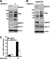
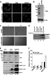
PML represses EBV reactivation. AGS-EBV (A-EBV) cells were infected with a lentivirus carrying shRNA against all PML isoforms and were selected for cells containing the lentivirus to generate AGS-EBVshPML cells (A-EBVshPML). (A) Cells were fixed and stained with antibodies against EBNA1 (right panels) and PML (middle panels). Images with the same antibody treatment were captured using the same exposure times. (B) Equal amounts of cell lysates from AGS-EBV (wt) and AGS-EBVshPML (shPML) were compared by Western blotting using an antibody that recognized all isoforms of PML or actin (loading control). (C) Light microscopy images of AGS-EBV and AGS-EBVshPML cells from two experiments (I and II). Examples of enlarged, rounded cells in the AGS-shPML cells are seen in the bottom panels. (D) Equal amounts of cell lysates from AGS-EBV (before and after NaB-TPA treatment) and AGS-EBVshPML (no NaB-TPA treatment) were compared by Western blotting using antibodies that recognize BMRF1, BZLF1, and actin. Lysates from AGS-EBV cells containing a negative-control lentivirus expressing shGFP are also shown (AGS-EBVshGFP, lane 2, no NaB-TPA treatment). (E) Equal amounts of cell lysates from AGS-EBV (PML+) and AGS-EBVshPML (PML−) were compared post-treatment with NaB, TPA, and NaB-TPA by Western blotting using antibodies against PML (all isoforms), EBNA1, BMRF1, BZLF1, and actin. Light (LE) and dark (DE) exposures of the same blot are shown for BMRF1. (F) EBV genomic DNA was quantified post-treatment with DMSO, NaB, TPA, or NaB-TPA as described for Fig. 1D. Average levels with standard deviations after each treatment are shown; values were normalized to AGS-EBV cells treated with DMSO (set to 1).
Fig 5
Fig 5
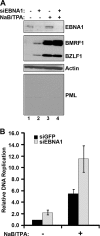
The positive role of EBNA1 in EBV lytic infection is not observed in the absence of PML. (A) AGS-EBVshPML cells were treated with siRNA against GFP (−siEBNA1) or EBNA (+siEBNA1) and then treated with NaB-TPA (+) or the DMSO control (−NaB-TPA). Equal amounts of cell lysates were compared by Western blotting using the indicated antibodies. (B) EBV genomic DNA was quantified in each sample as described for Fig. 1D. The AGS-EBVshPML sample treated with siGFP and DMSO was set to 1 and used to normalize all other samples.
Fig 6
Fig 6
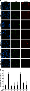
Effect of single PML isoforms on EBV reactivation and lytic infection. AGS-EBVshPML (shPML) cells were infected with lentivirus expressing individual PML isoforms I to VI. (A) The cells were fixed and stained 72 h postinfection for PML (green) and BZLF1 (red). (B) AGS-EBVshPML cells (−), parental AGS-EBV cells (+) and the single PML isoform cells (I to VI) were scored for whether or not they expressed BZLF1, and the percentages of BZLF1-positive cells were determined. For the single PML isoform samples, only the PML-positive cells were scored. Average values with standard deviations from three independent experiments are shown.
Fig 7
Fig 7
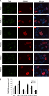
EBNA1 preferentially disrupts PML NBs formed by PML IV. CNE2 cells expressing single PML isoforms I to VI were generated as in the work of Sarkari et al. (55) and were transfected with an EBNA1 expression plasmid. (A) Cells were stained with antibodies against PML (green) and EBNA1 (red) and visualized by IF microscopy. These images were also merged with a DAPI counterstain (right panels). (B) The number of PML NBs per cell was counted for EBNA1-positive cells 48 h after EBNA1 transfection (white bars) and EBNA1-negative cells (control; black bars), and average values from three independent experiments (with standard deviations) are shown.
Fig 8
Fig 8
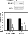
PML IV can suppress EBV reactivation in EBNA1-depleted cells. AGS-EBVshPML cells were treated with siRNA against EBNA1 (siEBNA1) or not treated (no siEBNA1), and then cells were reconstituted with PML IV or PML I or left with no PML (−). Cells were then imaged for BZLF1 and PML as described for Fig. 6A, and the percentage of cells expressing BZLF1 was plotted in the bar graph. The Western blot on the top was performed with AGS-EBVshPML cell extracts prior to introducing PML IV or PML I to confirm the EBNA1 depletion. **, P < 0.01.

Similar articles

Cited by

References

    1. Adams A. 1987. Replication of latent Epstein-Barr virus genomes. J. Virol. 61:1743–1746 - PMC - PubMed
    1. Adamson AL, Kenney S. 2001. Epstein-Barr virus immediate-early protein BZLF1 is SUMO-1 modified and disrupts promyelocytic leukemia bodies. J. Virol. 75:2388–2399 - PMC - PubMed
    1. Ahn JH, Hayward GS. 2000. Disruption of PML-associated nuclear bodies by IE1 correlates with efficient early stages of viral gene expression and DNA replication in human cytomegalovirus infection. Virology 274:39–55 - PubMed
    1. Ahsan N, Kanda T, Nagashima K, Takada K. 2005. Epstein-Barr virus transforming protein LMP1 plays a critical role in virus production. J. Virol. 79:4415–4424 - PMC - PubMed
    1. Bell P, Lieberman PM, Maul GG. 2000. Lytic but not latent replication of Epstein-Barr virus is associated with PML and induces sequential release of nuclear domain 10 proteins. J. Virol. 74:11800–11810 - PMC - PubMed

Publication types

MeSH terms