. 2012 May;13(5):449-56.
doi: 10.1038/ni.2263.

NLRC4-driven production of IL-1β discriminates between pathogenic and commensal bacteria and promotes host intestinal defense

Affiliations

NLRC4-driven production of IL-1β discriminates between pathogenic and commensal bacteria and promotes host intestinal defense

Luigi Franchi et al. Nat Immunol. 2012 May.

Abstract

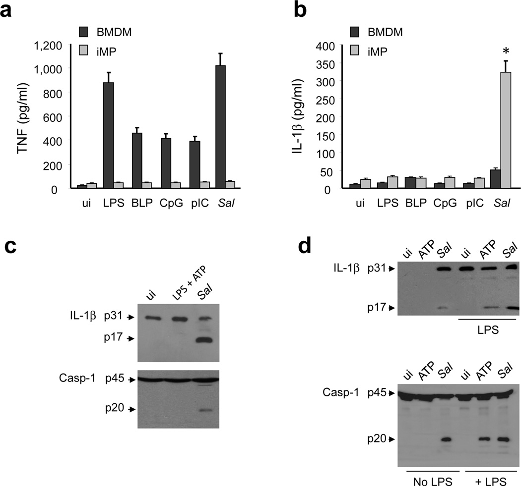
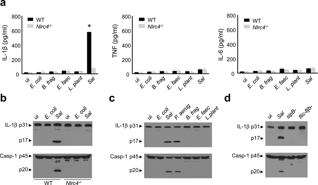
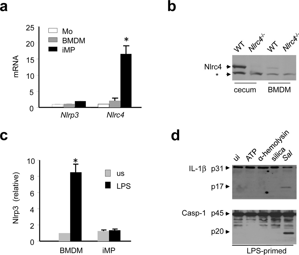
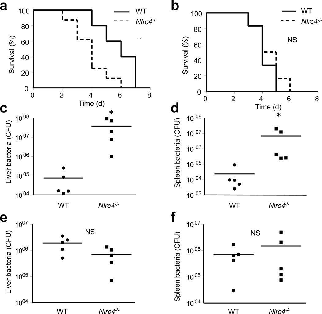
Intestinal phagocytes transport oral antigens and promote immune tolerance, but their role in innate immune responses remains unclear. Here we found that intestinal phagocytes were anergic to ligands for Toll-like receptors (TLRs) or commensals but constitutively expressed the precursor to interleukin 1β (pro-IL-1β). After infection with pathogenic Salmonella or Pseudomonas, intestinal phagocytes produced mature IL-1β through the NLRC4 inflammasome but did not produce tumor necrosis factor (TNF) or IL-6. BALB/c mice deficient in NLRC4 or the IL-1 receptor were highly susceptible to orogastric but not intraperitoneal infection with Salmonella. That enhanced lethality was preceded by impaired expression of endothelial adhesion molecules, lower neutrophil recruitment and poor intestinal pathogen clearance. Thus, NLRC4-dependent production of IL-1β by intestinal phagocytes represents a specific response that discriminates pathogenic bacteria from commensal bacteria and contributes to host defense in the intestine.


Figures

Figure 1
Figure 1. iMP express high amounts of pro-IL-1β, but are hyporesponsive to TLR stimulation
(a and b) BMDM, iMP isolated from specific pathogen free (SPF) or germ-free (GF) mice were stimulated (us) for 16 hrs with the indicated TLR agonists (LPS 10μg/ml; BLP 10μg/ml; CpG 10μg/ml; pIC10μg/ml). Cell-free supernatants were analyzed by ELISA for production of IL-β. Values represent mean ± s.d. of triplicate cultures. (c) Relative IL-1β levels in monocytes (Mo), BMDM, iMP and BMDM stimulated with LPS for 6 hrs as measured by quantitative RT-PCR. Relative expression was normalized to the expression of β-actin. Values represent mean ± s.d. of triplicate cultures. (d) IL-1β and Erk1-2 protein were measured by immunoblotting using protein extracts from iMP isolated from SPF or GF mice. (a-d) Results are representative of at least three separate experiments. * statistically significant; NS, not significant
Figure 2
Figure 2. Salmonella infection induces caspase-1 activation and IL-1β production in iMP
(a and b) BMDM and iMP were uninfected (ui) or stimulated with the indicated TLR agonists (LPS 10μg/ml; BLP 10μg/ml; CpG 10μg/ml; pIC 10μg/ml) or infected with Salmonella (Sal). After 16 hrs cell-free supernatants were analyzed for the production of TNF and IL-1β by ELISA. Values represent mean ± s.d. of triplicate cultures. (c and d) iMP (c) and BMDM (d) were uninfected (ui) or stimulated with LPS (1μg/ml) for 6 hrs , ATP (5 mM) for 30 minutes or infected with Sal. Cell extracts were immunoblotted with IL-1β antibody (arrows denote pro-IL-1β and the mature cytokine p17 subunit) or caspase-1 antibody (arrows denote procaspase-1 and its processed p20 subunit). Results are representative of at least three independent experiments. (e-f) Human peripheral blood (PB) monocytes and intestinal lamina propria (LP) macrophages were stimulated with LPS (100 ng/ml), commensal E. coli, wild-type Salmonella (WT) or Salmonella sipB-, flic-fljB-mutants for 2 hrs. Infection of monocytes and macrophages was performed at a cell/bacteria ratio of 1/10. The production of TNF and IL-1β in cell-free supernatants was measured by ELISA. Values represent mean ± s.d. of triplicate cultures of a representative individual. Cell extracts were immunoblotted with mature IL-1β antibody (p17 subunit) or caspase-1 antibody (arrows denote procaspase-1 and its processed p20 subunit). Results are representative of at least three independent experiments (a-d) and two experiments (e-f).* statistically significant, NS not significant.
Figure 3
Figure 3. Pathogenic, but not commensal bacteria, induce the activation of the NLRC4 - inflammasome in iMP
(a) iMP isolated from WT or Nlrc4−/− mice were uninfected (ui) or were infected with the indicated commensal bacteria or pathogenic Sal. Cell-free supernatants were analyzed by ELISA for the production of TNF, IL-6 and IL-1β. Values represent mean ± s.d. of triplicate cultures. (b-d) iMP isolated from WT or Nlrc4−/− mice were uninfected (ui) or were infected with E. coli, P. aeruginosa (P.aerug), B. fragilis (B. frag), E. faecalis (E. faec), L. plantarum (L. plant), Sal or Salmonella sipB- , flic-fljB- mutants as indicated in the figure. Cell extracts were immunoblotted with IL-1β antibody (arrows denote pro-IL-1β and the mature cytokine p17 subunit) or caspase-1 antibody (arrows denote procaspase-1 and its processed p20 subunit). Results are representative of at least three independent experiments (a-f). * statistically significant
Figure 4
Figure 4. Intestinal phagocytes express a functional NLRC4, but not NLRP3, inflammasome
(a) Relative mRNA expression of NLRP3 and NLRC4, normalized to β-actin, was measured in monocytes (Mo), BMDM or iMP by quantitative RT-PCR. Values represent mean ± s.d. of duplicate cultures. (b) NLRC4 protein levels were measured by immunoblotting of cecal or BMDM extracts from WT or Nlrc4−/− mice. * denotes non-specific protein band (c) Relative NLRP3 mRNA expression in unstimulated (us) cells and cells stimulated with 1 µg/ml of LPS for 6 hrs. Relative gene expression was normalized to the expression of β-actin. Values represent mean ± s.d.. (d) iMP were primed with LPS (1μg/ml) for 4 hrs and stimulated with ATP (5 mM) for 30 minutes, α-hemolysin (1μg/ml) for 3 hrs, silica (500 μg/ml) for 6 hrs or live Salmonella (Sal) for 30 minutes. IL-1β production and caspase-1 activation were measured by immunoblotting. Arrows denote pro-IL-1β (p31) and the mature p17 subunit or procaspase-1 (p45) and its processed p20 subunit. Results are representative of at least three separate experiments (a-d). * statistically significant
Figure 5
Figure 5. Nlrc4−/− mice are more susceptible to orogastric, but not intraperitoneal, Salmonella infection
(a and b) WT and Nlrc4−/− mice in the Balb/c background were infected orogastrically (a) or intraperitoneally (b) with Salmonella, and survival was monitored over time. (a) P = 0.028 (b) P = 0.44 (c-f) WT and Nlrc4−/− Balb/c mice were infected orogastrically (c and d) or intraperitoneally (e and f) with Salmonella and total bacterial loads per indicated organ were determined on day 3 post infection. Each symbol represents an individual mouse. Results are representative of at least three independent experiments with at least five mice per genotype (a-f). * significant, NS not significant
Figure 6
Figure 6. Il1r−/− mice are more susceptible to orogastric, but not intraperitoneal, Salmonella infection
(a-d) WT and Il1r−/− mice in the Balb/c background were infected orally (a) or intraperitoneally (b) with Salmonella, and survival was monitored over time. (a) P = 0.012 (b) P = 0.54 (c-f) WT and Il1r−/− Balb/c mice were infected orogastrically (c and d) or intraperitoneally (e and f) with Salmonella and bacterial loads per indicated organ were determined on day 3 post infection. Each symbol represents an individual mouse. Results are representative of at least three separate experiments with at least five mice per genotype (a-f). * significant, NS not significant
Figure 7
Figure 7. The NLRC4 inflammasome promotes host defense through neutrophil recruitment in the intestine
(a) WT, Nlrc4−/− and Il1r−/− mice were infected with Salmonella orogastrically and 20 hrs later intestinal tissue was harvested. Cecum sections were stained with hematoxylin and eosin and scored for the indicated pathological changes. Results are representative of three separate experiments with five mice for each group. (b) Lamina propria mononuclear cells were isolated from the large intestine of WT, Nlrc4−/− and Il1r−/− 20 hrs after orogastric Salmonella (Sal) infection, labeled with antibodies for indicated surface markers and analyzed by flow cytometry. The percentage of neutrophils (CD11b+Ly6G+ positive cells) from 4 different experiments was plotted. (c) Expression of indicated adhesion molecules by quantitative RT-PCR in cecal tissue 12 hrs after Salmonella infection. Gene expression was normalized to the expression of β-actin, and expressed as fold induction compared to uninfected mice. Values represent mean ± s.d. n=6 . (d and e). Mice were pretreated with anti Ly-6G antibody, or control rat IgG, and then infected orogastrically with 1 × 10⁵ Salmonella. Bacterial burden in the cecum was determined on day 3 post infection (d) and survival was monitored over time (e). (f and g) WT, Nlrc4−/− and Il1r−/− mice were infected orogastrically with Salmonella and the bacterial burden in the cecum was determined on day 2 (f) and on day 3 (g) post infection. Results are representative of three independent experiments. * statistically significant NS not significant.

Comment in

Similar articles

Cited by

References

    1. Kawai T, Akira S. Toll-like receptors and their crosstalk with other innate receptors in infection and immunity. Immunity. 2011;34:637–650. - PubMed
    1. Abraham C, Medzhitov R. Interactions between the host innate immune system and microbes in inflammatory bowel disease. Gastroenterology. 2011;140:1729–1737. - PMC - PubMed
    1. Franchi L, Munoz-Planillo R, Reimer T, Eigenbrod T, Nunez G. Inflammasomes as microbial sensors. Eur J Immunol. 2010;40:611–615. - PubMed
    1. Franchi L, et al. Intracellular NOD-like receptors in innate immunity, infection and disease. Cell Microbiol. 2008;10:1–8. - PubMed
    1. Franchi L, Eigenbrod T, Munoz-Planillo R, Nunez G. The inflammasome: a caspase-1-activation platform that regulates immune responses and disease pathogenesis. Nat Immunol. 2009;10:241–247. - PMC - PubMed

Publication types

MeSH terms