. 2012 Apr 6;90(4):661-74.
doi: 10.1016/j.ajhg.2012.02.026.

Attenuated BMP1 function compromises osteogenesis, leading to bone fragility in humans and zebrafish

Affiliations

Attenuated BMP1 function compromises osteogenesis, leading to bone fragility in humans and zebrafish

P V Asharani et al. Am J Hum Genet. .

Abstract

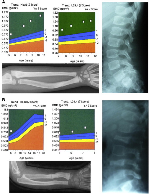
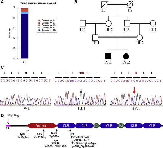
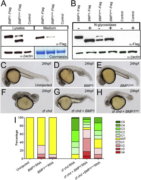
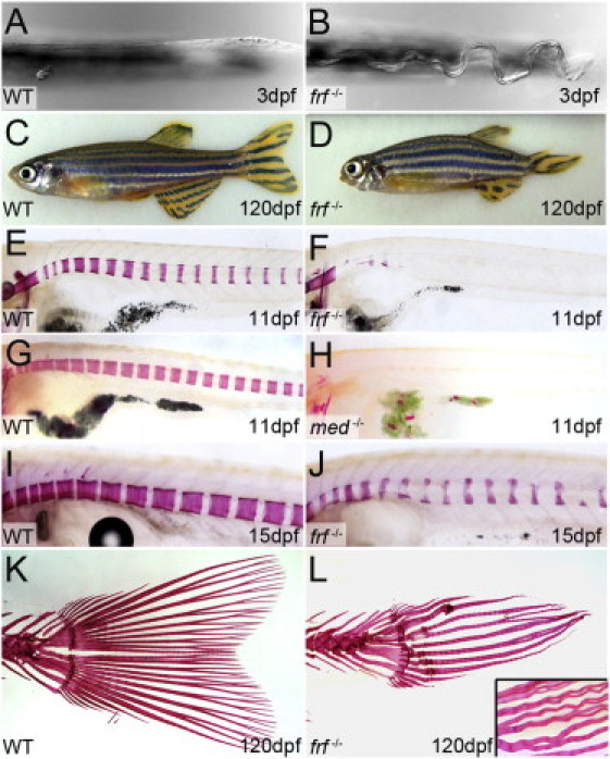
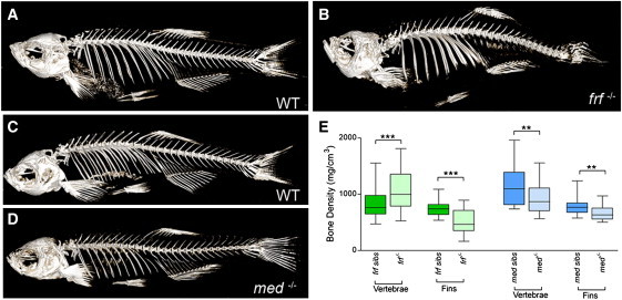
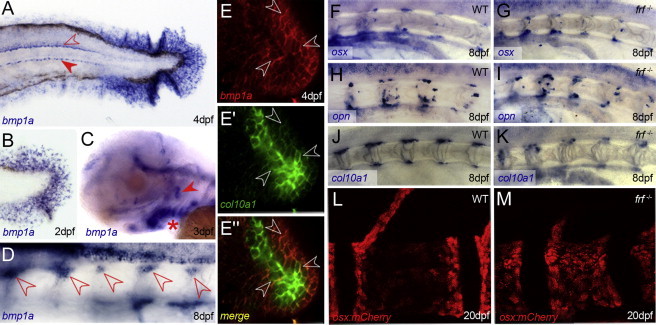
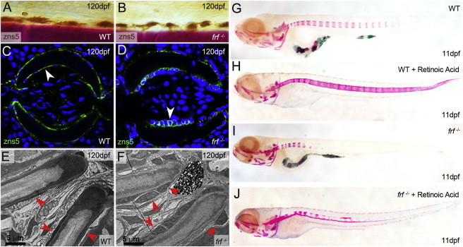
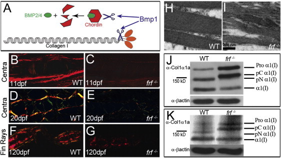
Bone morphogenetic protein 1 (BMP1) is an astacin metalloprotease with important cellular functions and diverse substrates, including extracellular-matrix proteins and antagonists of some TGFβ superfamily members. Combining whole-exome sequencing and filtering for homozygous stretches of identified variants, we found a homozygous causative BMP1 mutation, c.34G>C, in a consanguineous family affected by increased bone mineral density and multiple recurrent fractures. The mutation is located within the BMP1 signal peptide and leads to impaired secretion and an alteration in posttranslational modification. We also characterize a zebrafish bone mutant harboring lesions in bmp1a, demonstrating conservation of BMP1 function in osteogenesis across species. Genetic, biochemical, and histological analyses of this mutant and a comparison to a second, similar locus reveal that Bmp1a is critically required for mature-collagen generation, downstream of osteoblast maturation, in bone. We thus define the molecular and cellular bases of BMP1-dependent osteogenesis and show the importance of this protein for bone formation and stability.


Figures

Figure 1
Figure 1
Two Siblings with Autosomal-Recessive Bone Fragility and High-Bone-Mass Phenotype (A and B) Clinical data of both individuals are shown. Above, diagrams illustrate Z scores of bone-mineral-density measurements of the head and vertebrae L2–L4, respectively, indicating highly increased levels of bone mineral density. X-rays show fractured and bent forearms (below) and spinal columns (right) of individuals. High-radiation X-rays of the forearm and spinal column of individual 1 indicate an intense bone density. Vertebrae of individual 2 are flattened and irregularly formed (B).
Figure 2
Figure 2
Whole-Exome Sequencing and Filtering Identify Mutation in BMP1 (A) Statistical overview of target-base coverage during sequencing process. Over 90% of identified variations were covered more than 20×. (B) Pedigree structure of the consanguineous Turkish family. (C) Sequence chromatograms of the identified c.34G>C BMP1 mutation predicted to substitute the glycine at position 12 with arginine. The c.34G>C mutation was found to be heterozygous (middle panel) in both parents and homozygous in both individuals (right panel). (D) Schematic view of BMP1 domain structure. The locations of identified mutations in humans (above) and zebrafish (below) are shown. Note that the frf tf5 mutation generates multiple splice isoforms. The following abbreviation is used: SP, signal peptide.
Figure 3
Figure 3
A Signal-Peptide Substitution in BMP1 Causes Secretion and Glycosylation Defects In Vitro and Loss of Protease Activity In Vivo (A) Immunoblot of HEK 293T cells transfected with either Flag-tagged WT BMP1 (BMP1-Flag; lanes 1 and 4) or p.Gly12Arg-substituted BMP1 (BMP1mut-Flag; lanes 2 and 5) and untransfected control cells (control; lanes 3 and 6). Immunoblotting shows that the p.Gly12Arg protein (lanes 1–3) isolated from cell lysates had increased mobility; reduced amounts of this BMP1 protein were secreted into the medium (lanes 4–6). (B) Immunoblot of lysates of HEK 293T cells transfected with either Flag-tagged WT BMP1 (BMP1-Flag; lanes 1 and 2) or p.Gly12Arg-substituted BMP1 (BMP1mut-Flag; lanes 3 and 4) and untransfected controls (control; lanes 5 and 6). After being harvested, lysates were either treated with N-glycosidase (lanes 2, 4, and 6) or left untreated (lanes 1, 3, and 5). The predominant mutant-BMP1 band with increased mobility migrates at the same rate as deglycosylated WT BMP1. (C–I) The p.Gly12Arg-substituted BMP1 exhibits reduced Chordinase activity in vivo. Lateral views of uninjected 24 hpf zebrafish embryos (C) and embryos injected with RNA encoding either WT BMP1 (BMP1; D and G) or p.Gly12Arg signal-peptide variant BMP1 (BMP1mut; E and H). Chordinase activity was assessed by its ability to ventralize WT embryos (C–E) or rescue dorsalized (chordin RNA injected) embryos (F–H). In both assays, the mutant BMP1 showed reduced ability to counteract either the endogenous or exogenous Chordin (quantified in I).
Figure 4
Figure 4
The Zebrafish frilly fins Mutant Displays Larval Fin-Fold Ruffling and Osteogenesis Defects (A and B) Ventral view of the posterior medial fin fold at 3 dpf in a frilly fins (frf) (B) larva showing undulations in the fin fold, which normally has a linear morphology (A). (C and D) Compared to the siblings, 4-month-old frf−/− adults (D) are short and display axis defects, body curvature, and fin and craniofacial dysmorphogenesis. (E–L) Alizarin-red staining of frf−/− (F, J, and L), microwaved (med−/−) (H), and WT siblings (E, G, I, and K) at 11 dpf (E–H), 15 dpf (I and J), and 4 months (K and L). Both frf and med display reduced ossification of the vertebrae (F and H), whereas nascent vertebrae are osteopenic and dysmorphic (J). Tail fins in frf−/− have lost the WT fan shape (K) and display fracture calluses, reduced bifurcations (L), and crinkled lepidotrichia, which often fuse to each other (L; inset).
Figure 5
Figure 5
CT Analysis of frilly fins and microwaved Bone Reveals Altered Adult Bone Densities (A–D) microCT analysis of bone density in a 7-month-old frf−/− mutant (B) and its WT sibling (A) as well as a med−/− mutant (D) and its WT sibling (C). Note that although the med mutant looks overtly normal (D), the frf mutant skeleton displays axial curvature and defects in the head skeleton (B). (E) Box plots of density measurements derived from microCT analysis of mutants and sibling vertebrae and fin lepidotrichia. med mutants have osteopenia of both lepidotrichia in the fins and vertebrae (blue boxes). Although frf mutants also display osteopenia of the fins, they show an increased bone density in the vertebrae (green boxes). The Mann-Whitney U test was performed for comparing densities between mutants and siblings for each bone type (∗∗∗ denotes p < 0.001; ∗∗ denotes p < 0.01; and n = 8 for all data sets). Boxes indicate the median and the 25th and 75th percentiles, whereas the whiskers display the largest and smallest values.
Figure 6
Figure 6
bmp1a Is Expressed in Osteoblasts, which Appear Normally Differentiated in frilly fins (A–D) In situ hybridization of bmp1a at 4 dpf (A), 2 dpf (B), 3 dpf (C) and 8 dpf (D). Expression is seen in fin mesenchyme cells of the fin fold (A and B), floor plate and hypochord (A, open and filled arrowheads, respectively), branchial arches (C, asterisk), and operculum (C, arrowhead) and perichordal cells of the anterior notochord (D, arrowheads). (E–E″) Confocal images of double-fluorescent in situ hybridizations showing coexpression of bmp1a (E and E″; red) and the osteoblast marker collagen10a1 (E′ and E″; green) in osteoblasts on the operculum at 4 dpf. Most cells express both markers (three cases highlighted by arrowheads), and central, more mature osteoblasts express slightly higher levels of col10a1. (F–K) Perichordal expression of osteoblast markers is not disrupted in frf mutants. Lateral images of anterior notochord of 8 dpf WT (F, H, and J) and frf−/− (G, I, and K) larvae hybridized with probes for sp7 (F and G), osteopontin (H and I), and collagen10a1 (J and K). (L and M) Confocal images of sp7:mCherry-expressing osteoblasts on vertebrae of WT (L) and frf mutant (M) larvae at 20 dpf. There is no reduction in osteoblast numbers in the mutant.
Figure 7
Figure 7
Osteoblasts in frilly fins Mutants Have Altered Morphology but Cannot Hyperossify upon RA Treatment (A–D) Immunohistochemical (A and B) and immunofluorescent (C and D) staining of osteoblasts with the zns5 antibody (brown stain in A and B; green stain in C and D) in WT (A and C) and frf−/− (B and D) fins at 120 dpf. (A) and (B) display lateral views of fin rays counterstained with alizarin red (bone is in red), whereas (C) and (D) are transverse sections of fin rays counterstained with DAPI (blue). There is no loss in number of zns5+ osteoblasts in frf−/− mutants. However, the osteoblasts display an altered morphology when viewed in cross section; they appear more cuboidal where they are normally flat cells that maintain intimate contact with the bone surface (arrowheads in C and D). (E and F) Electron micrographs of transverse sections of adult fin rays. Osteoblasts are indicated with red arrowheads and appear flat in WT fins (E) yet more cuboidal in frf−/− fins (F). (G–J) Alizarin-red staining of 11 dpf WT (G and H) and frf−/− larvae (I and J) treated with (H and J) or without (G and I) RA for enhancing osteoblast activity. Despite normal numbers of differentiated osteoblasts in frf mutants, these cells are unable to mineralize the notochord efficiently upon RA stimulation; this suggests a defect downstream of osteoblast differentiation.
Figure 8
Figure 8
frf Displays Defects in Fibrillar Collagen Order and Col1a1a Processing (A) Major proteolytic roles of Bmp1 include removing the C-propeptide (orange ovals) of pro-Collagen I and cleaving the BMP2/4 inhibitor, Chordin (red hexagon), to release free BMP2/4 (green oval). (B–G) Picrosirius-red stained sagittal (B–E) and transverse (F and G) sections of WT (B, D, and F) and frf−/− (C, E, and G) larvae at 11 dpf (B and C), 20 dpf (D and E), and 4 months (F and G). Sections viewed under polarized light reveal the reduced collagen-fiber-associated birefringency in the Centra region (B–E) and fin rays (F and G) in frf−/− mutant (C, E, and G) and WT (B, D, and F) larvae. (H and I) Transmission electron micrographs of longitudinal sections of WT (H) and frf−/− (I) larval medial fins at 6 dpf show loss of structured collagen fibers in the mutant. (J and K) Immunoblots of protein extracted from WT (lane 1) and frf−/− mutant (lane 2) larvae probed with an antibody directed against zebrafish Collagen1α1a (upper panels in both J and K) or an antibody against β-actin as a loading control (lower panels). The four possible Collagen1α1 forms are indicated on the right; these forms include Procollagen1α1 retaining both C- and N- terminal propetides (Pro α1[I]), mature collagen α1(I) retaining neither propeptide (α1[I]), a form retaining only the N-propeptide (pN α1[I]), and a form retaining only the C-propeptide (pC α1[I]). In 6 dpf (J) and 4-month-old (K) frf−/− mutants, the two forms retaining the C-propeptide predominate.

Similar articles

Cited by

References

    1. Byers P.H., Cole W.G. Osteogenesis Imperfecta. In: Royce P., Steinmann B., editors. Connective Tissue and its Heritable Disorders: Molecular, Genetic, and Medical Aspects. Second Edition. John Wiley & Sons; Hoboken, NJ: 2002. pp. 385–430.
    1. Sillence D.O., Rimoin D.L. Classification of osteogenesis imperfect. Lancet. 1978;1:1041–1042. - PubMed
    1. Basel D., Steiner R.D. Osteogenesis imperfecta: Recent findings shed new light on this once well-understood condition. Genet. Med. 2009;11:375–385. - PubMed
    1. Rauch F., Glorieux F.H. Osteogenesis imperfecta. Lancet. 2004;363:1377–1385. - PubMed
    1. Marini J.C., Forlino A., Cabral W.A., Barnes A.M., San Antonio J.D., Milgrom S., Hyland J.C., Körkkö J., Prockop D.J., De Paepe A. Consortium for osteogenesis imperfecta mutations in the helical domain of type I collagen: Regions rich in lethal mutations align with collagen binding sites for integrins and proteoglycans. Hum. Mutat. 2007;28:209–221. - PMC - PubMed

Publication types

MeSH terms