. 2012 Jul 1;21(13):2899-911.
doi: 10.1093/hmg/dds116. Epub 2012 Mar 27.

Evaluating the role of the FUS/TLS-related gene EWSR1 in amyotrophic lateral sclerosis

Affiliations

Evaluating the role of the FUS/TLS-related gene EWSR1 in amyotrophic lateral sclerosis

Julien Couthouis et al. Hum Mol Genet. .

Abstract

Amyotrophic lateral sclerosis (ALS) is a fatal neurodegenerative disease affecting motor neurons. Mutations in related RNA-binding proteins TDP-43, FUS/TLS and TAF15 have been connected to ALS. These three proteins share several features, including the presence of a bioinformatics-predicted prion domain, aggregation-prone nature in vitro and in vivo and toxic effects when expressed in multiple model systems. Given these commonalities, we hypothesized that a related protein, EWSR1 (Ewing sarcoma breakpoint region 1), might also exhibit similar properties and therefore could contribute to disease. Here, we report an analysis of EWSR1 in multiple functional assays, including mutational screening in ALS patients and controls. We identified three missense variants in EWSR1 in ALS patients, which were absent in a large number of healthy control individuals. We show that disease-specific variants affect EWSR1 localization in motor neurons. We also provide multiple independent lines of in vitro and in vivo evidence that EWSR1 has similar properties as TDP-43, FUS and TAF15, including aggregation-prone behavior in vitro and ability to confer neurodegeneration in Drosophila. Postmortem analysis of sporadic ALS cases also revealed cytoplasmic mislocalization of EWSR1. Together, our studies highlight a potential role for EWSR1 in ALS, provide a collection of functional assays to be used to assess roles of additional RNA-binding proteins in disease and support an emerging concept that a class of aggregation-prone RNA-binding proteins might contribute broadly to ALS and related neurodegenerative diseases.


Figures

Figure 1.
Figure 1.
When expressed in yeast, the human RRM RNA-binding protein EWSR1 shows similar properties to FUS and TDP-43. (A) Localization patterns in yeast cells of human TDP-43–YFP, FUS–YFP and EWSR1–YFP fusion proteins, forming multiple cytoplasmic foci. When YFP alone is expressed, it is localized diffusely throughout the cytoplasm. (B) Spotting assays to assess the toxicity of human EWSR1–YFP protein. Transformants were grown on the synthetic media containing either glucose (control, protein expression ‘off’) or galactose (protein expression ‘on’). TDP-43–YFP and FUS–YFP proteins overexpression were very toxic, while EWSR1–YFP was moderately toxic and YFP alone is not toxic (control).
Figure 2.
Figure 2.
Missense mutations in EWSR1 in ALS patients. (A) Comparison of FUS, EWSR1 and TAF15 demonstrates similar domain architecture. All three proteins contain a single RRM, a glycine-rich domain, a predicted prion domain, RGG domains and a C-terminal PY-motif, which can function as an NLS. Mutations in FUS and TAF15 are examples of those similar to variants found in EWSR1. (B and C) DNA sequence analysis of EWSR1 in North American Caucasian ALS patients identified two missense mutations (shown are electropherograms highlighting the sequence variants). (B) A single base substitution (asterisk) changing the WT guanine at 1532 to cytosine (c.1532 G>C) predicted to lead to an alanine substituting for glycine (p.G511A). (C) Another EWSR1 variant in an ALS case: c.1655 C>T, predicted to lead to a leucine substituted for proline (p.P552L). (D) Sequence alignment of amino acids 505–564 of EWSR1 from diverse vertebrate species indicates that the mutated residues in EWSR1 are highly conserved. Identical amino acids have a black background, similar amino acids are gray and mutation sites are red.
Figure 3.
Figure 3.
ALS-linked EWSR1 mutations promote cytoplasmic localization in motor neurons with mislocalization to the neurites of primary neurons cultured from mouse spinal cord. Primary mouse neuron cultures were transfected with WT or mutant EWSR1, stained with a-EWSR1 (red) and a-doublecortin (DCX, green). G511A and P552L variants were only found in ALS patients, while R635C was only found in controls and G584S was found in both (and this ALS case also harbored a C9ORF72 hexanucleotide expansion). (A) Endogenous EWSR1 is almost exclusively localized within the nucleus of neurons. Overexpression of WT EWSR1, or variants found in controls, shows localization within the nucleus or cytoplasm of neurons, with rare neurites containing EWSR1. Only the ALS-linked mutant forms of EWSR1 showed increased mislocalization into the neurites, including dendrites and axons. Scale bar is 10 µm. (B) Quantitation of mislocalization of endogenous transfected, WT or mutant, EWSR1 into neuronal processes. *P = 0.0056 for comparing G511A to WT, and P = 0.0008 comparing P552L to WT (two-tailed unequal variance t-test). (C) ES cell-derived neurons were transduced with doxycycline (Dox) inducible lentiviruses expressing WT or ALS-linked mutants of EWSR1, each carrying FLAG and myc epitope tags in their amino- and carboxy-termini, respectively. Five days after induction of expression by Dox, the localization of the proteins was visualized by immunofluorescence microscopy with anti-FLAG antibody (red); nuclei were visualized by 4′,6-diamidino-2-phenylindole (DAPI) staining (blue). The localization of endogenous EWSR1 was performed with anti-EWSR1-specific antibodies (red) in non-transduced neurons. Induced expression of EWSR1 led to accumulation of proteins in the cytoplasm and neuronal processes of transduced cells and this effect was enhanced by the ALS-linked patient mutations. Expression levels and solubility of transduced proteins were determined by immunoblots with anti-FLAG antibodies of radioimmuno precipitation assay buffer (RIPA) and UREA fractions of cell lysates from EWSR1 WT and mutants 5 days after Dox induction. The expression levels of transduced proteins were comparable between WT and mutants with no apparent accumulation of proteins in the UREA fraction.
Figure 4.
Figure 4.
EWSR1 is intrinsically aggregation–prone and ALS-linked variants accelerate aggregation. (A) Following TEV protease cleavage to remove the N-terminal GST tag, FUS, EWSR1 and TDP-43 proteins were resolved by sodium dodecyl sulfate polyacrylamide gel electrophoresis (SDS–PAGE) and Coomassie stained to confirm purity and expected molecular weight. (B) GST-TDP-43, GST-FUS or GST-EWSR1 (3 µm) were incubated in the absence or presence of TEV protease at 25°C for 0–90 min with agitation. At the indicated times, the extent of aggregation was determined by measuring turbidity (absorbance at 395 nm). Note that very little aggregation occurs in the absence of TEV protease. Values represent means ± SEM (n = 3). A human RRM protein that lacks a prion-like domain, DND1, did not aggregate in this assay. (C) GST-TDP-43, GST-FUS or GST-EWSR1 (3 µm) was incubated in the presence of TEV protease at 25°C for 0–60 min with agitation. At the indicated times, reactions were processed for sedimentation analysis. Pellet and supernatant fractions were resolved by SDS–PAGE and stained with Coomassie Brilliant Blue. The amount of protein in the pellet fraction was determined by densitometry in comparison to known quantities of the appropriate protein. Values represent means ± SEM (n = 3). A human RRM protein that lacks a prion-like domain, DND1, did not aggregate in this assay. (D) GST-EWSR1 (3 µm) was incubated in the presence of TEV protease at 25°C for 0–60 min with agitation. At various times, reactions were processed for EM. Small arrows denote pore-shaped oligomers and large arrows denote linear polymers. Bar, 0.5 µm. (E) Gallery of EWSR1 oligomers formed after 10 min of aggregation. Bar, 50 nm. (F) Following TEV protease cleavage to remove the N-terminal GST tag, EWSR1 WT, G511A and P552L proteins were resolved by SDS–PAGE and Coomassie stained to confirm purity and expected molecular weight. (G) GST-EWSR1, GST-EWSR1 (G511A) or GST-EWSR1 (P552L) (3 µm) was incubated in the presence of TEV protease at 25°C for 0–75 min without agitation. At the indicated times, the extent of aggregation was determined by measuring turbidity (absorbance at 395 nm). Note that the ALS-linked EWSR1 variants, G511A and P552L, aggregated with accelerated kinetics. Values represent means ± SEM (n = 3).
Figure 5.
Figure 5.
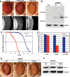
WT and disease-related mutants of EWSR1 lead to neural degeneration and dysfunction in Drosophila. (A) Expression of EWSR1 caused a dose-dependent disruption of retinal structure. (W) and (S) refer to weak and strong EWSR1 expression levels. (B) Western immunoblot showing the level of expression of EWSR1. β-Tubulin levels were used as the loading control. (A and B) Genotypes: control (Con) is driver line alone, gmr-GAL4(YH3)/+. EWSR1(W) is UAS-EWSR1(W)/gmr-GAL4(YH3). EWSR1(S) is UAS-EWSR1(S)/+; gmr-GAL4(YH3)/+. (C) Expression of EWSR1 in the nervous system reduces lifespan (red, compared with normal in blue). (D) EWSR1 caused progressive loss of climbing behavior when expressed in the nervous system. (C and D) Genotypes: elav is elav3A-GAL4/+. elav/EWSR1 is UAS-EWSR1(S)/+; elav3A-GAL4/+. (E) External eyes from 1 day adult flies expressing WT and mutant EWSR1.G511A under control of the gmr-GAL4 driver. (F) Immunoblot of head homogenates showing EWSR1 WT and mutant expression levels in transgenic flies. β-Tubulin served as loading control. (E and F) Genotypes: WT is UAS-EWSR1/+; gmr-GAL4(YH3)/+. G511A is UAS-EWSR1(G511A)/+; gmr-GAL4(YH3)/+. P552L is UAS-EWSR1(P552L)/gmr-GAL4(YH3).
Figure 6.
Figure 6.
Immunostaining to visualize localization of EWSR1 in spinal cord of control or ALS patients. (A and B), In control spinal cord neurons, EWSR1 is localized predominantly to the nucleus while in ALS spinal cord neurons, EWSR1 was present in cytoplasmic punctate granular structures (E, arrows) or in a diffuse pattern throughout the cytoplasm (CH). Scale bar is 25 µm.

Similar articles

Cited by

References

    1. Cleveland D.W., Rothstein J.D. From Charcot to Lou Gehrig: deciphering selective motor neuron death in ALS. Nat. Rev. Neurosci. 2001;2:806–819. doi:10.1038/35097565. - DOI - PubMed
    1. Andersen P.M., Al-Chalabi A. Clinical genetics of amyotrophic lateral sclerosis: what do we really know? Nat. Rev. Neurol. 2011;7:603–615. doi:10.1038/nrneurol.2011.150. - DOI - PubMed
    1. DeJesus-Hernandez M., Mackenzie I.R., Boeve B.F., Boxer A.L., Baker M., Rutherford N.J., Nicholson A.M., Finch N.A., Flynn H., Adamson J., et al. Expanded GGGGCC hexanucleotide repeat in noncoding region of C9ORF72 causes chromosome 9p-linked FTD and ALS. Neuron. 2011;72:245–256. doi:10.1016/j.neuron.2011.09.011. - DOI - PMC - PubMed
    1. Deng H.X., Chen W., Hong S.T., Boycott K.M., Gorrie G.H., Siddique N., Yang Y., Fecto F., Shi Y., Zhai H., et al. Mutations in UBQLN2 cause dominant X-linked juvenile and adult-onset ALS and ALS/dementia. Nature. 2011;477:211–215. doi:10.1038/nature10353. - DOI - PMC - PubMed
    1. Fecto F., Siddique T. Making connections: pathology and genetics link amyotrophic lateral sclerosis with frontotemporal lobe dementia. J. Mol. Neurosci. 2011;45:663–675. doi:10.1007/s12031-011-9637-9. - DOI - PubMed

Publication types

MeSH terms