. 2012 May;18(5):958-72.
doi: 10.1261/rna.032292.112. Epub 2012 Mar 22.

Maturation of mammalian H/ACA box snoRNAs: PAPD5-dependent adenylation and PARN-dependent trimming

Affiliations

Maturation of mammalian H/ACA box snoRNAs: PAPD5-dependent adenylation and PARN-dependent trimming

Heike Berndt et al. RNA. 2012 May.

Erratum in

  • RNA. 2014 Aug;20(8):1349

Abstract

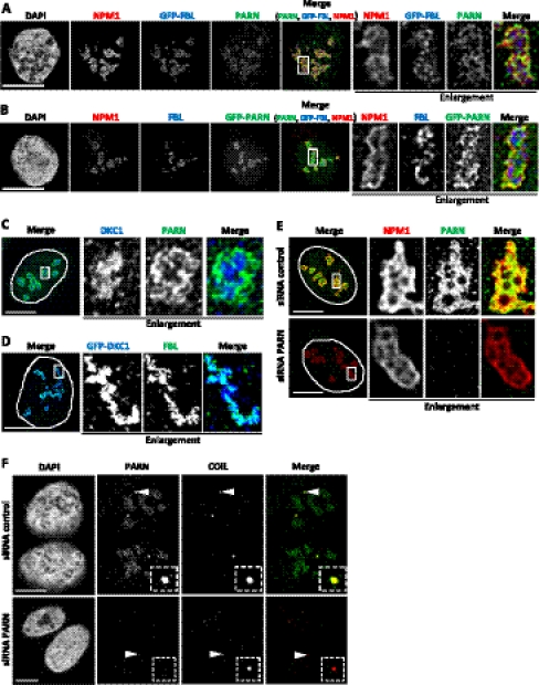
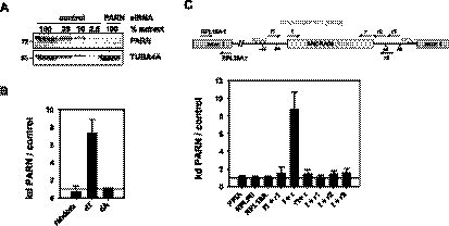
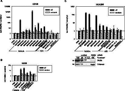
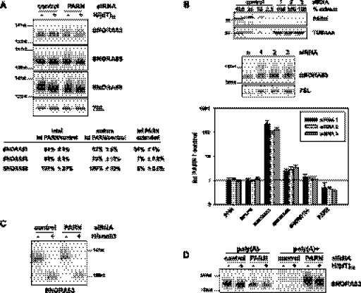
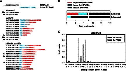
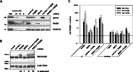
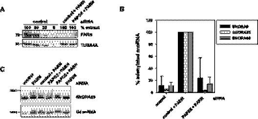
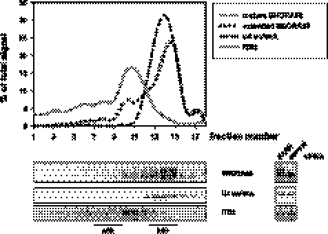
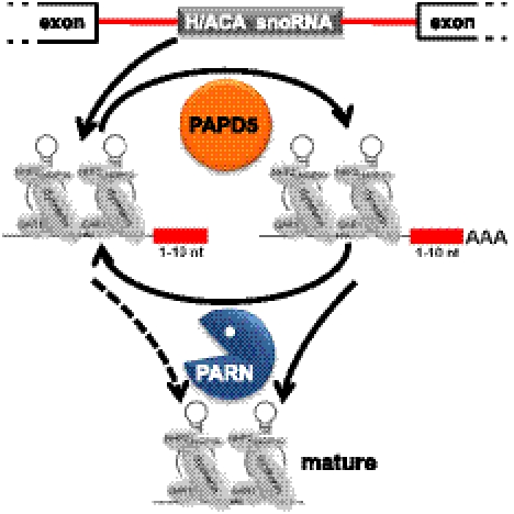
Small nucleolar and small Cajal body RNAs (snoRNAs and scaRNAs) of the H/ACA box and C/D box type are generated by exonucleolytic shortening of longer precursors. Removal of the last few nucleotides at the 3' end is known to be a distinct step. We report that, in human cells, knock-down of the poly(A) specific ribonuclease (PARN), previously implicated only in mRNA metabolism, causes the accumulation of oligoadenylated processing intermediates of H/ACA box but not C/D box RNAs. In agreement with a role of PARN in snoRNA and scaRNA processing, the enzyme is concentrated in nucleoli and Cajal bodies. Oligo(A) tails are attached to a short stub of intron sequence remaining beyond the mature 3' end of the snoRNAs. The noncanonical poly(A) polymerase PAPD5 is responsible for addition of the oligo(A) tails. We suggest that deadenylation is coupled to clean 3' end trimming, which might serve to enhance snoRNA stability.


Figures

FIGURE 1.
FIGURE 1.
PARN is localized in nucleoli and Cajal bodies. (A) Localization of endogenous PARN and nucleophosmin (NPM1) was analyzed by immunostaining in U2OS cells transfected with GFP-fibrillarin (GFP-FBL). All three proteins are localized in nucleoli. Significant colocalization of PARN with NPM1, but not with GFP-FBL, is observed within the nucleoli upon enlargement (2.5-fold magnification) of boxed region. (B) Localization of GFP-PARN was analyzed in U2OS cells counterstained for nucleophosmin (NPM1) and fibrillarin (FBL). Significant colocalization of GFP-PARN with NPM1, and to a much lesser extent with FBL, is observed in nucleoli upon enlargement of boxed region. (C,D) Localization of PARN, endogenous dyskerin (DKC1), GFP-dyskerin, and FBL was analyzed in U2OS cells. DKC1 colocalizes with FBL but not PARN at the interior of nucleoli, as observed upon enlargement of regions boxed in the left panel. (E) Localization of PARN in nucleoli is lost upon PARN knock-down, as indicated by counterstaining for nucleophosmin (NPM1). Enlargements (2.5-fold magnification) of boxed regions in left panel are shown in the right panels. (F) Localization of PARN was analyzed in cells counterstained for coilin (COIL). Both proteins colocalize in Cajal bodies in U2OS cells transfected with control siRNA. Upon PARN knock-down, the signal in nucleoli and some Cajal bodies is lost or severely diminished. Enlargements (2.5-fold magnification) of Cajal bodies indicated by arrow heads are shown as boxed insets. Note that PARN knock-down appears to affect Cajal body number and size; so far, this effect has not been further analyzed. In CE, nuclei are indicated by dashed lines. Bar indicates 10 μm.
FIGURE 2.
FIGURE 2.
Oligoadenylated SNORA68 is enriched in PARN knock-down cells. (A) U2OS cells were transfected with PARN siRNA 1 or control siRNA. PARN levels were determined by Western blotting. A fixed amount of the PARN knock-down extract was compared with a dilution series of control extract. Tubulin (TUBA4A) served as a loading control. This is one representative example of many knock-down experiments. (B) The level of SNORA68 was analyzed by qRT-PCR. Total RNA was reverse-transcribed with random, oligo(dT) or oligo(dA) primers as indicated. SNORA68 sequences were quantified by qPCR and cross-normalized to PPIA and RPLP0. The qPCR of the oligo(dA)-primed sample was normalized to PPIA only, which is amplified due to an internal oligo(U) stretch. Data are plotted as abundance of SNORA68 in knock-down over control cells. (C) Oligo(dT)-primed cDNA as in B was analyzed by qPCR with primer combinations covering different parts of the RPL18A pre-mRNA as indicated at the top. Data were cross-normalized to PPIA and RPLP0 and are plotted as abundance of each RNA species in knock-down cells over control cells. In B, error bars indicate the standard deviation of five independent biological experiments, except the oligo(dA)-primed sample (average of two experiments). In C, the standard deviation is based on two independent biological experiments with two technical replicates each.
FIGURE 3.
FIGURE 3.
H/ACA, but not C/D box, RNAs are enriched upon PARN knock-down. (A) Levels of snoRNAs were analyzed by reverse-transcription of total RNA prepared from control and PARN knock-down U2OS cells using random or oligo(dT) priming and gene-specific qPCR. Data were cross-normalized to PPIA and RPLP0 messages and are plotted as RNA level in knock-down over control cells. (B) Relative scaRNA levels were determined in U2OS cells by qRT-PCR as described in A. (C) Relative snoRNA levels in HEK293 cells were analyzed by qRT-PCR as in A. The Western blot control for the knock-down efficiency shown below the histogram is a representative example. In AC, data represent the average of at least three independent biological experiments; bars indicate standard deviations.
FIGURE 4.
FIGURE 4.
Extended H/ACA box RNAs are oligoadenylated. (A) U2OS cells were treated with siRNA against PARN or control siRNA. Nuclear RNA was hybridized to dT12 and treated with RNase H or not. RNAs were analyzed by Northern blotting, with 7SL RNA serving as a loading control. Size markers (in nucleotides) are indicated. Quantifications reported in the bottom panel (see Materials and Methods) are based on normalization to the 7SL RNA. For SNORA63 and 68, n = 4; for SNORA65, n = 3. (B) U2OS cells were transfected with PARN siRNA 1, 2, or 3, or control siRNA. PARN levels were determined by Western blotting (top panel). Total RNA was analyzed by Northern blotting with a SNORA63 probe (middle panel). In the same RNA preparations, levels of snoRNAs were analyzed by oligo(dT)-primed reverse-transcription and gene-specific qPCR (bottom panel). Data were cross-normalized to PPIA and RPLP0 messages and are plotted as RNA level in knock-down over control cells. Error bars indicate the standard deviation of two independent biological experiments, each with two technical repeats. (C) The same RNA preparations as in A were hybridized with a DNA oligonucleotide matching the 3′ end of SNORA63 and treated with RNAse H or not. RNAs were analyzed by Northern blotting with a probe directed against SNORA63. The experiment also served as a positive control for the RNase H digestion in A and D. (D) Total RNA was prepared from control and PARN knock-down U2OS cells and separated by oligo(dT) cellulose chromatography. 2 μg of poly(A) and 0.25 μg of poly(A)+ RNA were hybridized to oligo(dT)12 and treated with RNase H or not. The RNA was analyzed by Northern blotting and hybridization with a SNORA63 probe.
FIGURE 5.
FIGURE 5.
3′ extended H/ACA box RNAs carry intron nucleotides plus oligo(A) tails. (A) Nuclear RNA from control and PARN knock-down U2OS cells and poly(A)+ RNA from knock-down cells were the same preparations as in Figure 4. SNORA63 clones were prepared by linker ligation, cDNA synthesis, nested PCR, and cloning (Materials and Methods). Sequences of individual clones derived from each of the three RNA preparations are compiled. Mature snoRNA sequences are in black, remaining intron nucleotides are blue, post-transcriptionally added sequences are in red. Nucleotides of ambiguous origin are gray. Numbers on the left indicate how often each sequence was found. Unique sequences have no number. (B) Nuclear RNA from control and PARN knock-down U2OS cells was used to analyze SNORA68 by linker ligation, cDNA synthesis, PCR, and deep sequencing. The percentage of reads ending with the mature end (black), containing additional intron nucleotides (blue) or nonencoded oligo(A) sequences (red) are shown. 100% is the number of reads that matched the SNORA68 sequence and could be grouped as described in Materials and Methods. (C) The positions of nonencoded oligo(A) sequences found by deep sequencing of SNORA68 are indicated above the intron sequence. Position 133 corresponds to the last nucleotide of mature SNORA68. Oligo(A) sequences shown at, for example, position 138 were attached to nucleotide 137. Sequences from the PARN knock-down and the control sample are gray and black as indicated.
FIGURE 6.
FIGURE 6.
RRP6 plays a minor role in H/ACA snoRNA processing. U2OS cells were treated with control siRNA and siRNAs directed against PARN or RRP6 in combinations as indicated. (A) Western blot of nuclear extracts shows knock-down efficiency of PARN and RRP6. PABPN1 served as loading control. Size markers are given in kD. (B) Nuclear RNA was analyzed by Northern blotting with a SNORA63 probe. 5.8S rRNA served as a loading control. Size markers are given in nt. Numbers at the bottom indicate the fraction of extended SNORA63 (see Materials and Methods). (C) Total RNA was isolated, and relative levels of snoRNAs were determined by qRT-PCR, as in Figure 3A. Data represent the average of three independent biological experiments (except “random priming, control + PARN;” only two replicates); error bars correspond to the standard deviation. Some of these data are presented in greater detail in Supplemental Figure S4.
FIGURE 7.
FIGURE 7.
PAPD5 is responsible for SNORA63 oligoadenylation. U2OS cells were treated with control siRNA and/or siRNAs directed against PARN, PAPD2, or PAPD5 in combinations as indicated. (A) A representative Western blot shows the knock-down efficiency of PARN. (B) Total RNA was isolated, and relative levels of adenylated snoRNAs were determined by qRT-PCR, as in Figure 3. SnoRNA levels in cells treated with a combination of PARN and control siRNA were set to 100%. Data represent the average of five independent experiments with two different siRNAs against PAPD5 (two and three experiments, respectively); error bars correspond to the standard deviation. As determined by qRT-PCR, 17 ± 12% of PAPD5 mRNA remained in the double knock-down. (C) Nuclear RNA was analyzed by Northern blotting with a SNORA63 probe. Remaining mRNA levels were 23% for PAPD2, 20% for PADP5 in the double knock-down, and 15% for PADP5 in the single knock-down, as determined by qRT-PCR. U4 snRNA served as a loading control. Size markers (in nt) are indicated.
FIGURE 8.
FIGURE 8.
Oligoadenylated SNORA63 is associated with preribosomal particles. Nuclear extract was prepared from U2OS cells after PARN knock-down and fractionated by sucrose gradient centrifugation. The distribution of SNORA63, U4 snRNA, and ribosomal precursor RNA was analyzed by Northern blotting. The probe for the ribosomal precursor hybridized with the internal transcribed spacer (ITS2) 5′ of the mature 28S rRNA sequence. Signals were quantified and are given as line diagrams (upper panel). RNA from PARN knock-down cells was compared to RNA from a control knock-down (right panel).
FIGURE 9.
FIGURE 9.
A model for the roles of PAPD5 and PARN in the maturation of H/ACA box snoRNAs. A late intermediate in the 3′ shortening of the snoRNA precursors, retaining the last few nucleotides of the intron, is the substrate for adenylate addition by PAPD5. The oligo(A) tails are removed by PARN. We speculate that the RNAs may go through several cycles of oligoadenylation/deadenylation. At some point, or in several steps, PARN may also remove the remaining intron stub.

Similar articles

Cited by

References

    1. Allmang C, Kufel J, Chanfreau G, Mitchell P, Petfalski E, Tollervey D 1999. Functions of the exosome in rRNA, snoRNA, and snRNA synthesis. EMBO J 18: 5399–5410 - PMC - PubMed
    1. Andersen JS, Lam YW, Leung AKL, Ong S-E, Lyon CE, Lamond AI, Mann M 2005. Nucleolar proteome dynamics. Nature 433: 77–83 - PubMed
    1. Bachellerie J-P, Cavaillé J, Hüttenhofer A 2002. The expanding snoRNA world. Biochimie 84: 775–790 - PubMed
    1. Balakin AG, Smith L, Fournier MJ 1996. The RNA world of the nucleolus: Two major families of small RNAs defined by different box elements with related functions. Cell 86: 823–834 - PubMed
    1. Behrens S-E, Tyc K, Kastner B, Reichelt J, Lührmann R 1993. Small nuclear ribonucleoprotein (RNP) U2 contains numerous additional proteins and has a bipartite RNP structure under splicing conditions. Mol Cell Biol 13: 307–319 - PMC - PubMed

Publication types