. 2012 Jun;20(6):1158-66.
doi: 10.1038/mt.2012.41. Epub 2012 Mar 20.

Cell Membrane-associated heparan sulfate is a receptor for prototype foamy virus in human, monkey, and rodent cells

Affiliations

Cell Membrane-associated heparan sulfate is a receptor for prototype foamy virus in human, monkey, and rodent cells

Md Nasimuzzaman et al. Mol Ther. 2012 Jun.

Abstract

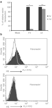
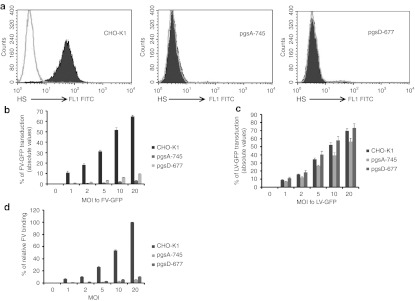
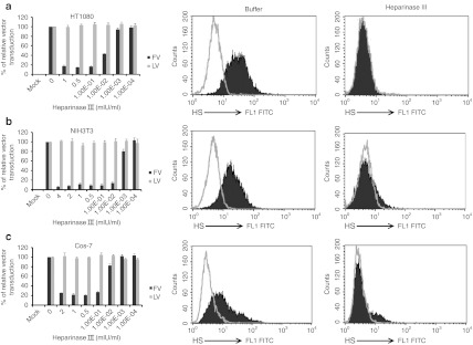
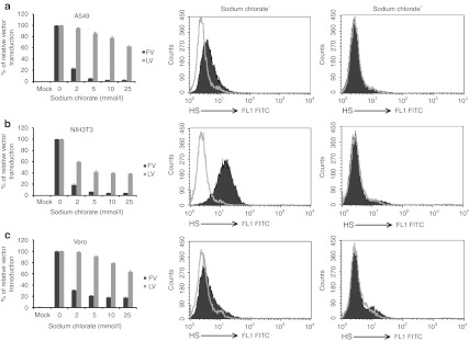
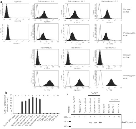
Foamy viruses (FVs) (spumaretroviruses) are good alternative to retroviruses as gene therapy vector. Despite four decades since the discovery of FV, its receptor molecule is still unknown. FV vector transduction of human CD34(+) cells was inhibited by culture with fibronectin. Because fibronectin contains heparin-binding domain, the interactions of fibronectin with heparan sulfate (HS) on cells might be inhibitory to FV transduction. These observations led us to investigate whether HS is a receptor for FV. Two mutant CHO cell lines (but not parental wild type) lacking cell surface HS but not chondroitin sulfate (CS) were largely resistant to FV attachment and transduction. Inhibition of HS expression using enzymes or chemicals greatly reduced FV transduction in human, monkey, and rodent cells. Raji cells, which lack HS and were largely resistant to FV, were rendered more permissive through ectopic expression of syndecan-1, which contains HS. In contrast, mutant syndecan-1-expressing cells were largely resistant to FV. Our findings indicate that cellular HS is a receptor for FV. Identifying FV receptor will enable better understanding of its entry process and optimal use as gene therapy vector to treat inherited and pathogenic diseases.


Figures

Figure 1
Figure 1
Fibronectin-inhibited FV transduction through downregulation of cell surface HS. (a) Human mobilized peripheral blood CD34+ cells were transduced with FV-GFP or VSV-G pseudotyped LV-GFP. Black bars (FN) showed CD34+ cells that were transduced with FV or LV vector overnight immediately after seeding on fibronectin-coated plates. Gray bars (FN+) showed cells were cultured overnight on fibronectin-coated plates and transduced with FV or LV vector overnight. Cells were analyzed for GFP expression by flow cytometry 3–7 days after transduction. For each FV and LV groups, the transduction of “FN” cells was converted into 100% and the transduction of “FN+” cells was calculated according to their proportional percentages. Data represented mean and standard error of the mean (SEM) of three independent experiments. * P = 1.6 × 10⁻⁵ and **P = 0.03. (b) CD34+ cells were stained with anti-HS antibody. Upper graph, HS expression in the absence of fibronectin; lower graph, inhibition of cell surface HS expression when cells were grown overnight on fibronectin. Light lines, isotype control; dark solid area, HS expression detected with anti-HS antibody staining. FITC, fluorescein isothiocyanate; FN, fibronectin; FV, foamy virus; GFP, green fluorescent protein; HS, heparan sulfate; LV, lentivirial vector; VSV, vesicular stomatitis virus.
Figure 2
Figure 2
HS-deficient CHO-K1 cells were largely resistant to FV transduction. FV and VSV-G LV transduction of wild-type CHO-K1 and mutant cells (pgsA-745 and pgsD-677) defective in HS synthesis were assessed. (a) Flow cytometric analyses of wild-type and mutant CHO cells were done to check for HS expression. Cells were stained with a monoclonal antibody (F58-10E4) specific for HS or its isotype control. (b) FV transduction of wild-type CHO-K1 and mutant cells was done at MOIs of 0, 1, 2, 5, 10, and 20 for 1 hour at 37 °C. Cells were harvested 3–7 days after transduction, and GFP analyses were done by flow cytometry. (c) As a control, LV-GFP vector transduction and analyses were done as for the FV group. (d) Binding of 35S-labeled FV to wild-type CHO-K1 and mutant lines, pgsA-745 and pgsD-677, was done at MOIs of 0, 1, 2, 5, 10, and 20 for 1.5 hours at 4 °C. After unbound virus particles were removed by washing, cells were lysed and radioactivity was counted. Counts of wild-type CHO-K1 cells were converted to 100% and counts of pgsA-745 and pgsD-677 cells were assessed according to their proportional percentages. Data represented the mean and standard error of the mean (SEM) of three independent experiments. FITC, fluorescein isothiocyanate; FV, foamy virus; GFP, green fluorescent protein; HS, heparan sulfate; LV, lentivirial vector; MOI, multiplicities of infection; VSV, vesicular stomatitis virus.
Figure 3
Figure 3
Heparinase III digestion of cell surface HS severely reduced FV transduction. Cells were incubated for 1 hour at 37 °C with the indicated concentrations of heparinase III. After washing, one group of cells was transduced with FV (black bars) or LV (gray bars) at MOI of 2 for 1 hour at 37 °C. Unbound virus was removed by extensive washing with PBS. Cells were analyzed for GFP expression 3–7 days after transduction. (a) HT1080, (b) NIH3T3, and (c) Cos-7. For each FV and LV groups, the transduction of untreated control cells was converted to 100% and the transduction of heparinase III-treated cells was calculated according to their proportional percentages. Data represented the mean and standard error of the mean (SEM) of three independent experiments. Another group of cells was fixed with paraformaldehyde and stained with anti-HS antibody to check HS expression. Middle and right panels showed the flow cytometric analysis of HS expression of buffer-treated control cells and heparinase III-treated cells, respectively. FITC, fluorescein isothiocyanate; FV, foamy virus; GFP, green fluorescent protein; HS, heparan sulfate; LV, lentivirial vector; MOI, multiplicities of infection; PBS, phosphate-buffered saline.
Figure 4
Figure 4
FV transduction was strongly dependent on cellular sulfation of HS. Cells were cultured in a low-sulfate F12 medium in the presence of increasing concentrations of sodium chlorate for 36 hours. One group of cells was transduced with FV (black bars) or LV (gray bars) at MOI of 2 for 1 hour at 37 °C, and another group of cells was fixed and stained with anti-HS antibody. (a) A549, (b) NIH3T3, and (c) Vero. Left graphs showed transduction efficiency. The transduction of untreated control cells was converted to 100% and the transduction of sodium chlorate-treated cells was assessed according to their proportional percentages. Data represented the mean and standard error of the mean (SEM) of three independent experiments. Middle and right graphs showed the flow cytometric analysis of HS expression of untreated control cells and sodium chlorate-treated cells, respectively. FITC, fluorescein isothiocyanate; FV, foamy virus; HS, heparan sulfate; LV, lentivirial vector; MOI, multiplicities of infection.
Figure 5
Figure 5
Ectopic expression of HS in Raji cells made them more permissive to FV transduction. (a) Raji cells transfected with syndecan-1 (first and second rows), and TDM-syndecan-1(third and fourth rows) expression plasmids were investigated for the cell surface expression of HS (first and third rows) and proteoglycan (second and fourth rows). (b) Mock, syndecan-1-, and TDM syndecan-1-transfected bulk cultures and cell clones were evaluated for FV transduction. Cells were transduced with FV at MOI of 2 overnight and checked for GFP expression 3–7 days after transduction. Data represented the absolute values of mean and standard error of the mean (SEM) of three independent experiments. (c) Southern blot analysis was done to check FV provirus DNA entry into the mock (untransfected), syndecan-1-, and TDM syndecan-1-transfected cells of both bulk cultures and cell clones. Lane 1, 3, and 7 were untransduced and lane 2, 4, 5, 6, 8, 9, and 10 were transduced with FV-GFP. Genomic DNA was extracted from cells and resolved on agarose gel. 32P-labeled FV pol sequence was used to probe the membranes. FITC, fluorescein isothiocyanate; FV, foamy virus; GFP, green fluorescent protein; HS, heparan sulfate; MOI, multiplicities of infection; TDM, triple deletion mutant.

Comment in

Similar articles

Cited by

References

    1. Linial M.2007Foamy viruses Fields BN, Knipe DM., and, Howley PM.eds). Fields Virology5th edn, vol. 1Lippincott Williams & Wilkins: Philadelphia; 2245–2262.
    1. Rethwilm A. Foamy virus vectors: an awaited alternative to gammaretro- and lentiviral vectors. Curr Gene Ther. 2007;7:261–271. - PubMed
    1. Rethwilm A. Molecular biology of foamy viruses. Med Microbiol Immunol. 2010;199:197–207. - PubMed
    1. Bauer TR, Jr, Allen JM, Hai M, Tuschong LM, Khan IF, Olson EM.et al. (2008Successful treatment of canine leukocyte adhesion deficiency by foamy virus vectors Nat Med 1493–97. - PMC - PubMed
    1. Cavazzana-Calvo M, Hacein-Bey S, de Saint Basile G, Gross F, Yvon E, Nusbaum P.et al. (2000Gene therapy of human severe combined immunodeficiency (SCID)-X1 disease Science 288669–672. - PubMed

Publication types

MeSH terms

LinkOut - more resources