

Independent evolution of macrophage-tropism and increased charge between HIV-1 R5 envelopes present in brain and immune tissue
- PMID: 22420378
- PMCID: PMC3362761
- DOI: 10.1186/1742-4690-9-20
Independent evolution of macrophage-tropism and increased charge between HIV-1 R5 envelopes present in brain and immune tissue
Abstract
Background: Transmitted HIV-1 clade B or C R5 viruses have been reported to infect macrophages inefficiently, while other studies have described R5 viruses in late disease with either an enhanced macrophage-tropism or carrying envelopes with an increased positive charge and fitness. In contrast, our previous data suggested that viruses carrying non-macrophage-tropic R5 envelopes were still predominant in immune tissue of AIDS patients. To further investigate the tropism and charge of HIV-1 viruses in late disease, we evaluated the properties of HIV-1 envelopes amplified from immune and brain tissues of AIDS patients with neurological complications.
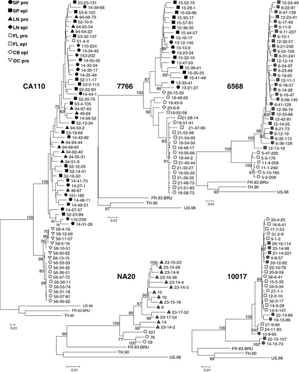
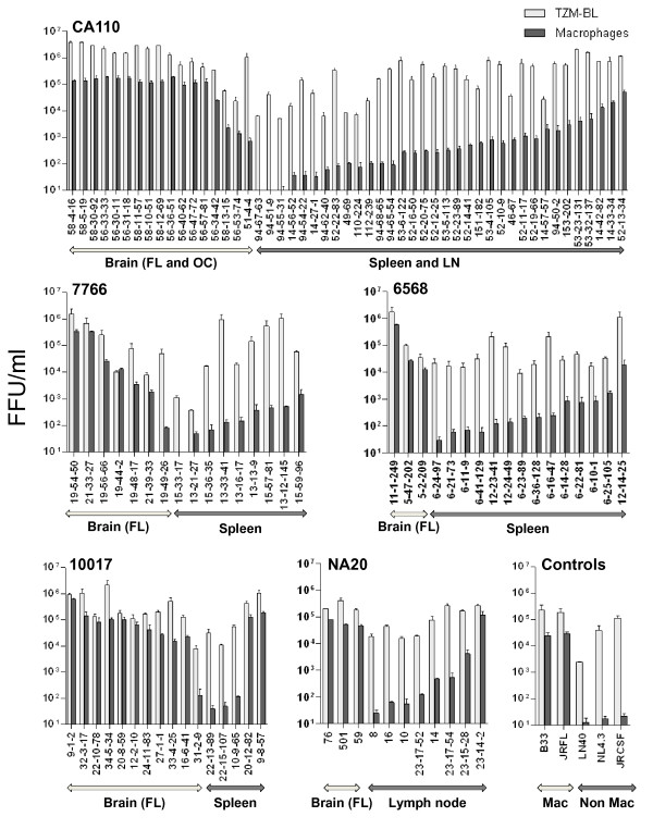
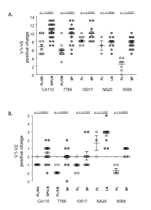
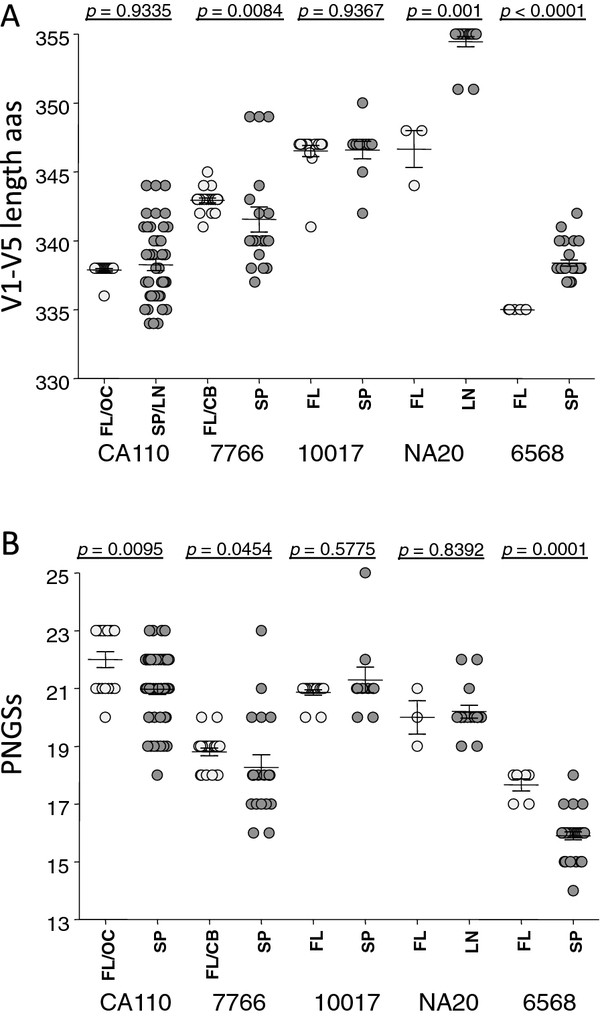
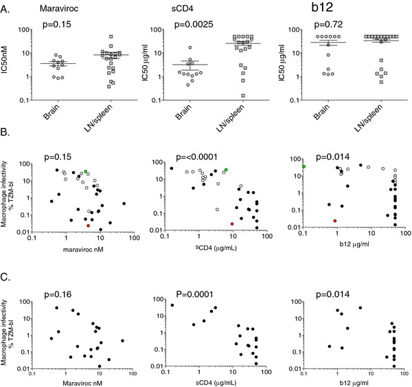
Results: Almost all envelopes amplified were R5. There was clear compartmentalization of envelope sequences for four of the five subjects. However, strong compartmentalization of macrophage-tropism in brain was observed even when brain and immune tissue envelope sequences were not segregated. R5 envelopes from immune tissue of four subjects carried a higher positive charge compared to brain envelopes. We also confirm a significant correlation between macrophage tropism and sensitivity to soluble CD4, a weak association with sensitivity to the CD4 binding site antibody, b12, but no clear relationship with maraviroc sensitivity.
Conclusions: Our study shows that non-macrophage-tropic R5 envelopes carrying gp120s with an increased positive charge were predominant in immune tissue in late disease. However, highly macrophage-tropic variants with lower charged gp120s were nearly universal in the brain. These results are consistent with HIV-1 R5 envelopes evolving gp120s with an increased positive charge in immune tissue or sites outside the brain that likely reflect an adaptation for increased replication or fitness for CD4+ T-cells. Our data are consistent with the presence of powerful pressures in brain and in immune tissues selecting for R5 envelopes with very different properties; high macrophage-tropism, sCD4 sensitivity and low positive charge in brain and non-macrophage-tropism, sCD4 resistance and high positive charge in immune tissue.
Figures
Similar articles
-
Variation in HIV-1 R5 macrophage-tropism correlates with sensitivity to reagents that block envelope: CD4 interactions but not with sensitivity to other entry inhibitors.Retrovirology. 2008 Jan 18;5:5. doi: 10.1186/1742-4690-5-5. Retrovirology. 2008. PMID: 18205925 Free PMC article.
-
HIV-1 R5 Macrophage-Tropic Envelope Glycoprotein Trimers Bind CD4 with High Affinity, while the CD4 Binding Site on Non-macrophage-tropic, T-Tropic R5 Envelopes Is Occluded.J Virol. 2018 Jan 2;92(2):e00841-17. doi: 10.1128/JVI.00841-17. Print 2018 Jan 15. J Virol. 2018. PMID: 29118121 Free PMC article.
-
Biological analysis of human immunodeficiency virus type 1 R5 envelopes amplified from brain and lymph node tissues of AIDS patients with neuropathology reveals two distinct tropism phenotypes and identifies envelopes in the brain that confer an enhanced tropism and fusigenicity for macrophages.J Virol. 2004 Jul;78(13):6915-26. doi: 10.1128/JVI.78.13.6915-6926.2004. J Virol. 2004. PMID: 15194768 Free PMC article.
-
Variation of macrophage tropism among HIV-1 R5 envelopes in brain and other tissues.J Neuroimmune Pharmacol. 2007 Mar;2(1):32-41. doi: 10.1007/s11481-006-9042-2. Epub 2006 Nov 7. J Neuroimmune Pharmacol. 2007. PMID: 18040824 Review.
-
Viral determinants of HIV-1 macrophage tropism.Viruses. 2011 Nov;3(11):2255-79. doi: 10.3390/v3112255. Epub 2011 Nov 15. Viruses. 2011. PMID: 22163344 Free PMC article. Review.
Cited by
-
Infection of ectocervical tissue and universal targeting of T-cells mediated by primary non-macrophage-tropic and highly macrophage-tropic HIV-1 R5 envelopes.Retrovirology. 2015 Jun 9;12:48. doi: 10.1186/s12977-015-0176-2. Retrovirology. 2015. PMID: 26055104 Free PMC article.
-
Efficiency of bridging-sheet recruitment explains HIV-1 R5 envelope glycoprotein sensitivity to soluble CD4 and macrophage tropism.J Virol. 2013 Jan;87(1):187-98. doi: 10.1128/JVI.01834-12. Epub 2012 Oct 10. J Virol. 2013. PMID: 23055568 Free PMC article.
-
HIV-1 non-macrophage-tropic R5 envelope glycoproteins are not more tropic for entry into primary CD4+ T-cells than envelopes highly adapted for macrophages.Retrovirology. 2015 Mar 14;12:25. doi: 10.1186/s12977-015-0141-0. Retrovirology. 2015. PMID: 25809903 Free PMC article.
-
No detection of CD4-independent human immunodeficiency virus 1 envelope glycoproteins in brain tissue of patients with or without neurological complications.Arch Virol. 2019 Feb;164(2):473-482. doi: 10.1007/s00705-018-4094-1. Epub 2018 Nov 10. Arch Virol. 2019. PMID: 30415390 Free PMC article.
-
Characterization of HIV variants from paired Cerebrospinal fluid and Plasma samples in primary microglia and CD4+ T-cells.J Neurovirol. 2024 Aug;30(4):380-392. doi: 10.1007/s13365-024-01207-w. Epub 2024 May 7. J Neurovirol. 2024. PMID: 38713307 Free PMC article.
References
-
- Salazar-Gonzalez JF, Salazar MG, Keele BF, Learn GH, Giorgi EE, Li H, Decker JM, Wang S, Baalwa J, Kraus MH, Parrish NF, Shaw KS, Guffey MB, Bar KJ, Davis KL, Ochsenbauer-Jambor C, Kappes JC, Saag MS, Cohen MS, Mulenga J, Derdeyn CA, Allen S, Hunter E, Markowitz M, Hraber P, Perelson AS, Bhattacharya T, Haynes BF, Korber BT, Hahn BH. et al.Genetic identity, biological phenotype, and evolutionary pathways of transmitted/founder viruses in acute and early HIV-1 infection. J Exp Med. 2009;206:1273–1289. doi: 10.1084/jem.20090378. - DOI - PMC - PubMed
-
- Asjo B, Morfeldt Manson L, Albert J, Biberfeld G, Karlsson A, Lidman K, Fenyo EM. Replicative capacity of human immunodeficiency virus from patients with varying severity of HIV infection. Lancet. 1986;2:660–662. - PubMed
Publication types
MeSH terms
Substances
Associated data
- Actions
- Actions
- Actions
- Actions
- Actions
- Actions
- Actions
- Actions
- Actions
- Actions
- Actions
- Actions
- Actions
- Actions
- Actions
- Actions
- Actions
- Actions
- Actions
- Actions
- Actions
- Actions
- Actions
- Actions
- Actions
- Actions
- Actions
- Actions
- Actions
- Actions
- Actions
- Actions
- Actions
- Actions
- Actions
- Actions
- Actions
- Actions
- Actions
- Actions
- Actions
- Actions
- Actions
- Actions
- Actions
- Actions
- Actions
- Actions
- Actions
- Actions
- Actions
- Actions
- Actions
- Actions
- Actions
- Actions
- Actions
- Actions
- Actions
- Actions
- Actions
- Actions
- Actions
- Actions
- Actions
- Actions
- Actions
- Actions
- Actions
- Actions
- Actions
- Actions
- Actions
- Actions
- Actions
- Actions
- Actions
- Actions
- Actions
- Actions
- Actions
- Actions
- Actions
- Actions
- Actions
- Actions
- Actions
- Actions
- Actions
- Actions
- Actions
- Actions
- Actions
- Actions
- Actions
- Actions
- Actions
- Actions
- Actions
- Actions
- Actions
- Actions
- Actions
- Actions
- Actions
- Actions
- Actions
- Actions
- Actions
- Actions
- Actions
- Actions
- Actions
- Actions
- Actions
- Actions
- Actions
- Actions
- Actions
- Actions
- Actions
- Actions
- Actions
- Actions
- Actions
- Actions
- Actions
- Actions
- Actions
- Actions
- Actions
- Actions
- Actions
- Actions
- Actions
- Actions
- Actions
- Actions
- Actions
- Actions
- Actions
- Actions
- Actions
- Actions
- Actions
- Actions
- Actions
- Actions
- Actions
- Actions
- Actions
- Actions
- Actions
- Actions
- Actions
- Actions
- Actions
- Actions
- Actions
- Actions
- Actions
- Actions
- Actions
- Actions
- Actions
- Actions
- Actions
- Actions
- Actions
- Actions
- Actions
- Actions
- Actions
- Actions
- Actions
- Actions
- Actions
- Actions
- Actions
- Actions
- Actions
- Actions
- Actions
- Actions
- Actions
- Actions
- Actions
Grants and funding
- R01 AI089334/AI/NIAID NIH HHS/United States
- R01 HD049273/HD/NICHD NIH HHS/United States
- R01 MH64408/MH/NIMH NIH HHS/United States
- P01 AI082274/AI/NIAID NIH HHS/United States
- R24MH59724/MH/NIMH NIH HHS/United States
- R24 NS45491/NS/NINDS NIH HHS/United States
- 095831/Wellcome Trust/United Kingdom
- U01MH083501/MH/NIMH NIH HHS/United States
- U01MH083507/MH/NIMH NIH HHS/United States
- P30-AI42845/AI/NIAID NIH HHS/United States
- R24MH59745/MH/NIMH NIH HHS/United States
- U01MH083506/MH/NIMH NIH HHS/United States
- U01 MH083545/MH/NIMH NIH HHS/United States
LinkOut - more resources
Full Text Sources
Medical
Molecular Biology Databases
Research Materials
