
Whole genome deep sequencing of HIV-1 reveals the impact of early minor variants upon immune recognition during acute infection

Matthew R Henn et al. PLoS Pathog. 2012.

Abstract

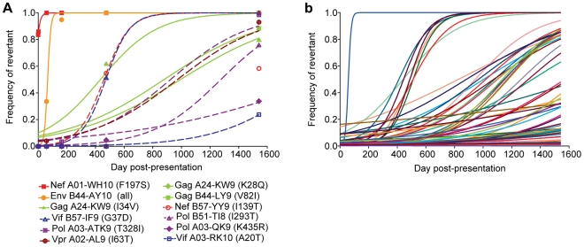
Deep sequencing technologies have the potential to transform the study of highly variable viral pathogens by providing a rapid and cost-effective approach to sensitively characterize rapidly evolving viral quasispecies. Here, we report on a high-throughput whole HIV-1 genome deep sequencing platform that combines 454 pyrosequencing with novel assembly and variant detection algorithms. In one subject we combined these genetic data with detailed immunological analyses to comprehensively evaluate viral evolution and immune escape during the acute phase of HIV-1 infection. The majority of early, low frequency mutations represented viral adaptation to host CD8+ T cell responses, evidence of strong immune selection pressure occurring during the early decline from peak viremia. CD8+ T cell responses capable of recognizing these low frequency escape variants coincided with the selection and evolution of more effective secondary HLA-anchor escape mutations. Frequent, and in some cases rapid, reversion of transmitted mutations was also observed across the viral genome. When located within restricted CD8 epitopes these low frequency reverting mutations were sufficient to prime de novo responses to these epitopes, again illustrating the capacity of the immune response to recognize and respond to low frequency variants. More importantly, rapid viral escape from the most immunodominant CD8+ T cell responses coincided with plateauing of the initial viral load decline in this subject, suggestive of a potential link between maintenance of effective, dominant CD8 responses and the degree of early viremia reduction. We conclude that the early control of HIV-1 replication by immunodominant CD8+ T cell responses may be substantially influenced by rapid, low frequency viral adaptations not detected by conventional sequencing approaches, which warrants further investigation. These data support the critical need for vaccine-induced CD8+ T cell responses to target more highly constrained regions of the virus in order to ensure the maintenance of immunodominant CD8 responses and the sustained decline of early viremia.


Conflict of interest statement

The authors have declared that no competing interests exist.

Figures

Figure 1
Figure 1. PCR amplification strategy and performance of novel assembly, read alignment, and variant detection algorithms.
(A) PCR amplification strategy using four ∼3.2 kb amplicons spanning gag through nef of the HIV-1 genome. Amplicons were then pooled, sheared, barcoded by patient or time point, and batched for library construction and single-molecule 454 pyrosequencing. (B) AssembleViral454 v1.0 outperforms other algorithms in its ability to assemble de novo continuous consensus contigs that span the complete target region. Results are shown for 67 acute, chronic, or controller patient samples that had successful amplification of all four amplicons and at least 10-fold sequence coverage (sequencing reads per site) across >70% of the target genome. Black lines denote the mean score for each assembler, red line the median, red box ends the 25th and 75th quantiles, and red box whiskers the upper and lower quartiles plus/minus 1.5 times the interquartile range, respectively. (C) ReadClean454 v1.0 corrects for read alignment errors due to various sequence error modes and significantly reduces process error rate. Results shown are for virus from two infectious clones, NL43 (WT) and NL43 (RKLM) containing two point mutations in Gag , sequenced independently to 417- and 189-fold average coverage, respectively. Errors are defined as base calls or InDels that differ from the assembled consensus at a given position, and the read error rate is the total number of errors per total number of NQS passing bases interrogated. Percentage of reads on which a correction was made at each step are shown in parentheses. A final average process error rate of 0.5×10⁻⁴ was achieved based on both infectious clones. (D) V-Phaser v1.0, utilizes phasing information to identify a variant pair found in 1.0% of the reads covering both loci when there are 200 such reads; without phase, a three-fold increase in coverage is required to achieve the same 1.0% detection threshold. A variant at a frequency of 0.1% can be detected when phased coverage is 2999-fold.
Figure 2
Figure 2. Comparison of sequence variant quantification by 454 deep sequencing and by PCR cloning/sequencing.
Orthogonal regression of variant frequency estimates obtained by 454 and clonal sequence data across the highly variable 1544 nucleotide region spanning Vif to Tat in subject 9213 (slope = 1.01; 95% CI, 0.73 to 1.40).
Figure 3
Figure 3. Clinical course and whole genome deep sequence coverage for subject 9213.
(A) Clinical course of infection in subject 9213 shown as days post-presentation. Plasma viral load (copies/ml) is shown in red and CD4+ T cell count (cells/ml) in blue. Estimated acute/early Fiebig stages are shown and arrows indicate time points sequenced on the deep sequencing platform. (B) High-quality sequencing reads per site across the HIV-1 genome for subject 9213 at six time points (days post presentation). Reads are aligned to the consensus assembly of their respective time point using Mosaik v1.0 (Table S9 in Text S1) and coverage (sequencing reads per site) calculated from bases that pass the defined Neighbor Quality Standard (NQS, see Supplementary Methods in Text S1) . PCR amplicon locations are denoted by horizontal bars under the x-axis.
Figure 4
Figure 4. Rapidly expanding sequence diversity during HIV-1 infection.
Heat maps illustrate sites exhibiting amino acid sequence diversity at days 0, 3, 59, 165, 476 and 1543 post-presentation. Plotted is the percentage of amino acid diversity at each position with respect to the dominant baseline (day 0) amino acid residue. All 3174 amino acids of HIV-1 are represented, with the first amino acid of Gag located in the top left corner of the grid and the last amino acid of Nef located in the bottom right corner. Completely conserved residues are dark blue, low-level variant residues (<10% divergent from baseline) are light blue, moderately variable residues (10–50%) in orange, and highly variant residues (>50%) in red. (A) 0 days p.p., (B) 3 days p.p., (C) 59 days p.p., (D) 165 days p.p., (E) 476 days p.p., (F) 1543 days p.p..
Figure 5
Figure 5. Limited evolution in the HIV-1 proteome prior to establishment of viral set point.
Sequence diversity is plotted for all evolving codons in each HIV-1 protein as the percent of sequences with an amino acid residue different from the dominant baseline residue. Colored lines denote individual evolving amino acid residues within each protein. The time of infection prior to the establishment of viral set point (day 165) is highlighted in grey.
Figure 6
Figure 6. Cellular immune responses drive early low-frequency quasispecies diversity.
(A) For each protein, the average frequency of non-dominant baseline residues of positions within the 19 described CD8 epitopes restricted by subject 9213's HLA alleles (left) and outside of the 19 described epitopes (right) is plotted for each time point sequenced. Colored lines denote the proteins for which diversity was substantially higher inside of CD8 epitopes versus outside CD8 epitopes. (B) To determine rates of viral escape for each epitope escape mutations were defined as any amino acid substitution within the epitope. Symbols denote the cumulative observed frequency of all escape mutations, and lines depict the best fit by non-linear regression of the observed frequency data to the CTL escape model of Asquith et al. . Open symbols and dashed lines denote epitopes for which evolution was consistent with reversion. Black symbols and dotted lines denote epitopes for which there was no evidence of escape. CD8 responses against each epitope are shown in parentheses in the legend and were measured by IFN-gamma Elispot assay (Spot Forming Cells/Mill PBMC (SFC)). (C) Frequency of wild-type (black) and variant (red) haplotypes of the Vif B38-WI9 epitope and flanking regions over time. Shown at the top is the clade B consensus sequence for reference. (D) Frequency of wild-type (black) and variant (red) haplotypes of the Nef A24-RW8 B38-WI9 epitope and flanking regions over time. Blue residues highlight differences between the day 0 transmitted sequence and HIV-1B consensus sequence.
Figure 7
Figure 7. Viral escape from acute and chronic phase CD8+ T cell responses.
Stacked heat-maps illustrate variant codon frequencies over time for each residue of the CD8 epitopes targeted by subject 9213. Shown are epitopes targeted during the acute (Day 59) and chronic (Day 476) phases of HIV-1 infection. The baseline sequence is shown at the top of each epitope, with non-HIV-1B consensus residues highlighted in blue. The magnitude of each response is shown in SFC per million PBMC.
Figure 8
Figure 8. Variant-specific CD8+ T cell Elispot responses.
Elispot responses in Spot Forming Cells (SFC) per million PBMC to wild-type and variant peptides for the two dominant epitopes (A) Vif B38-WI9 (WHLGQGVSI) and (B) Nef A24-RW8 (RYPLTFGW). Bars in black denote responses to clade B consensus epitopes. Bars in red, orange, and pink denote responses to epitopes containing escape variants.
Figure 9
Figure 9. Reversion of transmitted mutations over the course of infection.
(A) Rates of reversion of transmitted mutations within both restricted and unrestricted CD8 epitopes in subject 9213. Reversion was defined as the replacement of a transmitted non-consensus residue by the HIV-1B consensus residue. Symbols denote the observed frequency of viruses expressing the consensus residue and lines depict the best fit by non-linear regression of the observed frequency data to the CTL escape model of Asquith et al. . Closed symbols and solid lines denote epitopes restricted by subject 9213, while open symbols and dashed lines denote epitopes not restricted by 9213. Listed in parentheses are the mutations listed by the consensus residue, HXB2 position, followed by the transmitted mutation, i.e., F197S. (B) Rates of reversion of all transmitted mutations exhibiting sequence variation over the course of infection. Each line represents a different mutation.

Similar articles

Cited by

References

    1. Allen TM, O'Connor DH, Jing P, Dzuris JL, Mothe BR, et al. Tat-specific cytotoxic T lymphocytes select for SIV escape variants during resolution of primary viraemia. Nature. 2000;407:386–390. - PubMed
    1. Goulder PJ, Phillips RE, Colbert RA, McAdam S, Ogg G, et al. Late escape from an immunodominant cytotoxic T-lymphocyte response associated with progression to AIDS. Nat Med. 1997;3:212–217. - PubMed
    1. Timm J, Lauer GM, Kavanagh DG, Sheridan I, Kim AY, et al. CD8 epitope escape and reversion in acute HCV infection. J Exp Med. 2004;200:1593–1604. - PMC - PubMed
    1. Kwiatkowski D, Marsh K. Development of a malaria vaccine. Lancet. 1997;350:1696–1701. - PubMed
    1. Allen TM, Altfeld M, Geer SC, Kalife ET, Moore C, et al. Selective escape from CD8+ T-cell responses represents a major driving force of human immunodeficiency virus type 1 (HIV-1) sequence diversity and reveals constraints on HIV-1 evolution. J Virol. 2005;79:13239–13249. - PMC - PubMed

Publication types