. 2012 Jul;14(7):1122-34.
doi: 10.1111/j.1462-5822.2012.01784.x. Epub 2012 Apr 4.

Binding of Lassa virus perturbs extracellular matrix-induced signal transduction via dystroglycan

Affiliations

Binding of Lassa virus perturbs extracellular matrix-induced signal transduction via dystroglycan

Jillian M Rojek et al. Cell Microbiol. 2012 Jul.

Abstract

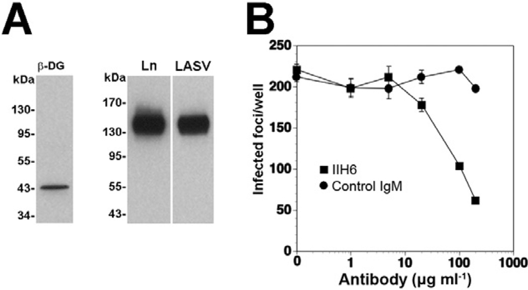
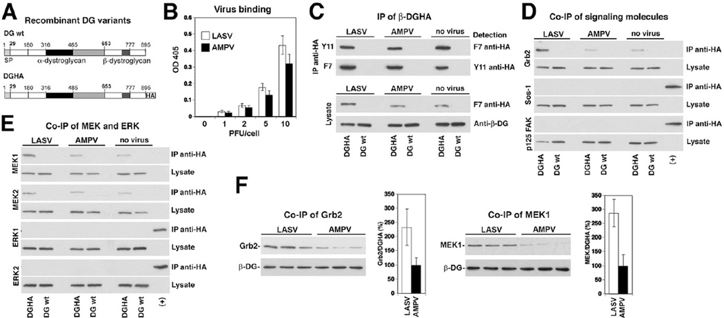
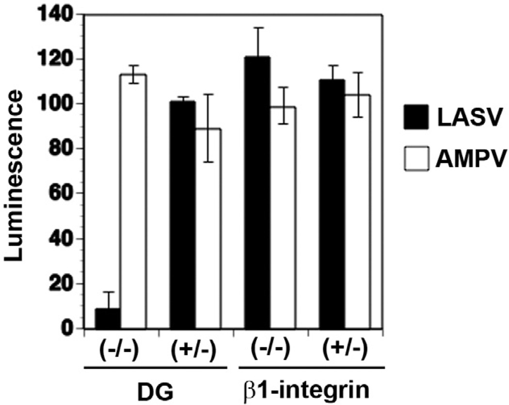
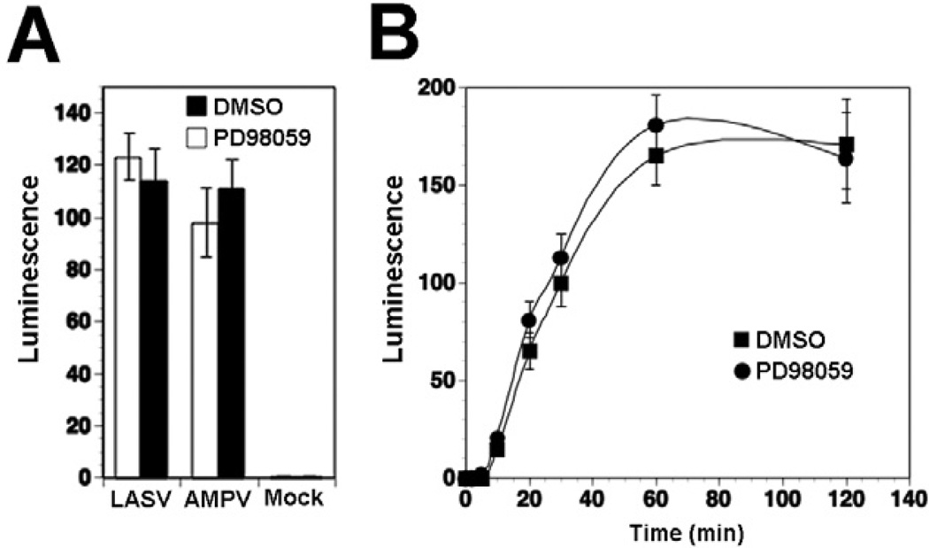
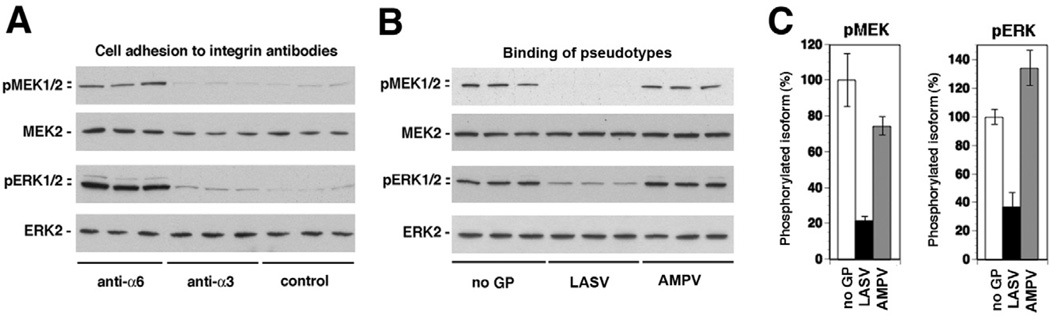
The arenavirus Lassa virus (LASV) causes a severe haemorrhagic fever with high mortality in man. The cellular receptor for LASV is dystroglycan (DG). DG is a ubiquitous receptor for extracellular matrix (ECM) proteins, which cooperates with β1 integrins to control cell-matrix interactions. Here, we investigated whether LASV binding to DG triggers signal transduction, mimicking the natural ligands. Engagement of DG by LASV resulted in the recruitment of the adaptor protein Grb2 and the protein kinase MEK1 by the cytoplasmic domain of DG without activating the MEK/ERK pathway, indicating assembly of an inactive signalling complex. LASV binding to cells however affected the activation of the MEK/ERK pathway via α6β1 integrins. The virus-induced perturbation of α6β1 integrin signalling critically depended on high-affinity LASV binding to DG and DG's cytoplasmic domain, indicating that LASV-receptor binding perturbed signalling cross-talk between DG and β1 integrins.


Figures

Fig. 1
Fig. 1. Cell entry of LASV pseudotypes in WI-26 VA4 human lung epithelial cells is mediated by DG
A. WI-26 VA4 cells express DG that is functional as LASV receptor. WI-26 VA4 cells were lysed and glycoproteins enriched by affinity purification with the lectin wheat germ agglutinin. Functional α-DG was detected by laminin overlay assay (Ln) and virus overlay protein binding assay using inactivated virus (LASV). B. Blocking of infection with mAb IIH6. Monolayers of WI-26 VA4 cells were blocked with mAb IIH6 or an unrelated mouse IgM (Control IgM) at the indicated concentrations for 2 h at 4°C. Next, LASV pseudotypes (200 PFU) were added for 45 min. Infection was assessed after 24 h by immunofluorescence staining for GFP. Infected foci were counted in each well (means ± SD, n = 3).
Fig. 2
Fig. 2. Binding of inactivated LASV to cells promotes association of β-DG with Grb2
A. Schematic representation of C-terminally tagged DG (DGHA). The N-terminal domain (white), the mucin-type domain (black) and the C-terminal domain (grey) of α-DG, β-DG and the C-terminal HA tag are indicated. B. Binding of inactivated LASV and AMPV to WI-26 VA4 cells: cell monolayers were incubated with the indicated concentrations of inactivated virus in the cold. Bound virus was detected with mAb 83.6 to arenavirus GP2, combined with a HRP-conjugated secondary antibody in a colour reaction (means ± SD, n = 3). C. IP of β-DGHA: WI-26 VA4 cells transiently transfected with either DGHA or wild-type DG (DG wt) were seeded on poly-l-lysine and incubated with inactivated LASV, AMPV, or no virus at a particle per cell ratio of 100. After 20 min, cells were lysed and DGHA precipitated with either a polyclonal rabbit antibody anti-HA Y11 or mouse mAb F7 anti-HA. Immunocomplexes were probed for HA in Western blot using the indicated antibodies. Total-cell lysates were probed for DGHA with mAb F7 anti-HA and for β-DG with pAb AP83 anti-β-DG. D and E. Co-immunoprecipitation (co-IP) of β-DGHA with signalling molecules: immunocomplexes and total lysates (C) were probed for the presence of Grb2, Sos-1, FAK, MEK1/2 and ERK1/2 in Western blot. In case of Sos-1, FAK and ERK1/2, a positive control corresponding to 0.1% of total-cell protein was included (+). F. Binding of inactivated LASV increases the association of β-DG with Grb2 and MEK1: triplicate specimens of WI-26 VA4 cells transiently transfected with DGHA were exposed to inactivated viruses, lysed and DGHA precipitated as in (C). DGHA, Grb2 and MEK1 were detected in Western blot as in (D) and (E). For quantitative analysis, X-ray films were scanned with a densitometer and the ratios of Grb2/DGHA and MEK1/Grb2 calculated. For normalization, signals obtained with the AMPV-negative control were defined as 100% (n = 3, ±SD).
Fig. 3
Fig. 3
Infection of LASV pseudotypes is independent on β1 integrins. DG (−/−) and DG (+/−), β1 integrin (−/−) and β1 integrin (+/−) mouse ES cells cultured in 96-well plates were infected with the retroviral pseudotypes bearing the GPs of LASV or AMPV (moi = 10). Infection was assessed after 48 h by luciferase assay (n = 3, ±SD).
Fig. 4
Fig. 4. Binding of inactivated LASV to cells perturbs laminin-induced activation of the ERK-MAP kinase pathway
A. Phosphorylation of MEK and ERK in WI-26 VA4 cells in response to cell adhesion to laminin-1. Serum-starved cells were detached and seeded onto wells coated with laminin-1 or kept in suspension. At the indicated time points, total-cell lysates were prepared and subsequently analysed for the presence of MEK1/2 and ERK1/2 as well as the phosphorylated forms of the kinases using specific antibodies. B. ERK is phosphorylated via MEK: suspensions of serum-starved cells were incubated with the MEK-specific inhibitor PD98059 (50 µM) or solvent control for 30 min and then plated onto wells coated with laminin-1 for 40 min. The phosphorylated forms of MEK1/2 and ERK1/2, MEK2 and ERK2 were detected in total-cell lysates. C. Binding of inactivated LASV to cells reduces laminin-induced activation of MEK and ERK: serum-starved WI-26 VA4 cells were detached, mixed with inactivated LASV or AMPV (10 particles per cell), or no virus, and plated onto wells coated with laminin-1. After 40 min of cell adhesion, total-cell lysates were prepared and analysed for the presence of phosphorylated MEK and ERK as in (A). D. Quantification of (C): the signal for phosphorylated MEK1/2 and ERK1/2 in the control samples set as 100% (n = 3, ±SD). E and F. Binding of LASV pseudotypes reduces laminin-induced activation of MEK and ERK: experiment was performed as in (C) and (D) using retroviral pseudotypes of LASV and AMPV (10 PFU per cell) and pseudotypes without GP (no GP).
Fig. 5
Fig. 5. MEK1 activity is dispensable for cell entry of LASV pseudotypes
A. Infection of LASV pseudotypes is not affected by the MEK inhibitor PD98059. Monolayers of WI-26 VA4 cells were pre-treated with PD98059 (50 µM) or solvent control (DMSO) for 30 min, followed by infection with LASV and AMPV pseudotypes (moi = 0.1). Infection was detected by luciferase assay and fold increase of luminescence above background is given (means ± SD, n = 3). B. The MEK inhibitor PD98059 does not affect LASV cell entry kinetics. Monolayers of WI-26 VA4 cells were pre-treated with PD98059 as in (A), followed by incubation with LASV pseudotypes (moi = 0.1) in the cold in presence of the drug. After 1 h, unbound virus was removed and pre-warmed (37°C) medium containing the drug added. At the indicated time points, 20 mM ammonium chloride was added and left throughout the experiment. At 24 h post infection was detected by luciferase assay as in (A) (means ± SD, n = 3). Please note that the apparent differences in infection at 60 min were not statistically significant.
Fig. 6
Fig. 6. Binding of LASV pseudotypes to cells blocks α6β1 integrin-mediated activation of MEK and ERK
A. Induction of MEK and ERK phosphorylation by anti-α6 integrin antibody. Serum-starved WI-26 VA4 cells were detached and plated onto wells coated with antibodies to α6 and α3 integrin or an isotype control antibody for 40 min. Activation of MEK and ERK was determined by Western blot. B. Binding of LASV pseudotypes reduces MEK and ERK phosphorylation induced by anti-α6 integrin antibody: serum-starved WI-26 VA4 cell suspensions were mixed with pseudotypes of LASV and AMPV at 10 particles per cell or control pseudotypes containing no GP (no GP) and plated onto wells coated with antibodies to α6. After 40 min of cell adhesion total-cell lysate were prepared and phosphorylation of MEK and ERK analysed by Western blot. C. Quantification of (B), setting the signal for phosphorylated MEK1/2 and ERK1/2 in the control sample (no GP) as 100% (n = 3, ±SD).
Fig. 7
Fig. 7. Functional α-DG and the cytoplasmic domain of β-DG are involved in LASV pseudotype-induced inhibition of α6 integrin-mediated MEK/ERK activation
A. Schematic representation of the DG mutants. B. Detection of the α-DG parts of the DG variants. DGE, DGΔCyt, wild-type DG and GFP were expressed in DG (−/−) ES cells using AdV vectors. After 48 h, total membrane extracts were prepared and probed with an antibody to HA and in virus overlay protein binding assay (VOPBA). C. Overexpression of DG DN mutants releases LASV pseudotype-induced inhibition of α6 integrin mediated MEK/ERK activation. WI-26 VA4 cells were infected with AdV expressing the DG variants. After 48 h, cells were serum-starved and detached. Single-cell suspensions were mixed with LASV and AMPV pseudotypes and plated onto wells coated with antibody to α6. Activation of ERK was determined by Western blot. D. Quantification of (B), setting the signal for phosphorylated MEK1/2 and ERK1/2 in the control sample (no GP) as 100% (n = 3, ±SD).
Fig. 8
Fig. 8. Working model: binding of LASV to DG perturbs signalling cross-talk with β1 integrins
A. Binding of laminin to cellular DG modulates MEK/ERK signalling through α6β1 integrins: laminin engages cellular DG via the LG domains 4 and 5 of the α1 chain (Hohenester et al., 1999) and binding critically depends on α-DG-linked O-glycans, in particular sugar polymers attached by LARGE (Kanagawa et al., 2004). Binding of laminin to α6β1 integrins involves the LG domains 1–3 of α1 (Hohenester et al., 1999) and results in activation of MEK/ERK signalling (Ferletta et al., 2003). Simultaneous binding of laminin to DG via LG4/5 inhibits activation of MEK/ERK via α6β1 integrins (Ferletta et al., 2003). B. High-affinity binding of LASV to cellular α-DG perturbs the signalling cross-talk between DG and α6β1 integrins (this study): binding of inactivated LASV and LASV pseudotypes to cellular DG inhibits activation of α6β1 integrins via laminin or a signalling-inducing antibody to α6. The effect of LASV on α6β1 integrin signalling depends on the functional O-glycosylation of α-DG and the cytoplasmic domain of β-DG. The nature of the inhibitory signal induced by binding of laminin LG4/5 and LASV is currently unknown.

Similar articles

Cited by

References

    1. Baranowski E, Ruiz-Jarabo CM, Pariente N, Verdaguer N, Domingo E. Evolution of cell recognition by viruses: a source of biological novelty with medical implications. Adv Virus Res. 2003;62:19–111. - PMC - PubMed
    1. Barresi R, Campbell KP. Dystroglycan: from biosynthesis to pathogenesis of human disease. J Cell Sci. 2006;119:199–207. - PubMed
    1. Barresi R, Michele DE, Kanagawa M, Harper HA, Dovicio SA, Satz JS, et al. LARGE can functionally bypass alpha-dystrolycan glycosylation defects in distinct congential muscular dystrophy. Nat Med. 2004;10:696–703. - PubMed
    1. Beyer WR, Popplau D, Garten W, von Laer D, Lenz O. Endoproteolytic processing of the lymphocytic choriomeningitis virus glycoprotein by the subtilase SKI-1/S1P. J Virol. 2003;77:2866–2872. - PMC - PubMed
    1. Borrow P, Oldstone MB. Mechanism of lymphocytic choriomeningitis virus entry into cells. Virology. 1994;198:1–9. - PubMed

Publication types