

Cutting edge: dendritic epidermal γδ T cell ligands are rapidly and locally expressed by keratinocytes following cutaneous wounding
- PMID: 22393149
- PMCID: PMC3311739
- DOI: 10.4049/jimmunol.1100887
Cutting edge: dendritic epidermal γδ T cell ligands are rapidly and locally expressed by keratinocytes following cutaneous wounding
Abstract
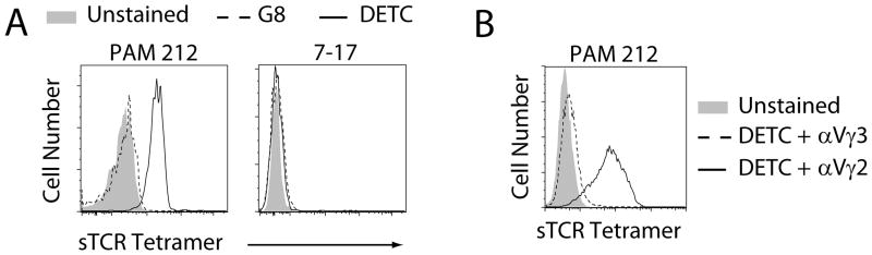
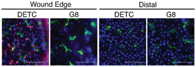
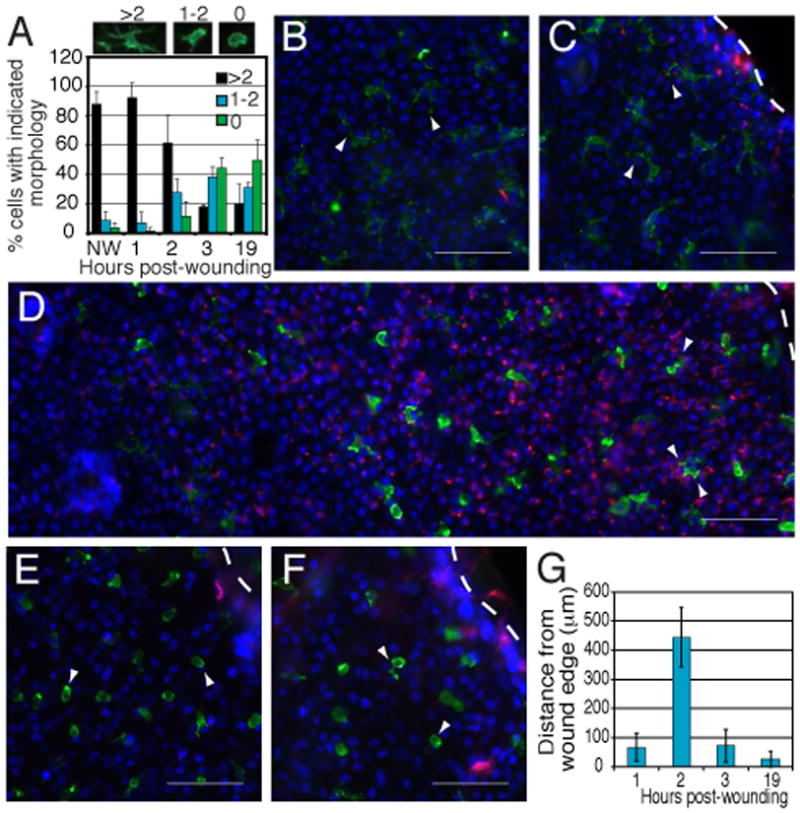
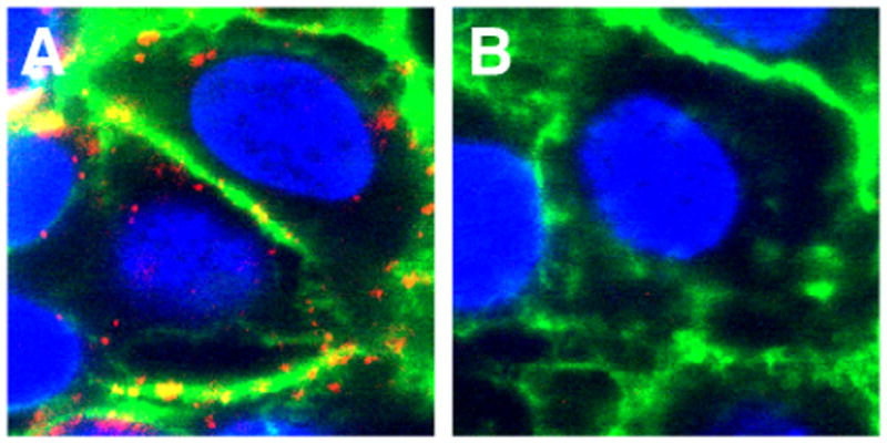
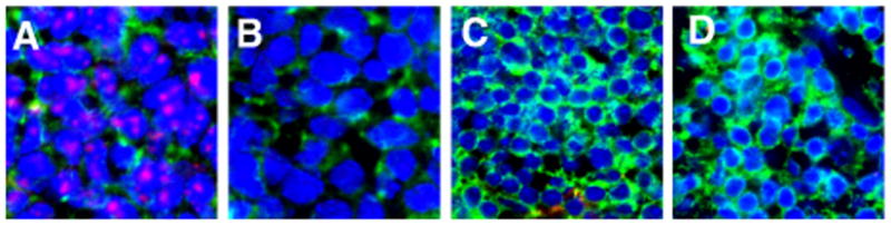
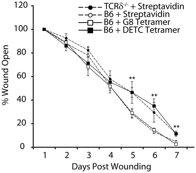
TCR-specific activation is pivotal to dendritic epidermal T cell (DETC) function during cutaneous wound repair. However, DETC TCR ligands are uncharacterized, and little is known about their expression patterns and kinetics. Using soluble DETC TCR tetramers, we demonstrate that DETC TCR ligands are not constitutively expressed in healthy tissue but are rapidly upregulated following wounding on keratinocytes bordering wound edges. Ligand expression is tightly regulated, with downmodulation following DETC activation. Early inhibition of TCR-ligand interactions using DETC TCR tetramers delays wound repair in vivo, highlighting DETC as rapid responders to injury. To our knowledge, this is the first visualization of DETC TCR ligand expression, which provides novel information about how ligand expression impacts early stages of DETC activation and wound repair.
Figures
Similar articles
-
A keratinocyte-responsive gamma delta TCR is necessary for dendritic epidermal T cell activation by damaged keratinocytes and maintenance in the epidermis.J Immunol. 2004 Mar 15;172(6):3573-9. doi: 10.4049/jimmunol.172.6.3573. J Immunol. 2004. PMID: 15004158
-
Dendritic epidermal T cells regulate skin antimicrobial barrier function.J Clin Invest. 2013 Oct;123(10):4364-74. doi: 10.1172/JCI70064. Epub 2013 Sep 24. J Clin Invest. 2013. PMID: 24051381 Free PMC article.
-
Differential roles of cytokine receptors in the development of epidermal gamma delta T cells.J Immunol. 2001 Aug 15;167(4):1929-34. doi: 10.4049/jimmunol.167.4.1929. J Immunol. 2001. PMID: 11489972
-
Functions of skin-resident γδ T cells.Cell Mol Life Sci. 2011 Jul;68(14):2399-408. doi: 10.1007/s00018-011-0702-x. Epub 2011 May 11. Cell Mol Life Sci. 2011. PMID: 21560071 Free PMC article. Review.
-
Functions of Vγ4 T Cells and Dendritic Epidermal T Cells on Skin Wound Healing.Front Immunol. 2018 Jun 4;9:1099. doi: 10.3389/fimmu.2018.01099. eCollection 2018. Front Immunol. 2018. PMID: 29915573 Free PMC article. Review.
Cited by
-
γδ T Cells Mediate a Requisite Portion of a Wound Healing Response Triggered by Cutaneous Poxvirus Infection.Viruses. 2024 Mar 10;16(3):425. doi: 10.3390/v16030425. Viruses. 2024. PMID: 38543790 Free PMC article.
-
Tissue Adaptations of Memory and Tissue-Resident Gamma Delta T Cells.Front Immunol. 2018 Nov 27;9:2636. doi: 10.3389/fimmu.2018.02636. eCollection 2018. Front Immunol. 2018. PMID: 30538697 Free PMC article. Review.
-
Mechanisms of T cell organotropism.Cell Mol Life Sci. 2016 Aug;73(16):3009-33. doi: 10.1007/s00018-016-2211-4. Epub 2016 Apr 1. Cell Mol Life Sci. 2016. PMID: 27038487 Free PMC article. Review.
-
Emerging Skin T-Cell Functions in Response to Environmental Insults.J Invest Dermatol. 2017 Feb;137(2):288-294. doi: 10.1016/j.jid.2016.08.013. Epub 2016 Oct 23. J Invest Dermatol. 2017. PMID: 27784595 Free PMC article. Review.
-
Chronic Inflammation and γδ T Cells.Front Immunol. 2016 May 27;7:210. doi: 10.3389/fimmu.2016.00210. eCollection 2016. Front Immunol. 2016. PMID: 27303404 Free PMC article. Review.
References
-
- Garcia KC, Teyton L, Wilson IA. Structural basis of T cell recognition. Annu Rev Immunol. 1999;17:369–397. - PubMed
-
- Havran WL, Chien YH, Allison JP. Recognition of self antigens by skin-derived T cells with invariant γδ antigen receptors. Science. 1991;252:1430–1432. - PubMed
-
- Girardi M, Lewis JM, Filler RB, Hayday AC, Tigelaar RE. Environmentally responsive and reversible regulation of epidermal barrier function by γδ T cells. J Invest Dermatol. 2006;126:808–814. - PubMed
Publication types
MeSH terms
Substances
Grants and funding
- R01 AI036964/AI/NIAID NIH HHS/United States
- R01 AI064811-03/AI/NIAID NIH HHS/United States
- T32AI007244/AI/NIAID NIH HHS/United States
- T32 AI007244/AI/NIAID NIH HHS/United States
- AI36964/AI/NIAID NIH HHS/United States
- A142267/PHS HHS/United States
- R01 AI036964-13/AI/NIAID NIH HHS/United States
- R01 AI042267/AI/NIAID NIH HHS/United States
- R21 AI095823/AI/NIAID NIH HHS/United States
- AI064811/AI/NIAID NIH HHS/United States
- R01 AI042267-10/AI/NIAID NIH HHS/United States
- T32 AI007244-27/AI/NIAID NIH HHS/United States
- R01 DK080048/DK/NIDDK NIH HHS/United States
- R01 AI064811/AI/NIAID NIH HHS/United States
LinkOut - more resources
Full Text Sources
Molecular Biology Databases