. 2012 Apr 27;287(18):14827-36.
doi: 10.1074/jbc.M112.344986. Epub 2012 Mar 5.

Phosphorylation by protein kinase Cα regulates RalB small GTPase protein activation, subcellular localization, and effector utilization

Affiliations

Phosphorylation by protein kinase Cα regulates RalB small GTPase protein activation, subcellular localization, and effector utilization

Timothy D Martin et al. J Biol Chem. .

Abstract

Ras-like (Ral) small GTPases are regulated downstream of Ras and the noncanonical Ral guanine nucleotide exchange factor (RalGEF) effector pathway. Despite RalA and RalB sharing 82% sequence identity and utilization of shared effector proteins, their roles in normal and neoplastic cell growth have been shown to be highly distinct. Here, we determined that RalB function is regulated by protein kinase Cα (PKCα) phosphorylation. We found that RalB phosphorylation on Ser-198 in the C-terminal membrane targeting sequence resulted in enhanced RalB endomembrane accumulation and decreased RalB association with its effector, the exocyst component Sec5. Additionally, RalB phosphorylation regulated vesicular trafficking and membrane fusion by regulating v- and t-SNARE interactions. RalB phosphorylation regulated vesicular traffic of α5-integrin to the cell surface and cell attachment to fibronectin. In summary, our data suggest that phosphorylation by PKCα is critical for RalB-mediated vesicle trafficking and exocytosis.


Figures
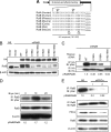
FIGURE 1.
FIGURE 1.
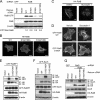
RalB is phosphorylated on serines 192 and 198 and PKCα is necessary for phosphorylation. A, serines 192 and 198 are evolutionarily conserved and include consensus PKC substrate motifs. Scansite high stringency analysis identified Ser-198 as a putative phosphorylation site for PKC isoforms α, β, γ, δ, and ϵ. Low stringency analysis identified Ser-192 as a putative phosphorylation site for PKC isoforms α, β, and γ and additionally Ser-192 and Ser-198 as a putative phosphorylation site for PKCζ. Ser-198 and to a lesser degree Ser-192 display similarity to the PKC consensus substrate motif of (R/K)(R/K)pSX(R/K), where pS is the phosphorylated serine residue, R/K is an arginine or lysine, and X is any amino acid. The C-terminal membrane-targeting regions of RalA and RalB were aligned using ClustalW. Phosphorylation sites are in boldface type, and the CAAX motif is underlined. B, ectopic restoration of endogenous RalB expression with putative phosphodeficient or phosphomimetic mutants of Ser-192 and Ser-198. Endogenous RalB expression was stably suppressed by pSuper retrovirus expression of control (GFP) or RalB shRNA in SW480 cells and then stably infected with empty pBabe-puro retrovirus vector or containing RNAi-insensitive cDNA sequences encoding HA epitope-tagged RalB WT or putative phosphorylation site mutants. Western blot analyses of total cell lysates were performed with anti-HA and anti-RalB and with anti-actin antibody to verify equivalent loading of protein. NS, nonspecific. C, Ser-192 and Ser-198 are the primary sites of RalB phosphorylation in vivo. HA-tagged WT or phosphodeficient RalB mutants were immunoprecipitated (IP) with an anti-HA antibody followed by separation using SDS-PAGE and by Western blot analyses with a pan-phosphoserine-specific antibody to determine RalB serine phosphorylation. IB, immunoblot. D, treatment with the bryostatin-1 (Bryo) PKC activator causes rapid and transient RalB serine phosphorylation. SW480 cells were stimulated with vehicle (DMSO) or 100 nm bryostatin-1 for the indicated times. Endogenous RalB was immunoprecipitated from harvested cell lysates, and RalB serine phosphorylation was determined by Western blotting with anti-Ser(P). Numbers represent the amount of Ser(P)-RalB normalized to time 0. Blot analyses for total RalB and β-actin were done to verify equivalent total protein loading. E, knockdown of endogenous PKCα inhibits RalB steady-state phosphorylation. Two independent shRNAs were used to knock down endogenous PKCα in SW480 cells. Endogenous RalB was immunoprecipitated; immune complexes were separated by SDS-PAGE, and Western blot analyses were performed with an anti-Ser(P) or anti-RalB antibody. Total cell lysates were blotted with anti-PKCα or anti-RalB or with anti-β-actin to verify equivalent protein loading.
FIGURE 2.
FIGURE 2.
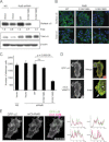
Ser-198 phosphorylation regulates RalB GTP-bound state, subcellular localization, and effector interaction. A, phosphodeficient S198A exhibits reduced GTP loading. SW480 cells stably infected with pSuper retrovirus vector expressing control (GFP) or RalB shRNA as well as the pBabe-puro empty vector or pBabe encoding HA-tagged RalB phosphorylation mutants were subjected to GST-Sec5-RBD pulldown analysis to monitor RalB-GTP formation. Total cell lysates were Western-blotted with anti-RalB or with anti-β-actin antibody to verify equivalent loading of protein. Numbers represent the amount of RalB-GTP normalized to WT RalB. B, bryostatin-1 treatment results in RalB endomembrane localization. SW480 cells stably expressing RalB shRNA as well as HA-tagged RalB WT or S192A/S198A were stimulated with DMSO or 100 nm bryostatin-1 for 10 min. RalB localization was visualized by confocal microscopy with anti-HA antibody. Scale bar, 20 μm. C, phosphorylation mutants of RalB exhibit different subcellular localizations. SW480 CRC cells from A were fixed, and RalB localization was visualized by confocal microscopy with anti-HA antibody. Scale bar, 20 μm. D, phosphorylation of Ser-198 regulates RalB endomembrane localization. SW480 cells transiently expressing GFP-tagged RalB WT or S198A were stimulated with DMSO (vehicle control) or 100 nm bryostatin-1 for 10 min. RalB localization was visualized by confocal microscopy. Scale bar, 20 μm. E, phosphorylation of RalB enhances RalBP1 association. SW480 cells from A were transiently transfected with GFP-RalBP1. HA-RalB was immunoprecipitated (IP), and immune complexes were separated by SDS-PAGE, and Western blot analyses were performed with anti-GFP to determine association with RalB, or with anti-HA and anti-β-actin antibodies to verify equivalent total protein loading. Data shown are representative of three independent experiments. F, phosphorylation of RalB is associated with decreased Sec5 association. SW480 cells from A were transiently transfected with GFP-Sec5. HA-RalB was immunoprecipitated, and immune complexes were separated by SDS-PAGE, and Western blot analyses were performed with anti-GFP antibody to determine co-precipitation with RalB or anti-HA and with anti-β-actin to verify equivalent total protein loading. G, phosphorylation of RalB is associated with decreased Sec8 association. HA-tagged RalB was immunoprecipitated from SW480 CRC cells from A. Immune complexes were separated by SDS-PAGE, and Western blot analyses were performed with anti-Sec8 to determine co-precipitation or anti-HA to verify equivalent protein loading. Total cell lysates were blotted with anti-HA, anti-Sec8, and anti-β-actin to verify equivalent protein expression. IB, immunoblot.
FIGURE 3.
FIGURE 3.
RalB phosphorylation regulates vesicular fusion. A, phosphorylation regulates the co-localization of RalB with the v-SNARE VAMP3. SW480 cells were transiently transfected with GFP-RalB WT or phosphorylation mutants and mCh-VAMP3. Insets indicate co-localization of RalB with VAMP3 at the plasma membrane (1) and on internal membranes (2). Scale bar represents 20 μm. B, phosphorylation of RalB regulates the association of VAMP3 and SNAP-23. SNAP-23 was immunoprecipitated (IP) from SW480 cells expressing RalB shRNA as well as empty vector or HA-tagged RalB phosphorylation mutants. Immune complexes were separated by SDS-PAGE, and Western blot analyses were performed with anti-VAMP3 to determine SNARE complex formation and anti-SNAP-23 to verify equivalent loading. C, phosphorylation of RalB regulates vesicular fusion. SW480 cells from Fig. 2A were transiently transfected with the TfR-mCh-SEP expression construct to monitor vesicular fusion. Fusions were determined by an increase in SEP signal due to surface delivery (n = 10 cells). Images shown are representative of independent fusion events denoted by the arrows. Values shown are means ± S.E., and an unpaired t test was used to determine significance (p < 0.05). NS, nonspecific.
FIGURE 4.
FIGURE 4.
Phosphorylation of RalB regulates the trafficking of α5-integrin. A, RalB phosphorylation alters α5 surface expression. Surface proteins were labeled with biotin in SW480 cells prepared as in Fig. 2A. Biotinylated proteins were isolated on neutravidin resin and resolved by SDS-PAGE. Western blot analyses were performed with anti-α5 antibody to determine the amount of biotinylated, surface-exposed α5. Total cell lysate was blotted with anti-α5 and with anti-β-actin to verify equivalent total protein loading. B, RalB phosphorylation regulates the surface expression of endogenous α5. α5 expression was determined in endogenous RalB-depleted SW480 cells as in Fig. 2A. Representative images are shown of cells that were either nonpermeabilized (for surface α5) or permeabilized (for total α5) and were stained with an Alexa 488-conjugated α5 antibody. C, cellular attachment to fibronectin is reduced in cells expressing phosphomimetic RalB. SW480 cells from Fig. 2A were allowed to attach to fibronectin. Nonadherent cells were removed, and attached cells were quantitated. Values shown are means ± S.E., and an unpaired t test was used to determine significance (p < 0.05). n.s., not significant. D, VAMP3 and α5 co-localize on endosomes and at the plasma membrane. SW480 cells were transiently transfected with mCh-VAMP3 and GFP-α5. Live cells were imaged, and co-localization of VAMP3 with α5 at the plasma membrane (arrow) and on endosomes (arrowhead) was seen. E, RalB and α5 co-localize on endosomes and at the plasma membrane. SW480 cells were transiently transfected with mCh-RalB and GFP-α5. Cells were seeded on glass coverslips for live cell imaging 48 h after transfection. Line scans of the merged image indicate co-localization of RalB with α5 on both internal vesicles and at the plasma membrane. Ten different images were quantitated and a Pearson's correlation coefficient of 0.83 ± 0.001 (S.E.) was calculated. Data are representative of at least two independent experiments. Scale bar, 20 μm.
FIGURE 5.
FIGURE 5.
Model for RalB-regulated vesicular fusion. Based on our observations that PKC-dependent phosphorylation promotes RalB translocation from the plasma membrane to endosomes and is associated with altered association with Sec5, we propose the following model for how these changes may then alter exocytosis, as shown by our tracking of α5-integrin to the cell surface. It is known that RalB is associated with the exocyst through its interaction with Sec5. An exocyst subcomplex consisting of Sec5, Exo84, Sec6, Sec8, Sec10, and Sec15 is docked to specific sites of vesicle fusion on the plasma membrane marked by the two additional exocyst components, Sec3 and Exo70. We propose that RalB-regulated exocyst docking occurs at fusion sites (Step 1). PKCα-mediated phosphorylation of RalB is then sufficient to disengage RalB from Sec5 and presumably the exocyst complex (Step 2). Next, we speculate that the v-SNARE VAMP3 engages its t-SNARE SNAP-23 (Step 3) leading to vesicular fusion (Step 4) and delivery of cargo proteins like α5-integrin (Step 5). Phosphorylated RalB is internalized where it may be dephosphorylated to participate in a new round of vesicle delivery.

Similar articles

Cited by

References

    1. Bodemann B. O., White M. A. (2008) Ral GTPases and cancer. Linchpin support of the tumorigenic platform. Nat. Rev. Cancer 8, 133–140 - PubMed
    1. Neel N. F., Martin T. D., Stratford J. K., Zand T. P., Reiner D. J., Der C. J. (2011) The RalGEF-Ral effector signaling network. The road less traveled for anti-Ras drug discovery. Genes Cancer 2, 275–287 - PMC - PubMed
    1. Lim K. H., O'Hayer K., Adam S. J., Kendall S. D., Campbell P. M., Der C. J., Counter C. M. (2006) Divergent roles for RalA and RalB in malignant growth of human pancreatic carcinoma cells. Curr. Biol. 16, 2385–2394 - PubMed
    1. Martin T. D., Samuel J. C., Routh E. D., Der C. J., Yeh J. J. (2011) Activation and involvement of Ral GTPases in colorectal cancer. Cancer Res. 71, 206–215 - PMC - PubMed
    1. Lim K. H., Baines A. T., Fiordalisi J. J., Shipitsin M., Feig L. A., Cox A. D., Der C. J., Counter C. M. (2005) Activation of RalA is critical for Ras-induced tumorigenesis of human cells. Cancer Cell 7, 533–545 - PubMed

Publication types