

B cell maintenance of subcapsular sinus macrophages protects against a fatal viral infection independent of adaptive immunity
- PMID: 22386268
- PMCID: PMC3359130
- DOI: 10.1016/j.immuni.2012.01.013
B cell maintenance of subcapsular sinus macrophages protects against a fatal viral infection independent of adaptive immunity
Abstract
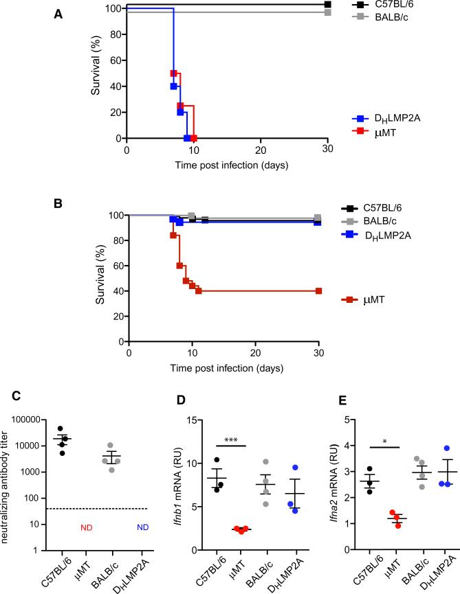
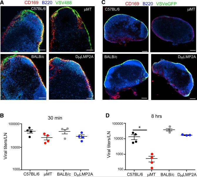
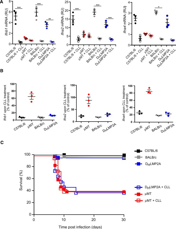
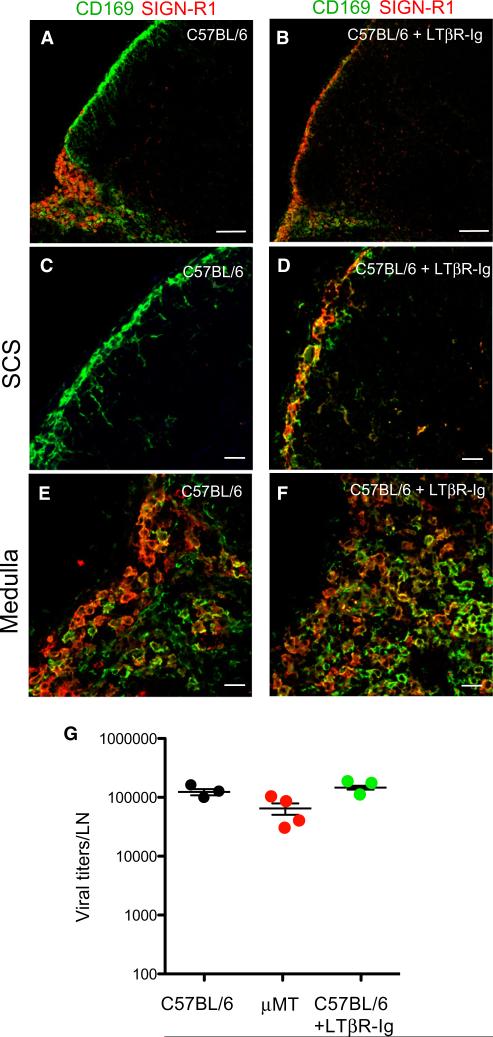
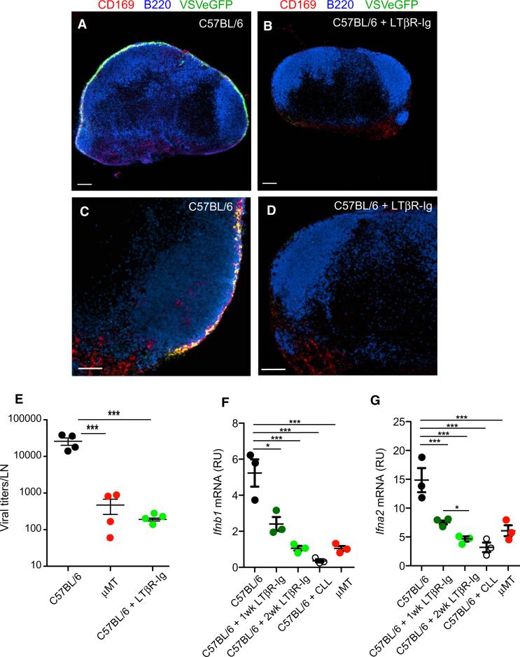
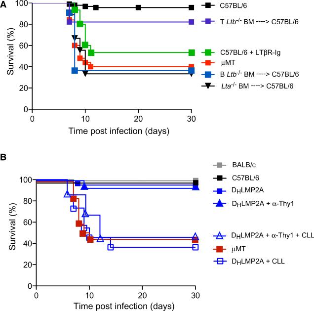
Neutralizing antibodies have been thought to be required for protection against acutely cytopathic viruses, such as the neurotropic vesicular stomatitis virus (VSV). Utilizing mice that possess B cells but lack antibodies, we show here that survival upon subcutaneous (s.c.) VSV challenge was independent of neutralizing antibody production or cell-mediated adaptive immunity. However, B cells were absolutely required to provide lymphotoxin (LT) α1β2, which maintained a protective subcapsular sinus (SCS) macrophage phenotype within virus draining lymph nodes (LNs). Macrophages within the SCS of B cell-deficient LNs, or of mice that lack LTα1β2 selectively in B cells, displayed an aberrant phenotype, failed to replicate VSV, and therefore did not produce type I interferons, which were required to prevent fatal VSV invasion of intranodal nerves. Thus, although B cells are essential for survival during VSV infection, their contribution involves the provision of innate differentiation and maintenance signals to macrophages, rather than adaptive immune mechanisms.
Copyright © 2012 Elsevier Inc. All rights reserved.
Figures

Comment in
-
B cells: An innate talent uncovered.Nat Rev Immunol. 2012 Mar 22;12(4):228-9. doi: 10.1038/nri3200. Nat Rev Immunol. 2012. PMID: 22437927 No abstract available.
-
B cells, not just for antibody anymore.Immunity. 2012 Mar 23;36(3):315-7. doi: 10.1016/j.immuni.2012.02.011. Immunity. 2012. PMID: 22444627 Free PMC article.
Similar articles
-
Subcapsular sinus macrophages prevent CNS invasion on peripheral infection with a neurotropic virus.Nature. 2010 Jun 24;465(7301):1079-83. doi: 10.1038/nature09118. Nature. 2010. PMID: 20577213 Free PMC article.
-
Tumor Necrosis Factor-Mediated Survival of CD169+ Cells Promotes Immune Activation during Vesicular Stomatitis Virus Infection.J Virol. 2018 Jan 17;92(3):e01637-17. doi: 10.1128/JVI.01637-17. Print 2018 Feb 1. J Virol. 2018. PMID: 29142134 Free PMC article.
-
Deficiency of the B cell-activating factor receptor results in limited CD169+ macrophage function during viral infection.J Virol. 2015 May;89(9):4748-59. doi: 10.1128/JVI.02976-14. Epub 2015 Feb 11. J Virol. 2015. PMID: 25673724 Free PMC article.
-
Subcapsular Sinus Macrophages: The Seat of Innate and Adaptive Memory in Murine Lymph Nodes.Trends Immunol. 2019 Jan;40(1):35-48. doi: 10.1016/j.it.2018.11.004. Epub 2018 Nov 27. Trends Immunol. 2019. PMID: 30502023 Review.
-
Lymph Node Subcapsular Sinus Macrophages as the Frontline of Lymphatic Immune Defense.Front Immunol. 2019 Feb 28;10:347. doi: 10.3389/fimmu.2019.00347. eCollection 2019. Front Immunol. 2019. PMID: 30891035 Free PMC article. Review.
Cited by
-
Peripheral prepositioning and local CXCL9 chemokine-mediated guidance orchestrate rapid memory CD8+ T cell responses in the lymph node.Immunity. 2013 Mar 21;38(3):502-13. doi: 10.1016/j.immuni.2012.11.012. Epub 2013 Jan 24. Immunity. 2013. PMID: 23352234 Free PMC article.
-
Lymph node macrophages drive innate immune responses to enhance the anti-tumor efficacy of mRNA vaccines.Mol Ther. 2024 Mar 6;32(3):704-721. doi: 10.1016/j.ymthe.2024.01.020. Epub 2024 Jan 18. Mol Ther. 2024. PMID: 38243602
-
Tumor-Draining Lymph Node Reconstruction Promotes B Cell Activation During E0771 Mouse Breast Cancer Growth.Front Pharmacol. 2022 Mar 28;13:825287. doi: 10.3389/fphar.2022.825287. eCollection 2022. Front Pharmacol. 2022. PMID: 35418862 Free PMC article.
-
The identification and developmental requirements of colonic CD169⁺ macrophages.Immunology. 2014 Jun;142(2):269-78. doi: 10.1111/imm.12251. Immunology. 2014. PMID: 24883436 Free PMC article.
-
Linking the microbiota and metabolic disease with lymphotoxin.Int Immunol. 2013 Jul;25(7):397-403. doi: 10.1093/intimm/dxt018. Epub 2013 May 8. Int Immunol. 2013. PMID: 23657002 Free PMC article. Review.
References
-
- Agyekum S, Church A, Sohail M, Krausz T, Van Noorden S, Polak J, Cohen J. Expression of lymphotoxin-beta (LT-beta) in chronic inflammatory conditions. J. Pathol. 2003;199:115–121. - PubMed
-
- Rabies vaccine failures. Lancet. 1988;1:917–918. Anonymous. - PubMed
-
- Bach P, Kamphuis E, Odermatt B, Sutter G, Buchholz CJ, Kalinke U. Vesicular stomatitis virus glycoprotein displaying retrovirus-like particles induce a type I IFN receptor-dependent switch to neutralizing IgG antibodies. J. Immunol. 2007;178:5839–5847. - PubMed
-
- Bachmann MF, Kündig TM, Hengartner H, Zinkernagel RM. Regulation of IgG antibody titers by the amount persisting of immune-complexed antigen. Eur. J. Immunol. 1994;24:2567–2570. - PubMed
-
- Bachmann MF, Hengartner H, Zinkernagel RM. T helper cell-independent neutralizing B cell response against vesicular stomatitis virus: role of antigen patterns in B cell induction? Eur. J. Immunol. 1995;25:3445–3451. - PubMed
Publication types
MeSH terms
Substances
Grants and funding
- P01 CA071932/CA/NCI NIH HHS/United States
- AI078897/AI/NIAID NIH HHS/United States
- 5T32-HL07623-20/HL/NHLBI NIH HHS/United States
- R01 AI069259-08/AI/NIAID NIH HHS/United States
- P01 AI078897/AI/NIAID NIH HHS/United States
- AI069259/AI/NIAID NIH HHS/United States
- T32 HL007623/HL/NHLBI NIH HHS/United States
- AI072252/AI/NIAID NIH HHS/United States
- P30 AI060354/AI/NIAID NIH HHS/United States
- P01 AI078897-05/AI/NIAID NIH HHS/United States
- R01 AI072252/AI/NIAID NIH HHS/United States
- R01 AI069259/AI/NIAID NIH HHS/United States
LinkOut - more resources
Full Text Sources
Other Literature Sources
Molecular Biology Databases
