. 2012 Mar 2;148(5):947-57.
doi: 10.1016/j.cell.2012.01.045.

Order out of disorder: working cycle of an intrinsically unfolded chaperone

Affiliations

Order out of disorder: working cycle of an intrinsically unfolded chaperone

Dana Reichmann et al. Cell. .

Abstract

The redox-regulated chaperone Hsp33 protects organisms against oxidative stress that leads to protein unfolding. Activation of Hsp33 is triggered by the oxidative unfolding of its own redox-sensor domain, making Hsp33 a member of a recently discovered class of chaperones that require partial unfolding for full chaperone activity. Here we address the long-standing question of how chaperones recognize client proteins. We show that Hsp33 uses its own intrinsically disordered regions to discriminate between unfolded and partially structured folding intermediates. Binding to secondary structure elements in client proteins stabilizes Hsp33's intrinsically disordered regions, and this stabilization appears to mediate Hsp33's high affinity for structured folding intermediates. Return to nonstress conditions reduces Hsp33's disulfide bonds, which then significantly destabilizes the bound client proteins and in doing so converts them into less-structured, folding-competent client proteins of ATP-dependent foldases. We propose a model in which energy-independent chaperones use internal order-to-disorder transitions to control substrate binding and release.


Figures

Figure 1
Figure 1. Peptide-Binding Specificity of Active Hsp33ox
(A) A peptide array was incubated with inactive Hsp33 (Hsp33red) or active Hsp33 (Hsp33ox), and Hsp33 was visualized with Cy5-fluorescently labeled anti-Hsp33 antibodies. False-color imaging of the signal intensities ranges from blue (no binding) to red (maximal binding). The order of overlapping peptides in the array, including normalized binding scores, is shown for select NuoCD peptides. Sequences of peptides that comprise the good binder PepNuoC are shown in bold red. (B–D) Relative amino acid occurrence (B), net charge (C), and hydrophilicity (D) in good binders (red) and nonbinders (blue) were calculated and normalized to the occurrence of the same amino acid or feature distribution in the entire library. A value of 100% indicates that occurrence of a specific amino acid is identical to occurrence in the total peptide library. See also Figure S1 and Table S1.
Figure 2
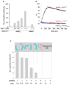
Figure 2. Validation of Peptide Binding in Solution and KD Measurements
(A) Competitor activity of PepNuoC was assessed by determining its ability to compete with 0.075 μM chemically unfolded CS for binding to 0.15 μM Hsp33ox or 0.375 μM DnaK/0.075 μM DnaJ. Complexes between chaperones and competitors were allowed to form for 4 min before chemically denatured CS was added. Light-scattering measurements of CS at 30°C were conducted as read-out for competitor activity. The light-scattering signal of CS in the presence of fully active chaperones (i.e., absence of competitor) was set to 0% competitor activity, whereas the light-scattering signal of CS in the absence of functional chaperone (i.e., full competition) was set to 100%. Presence of PepNuoC had no significant influence on the aggregation behavior of CS in the absence of chaperones. (B) SPR sensorgrams to monitor interactions between Hsp33ox or Hsp33red and immobilized PepNuoC or oxidized Hsp33C-term. The data were fitted (black symbols) using BIAevaluation software. (C) Competitor activity of predicted Hsp33 good binder peptides and nonbinder peptides was assessed by determining their ability to compete with 0.075 μM chemically unfolded CS for binding to 0.15 μM Hsp33ox. Light-scattering measurements were conducted as described above. The upper panel illustrates the NMR structures of the respective peptides according to PDB (Table S2B). Far-UV CD spectroscopy revealed an α helix for Pep1 and random coils for Pep6–8 (see Figure S2D). See also Figure S2 and Table S2.
Figure 3
Figure 3. Hsp33 and DnaK Show Distinct Peptide-Binding Specificity
(A) Comparison of secondary structure features in good binders and nonbinders of Hsp33ox with good binders of DnaK. Analysis of DnaK’s non-binders was not possible due to the semi-quantitative nature of the cellulose peptide array used to collect this data (Rüdiger et al., 1997). (B) Influence of 0.325 μM Hsp33C-term or α-casein on the ability of 0.13 μM active Hsp33ox or 0.65 μM DnaK /0.13 μM DnaJ to suppress the aggregation of 0.065 μM chemically denatured luciferase at 30°C. Competitor activity was determined as described above. (C) Time course of Arc unfolding and competitor activity. Arc unfolding was initiated by diluting Arc (0.75 μM) into HEPES buffer (pH 7.5) at 30°C. At distinct time points after start of the unfolding process, 0.15 μM active Hsp33ox or 0.187 μM DnaK/0.075 μM DnaJ was added to allow complex formation. After 10 s, 0.075 μM chemically unfolded CS was added. Light scattering of CS was monitored and competitor activity of Arc was determined as described above. Inset: Unfolding of 0.75 μM Arc was monitored by following the fluorescence of its single tryptophan residue located at the monomer-monomer interface upon dilution into buffer. Arrows indicate the time points at which Arc was analyzed for competitor activity. See also Figure S3.
Figure 4
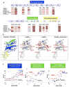
Figure 4. Hsp33’s Intrinsically Unfolded Linker Region Is involved in Substrate Binding
(A) Inactive Hsp33red, active Hsp33ox, and Hsp33ox in complex with thermally unfolded luciferase [Hsp33ox-S] before or after reduction with DTT ([Hsp33ox-S]red) were digested with trypsin for 5, 10, or 15 min, and cleavage products were analyzed by LC-MS. The most accessible sites (cleaved within 5 min) are indicated in dark red, and sites that were not cleaved within 15 min are indicated in gray. Peptides with very low abundance are indicated with a star. Color-coded secondary structure elements of Hsp33 are shown for reference. (B) Differences in the proteolytic patterns of Hsp33red, Hsp33ox, and Hsp33ox-substrate were mapped onto the modeled E. coli Hsp33 structure. Relative accessibility of the individual sites is indicated by color intensity, with the most accessible residues shown in dark red and the least accessible shown in gray. Unaltered trypsin cleavage sites are indicated in light blue. The color-coded domain structure of Hsp33 with labeled secondary structure elements is shown for reference. (C) Representative SUPREX curves of two fragments from the flexible linker region (aa 174–192, aa 203–221, after 5 min H/D-exchange) and one peptide from the stable N-terminal region (aa 26–47, after 2 min H/D exchange) obtained from Hsp33red (black), Hsp33ox (red), or the Hsp33ox-CS complex (blue). Solid lines are best fits of the data to a four-parameter sigmoidal equation, used to obtain C1/2 values (Table S3). See also Figure S4 and Table S3.
Figure 5
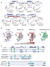
Figure 5. Reduction of Hsp33-Substrate Complexes Destabilizes Bound Substrate Proteins
(A) Representative SUPREX curves of CS fragments obtained from native CS (black) or thermally unfolded CS in complex with Hsp33ox before (red) or after (blue) complex reduction with 5 mM DTT. Solid lines represent best fits of the data. Secondary structure elements of CS are shown for reference. Peptide regions that show a significant change in relative thermodynamic stability are indicated by red bars, whereas regions that do not significantly change are indicated by pink bars (see also Table S4). Regions of CS defined as good binders by peptide array are shown by dark cyan bars, whereas regions of CS involved in monomer-monomer interactions are indicated by purple bars. Stars depict active site residues. (B) Comparison of the relative thermodynamic stability profile of CS peptides obtained from SUPREX analysis in native CS, in thermally unfolded CS in complex with Hsp33ox ([Hsp33ox-CS]), or upon DTT reduction ([Hsp33-CS]red). All identified peptides were mapped onto the CS structure (PDB: 2CTS), and changes in relative thermodynamic stability are depicted as color changes from pink to red as stability decreases. Right panel: Localization of Hsp33’s good binders in CS structure as defined by peptide array is depicted in green. (C) Comparison of limited proteolysis profiles of CS, MDH, or luciferase either in native form or upon thermal unfolding in the presence of Hsp33ox before or after reduction with DTT. Peptides detected in native form and upon complex formation are depicted in green, peptides found in complex both before and after reduction are shown in blue, and peptides that are only detected after complex reduction are indicated in orange. Regions of CS defined as good binders by peptide array are indicated by dark cyan bars. Peptides of CS identified in SUPREX analysis are indicated by red and pink bars, respectively (see Figure 5A for reference). See also Figure S5 and Table S4.
Figure 6
Figure 6. Schematic Model of Hsp33 as an Example of Stress-Specific Intrinsically Disordered Chaperones
Hsp33 senses unfolding conditions by converting stably folded regions into intrinsically disordered segments (green and cyan), which participate directly in the highly flexible binding of a wide variety of early unfolding intermediates. Client protein binding confers stability to intrinsically disordered regions of the chaperone. Return to nonstress conditions further stabilizes the chaperone (cyan regions), leading to destabilization/unfolding of the bound client protein, which appears to be necessary for substrate release and refolding by foldases. To simplify the model, the dimerization equilibrium of active Hsp33 was omitted.

Comment in

Similar articles

Cited by

References

    1. Bowie JU, Sauer RT. Equilibrium dissociation and unfolding of the Arc repressor dimer. Biochemistry. 1989;28:7139–7143. - PubMed
    1. Chen Z, Kurt N, Rajagopalan S, Cavagnero S. Secondary structure mapping of DnaK-bound protein fragments: chain helicity and local helix unwinding at the binding site. Biochemistry. 2006;45:12325–12333. - PubMed
    1. Cremers CM, Reichmann D, Hausmann J, Ilbert M, Jakob U. Unfolding of metastable linker region is at the core of Hsp33 activation as a redox-regulated chaperone. J Biol Chem. 2010;285:11243–11251. - PMC - PubMed
    1. Deuerling E, Schulze-Specking A, Tomoyasu T, Mogk A, Bukau B. Trigger factor and DnaK cooperate in folding of newly synthesized proteins. Nature. 1999;400:693–696. - PubMed
    1. Gaspar AM, Appavou MS, Busch S, Unruh T, Doster W. Dynamics of well-folded and natively disordered proteins in solution: a time-of-flight neutron scattering study. Eur Biophys J. 2008;37:573–582. - PubMed

Publication types

MeSH terms

LinkOut - more resources