

Neuroimaging signatures of frontotemporal dementia genetics: C9ORF72, tau, progranulin and sporadics
- PMID: 22366795
- PMCID: PMC3286334
- DOI: 10.1093/brain/aws001
Neuroimaging signatures of frontotemporal dementia genetics: C9ORF72, tau, progranulin and sporadics
Abstract
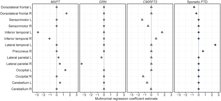
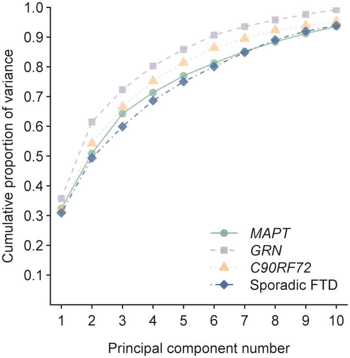
A major recent discovery was the identification of an expansion of a non-coding GGGGCC hexanucleotide repeat in the C9ORF72 gene in patients with frontotemporal dementia and amyotrophic lateral sclerosis. Mutations in two other genes are known to account for familial frontotemporal dementia: microtubule-associated protein tau and progranulin. Although imaging features have been previously reported in subjects with mutations in tau and progranulin, no imaging features have been published in C9ORF72. Furthermore, it remains unknown whether there are differences in atrophy patterns across these mutations, and whether regional differences could help differentiate C9ORF72 from the other two mutations at the single-subject level. We aimed to determine the regional pattern of brain atrophy associated with the C9ORF72 gene mutation, and to determine which regions best differentiate C9ORF72 from subjects with mutations in tau and progranulin, and from sporadic frontotemporal dementia. A total of 76 subjects, including 56 with a clinical diagnosis of behavioural variant frontotemporal dementia and a mutation in one of these genes (19 with C9ORF72 mutations, 25 with tau mutations and 12 with progranulin mutations) and 20 sporadic subjects with behavioural variant frontotemporal dementia (including 50% with amyotrophic lateral sclerosis), with magnetic resonance imaging were included in this study. Voxel-based morphometry was used to assess and compare patterns of grey matter atrophy. Atlas-based parcellation was performed utilizing the automated anatomical labelling atlas and Statistical Parametric Mapping software to compute volumes of 37 regions of interest. Hemispheric asymmetry was calculated. Penalized multinomial logistic regression was utilized to create a prediction model to discriminate among groups using regional volumes and asymmetry score. Principal component analysis assessed for variance within groups. C9ORF72 was associated with symmetric atrophy predominantly involving dorsolateral, medial and orbitofrontal lobes, with additional loss in anterior temporal lobes, parietal lobes, occipital lobes and cerebellum. In contrast, striking anteromedial temporal atrophy was associated with tau mutations and temporoparietal atrophy was associated with progranulin mutations. The sporadic group was associated with frontal and anterior temporal atrophy. A conservative penalized multinomial logistic regression model identified 14 variables that could accurately classify subjects, including frontal, temporal, parietal, occipital and cerebellum volume. The principal component analysis revealed similar degrees of heterogeneity within all disease groups. Patterns of atrophy therefore differed across subjects with C9ORF72, tau and progranulin mutations and sporadic frontotemporal dementia. Our analysis suggested that imaging has the potential to be useful to help differentiate C9ORF72 from these other groups at the single-subject level.
Figures


Comment in
-
The C9ORF72 syndrome: implications for clinical practice.J Neurol. 2012 Apr;259(4):794-6. doi: 10.1007/s00415-012-6479-5. J Neurol. 2012. PMID: 22430253 No abstract available.
Similar articles
-
The clinical and pathological phenotype of C9ORF72 hexanucleotide repeat expansions.Brain. 2012 Mar;135(Pt 3):723-35. doi: 10.1093/brain/awr353. Epub 2012 Feb 1. Brain. 2012. PMID: 22300876
-
Brain atrophy over time in genetic and sporadic frontotemporal dementia: a study of 198 serial magnetic resonance images.Eur J Neurol. 2015 May;22(5):745-52. doi: 10.1111/ene.12675. Epub 2015 Feb 12. Eur J Neurol. 2015. PMID: 25683866 Free PMC article.
-
Characterization of frontotemporal dementia and/or amyotrophic lateral sclerosis associated with the GGGGCC repeat expansion in C9ORF72.Brain. 2012 Mar;135(Pt 3):765-83. doi: 10.1093/brain/aws004. Brain. 2012. PMID: 22366793 Free PMC article.
-
Phenotypic signatures of genetic frontotemporal dementia.Curr Opin Neurol. 2011 Dec;24(6):542-9. doi: 10.1097/WCO.0b013e32834cd442. Curr Opin Neurol. 2011. PMID: 21986680 Review.
-
Frontotemporal dementia.Handb Clin Neurol. 2018;148:409-430. doi: 10.1016/B978-0-444-64076-5.00027-2. Handb Clin Neurol. 2018. PMID: 29478591 Review.
Cited by
-
Divergent brain functional network alterations in dementia with Lewy bodies and Alzheimer's disease.Neurobiol Aging. 2015 Sep;36(9):2458-67. doi: 10.1016/j.neurobiolaging.2015.05.015. Epub 2015 Jun 2. Neurobiol Aging. 2015. PMID: 26115566 Free PMC article.
-
A pan-European study of the C9orf72 repeat associated with FTLD: geographic prevalence, genomic instability, and intermediate repeats.Hum Mutat. 2013 Feb;34(2):363-73. doi: 10.1002/humu.22244. Epub 2013 Jan 4. Hum Mutat. 2013. PMID: 23111906 Free PMC article.
-
Diffusion tensor tractography versus volumetric imaging in the diagnosis of behavioral variant frontotemporal dementia.PLoS One. 2013 Jul 18;8(7):e66932. doi: 10.1371/journal.pone.0066932. Print 2013. PLoS One. 2013. PMID: 23874403 Free PMC article.
-
Assessment of psychiatric changes in C9ORF72 frontotemporal dementia.Alzheimers Res Ther. 2012 Dec 24;4(6):49. doi: 10.1186/alzrt152. eCollection 2012. Alzheimers Res Ther. 2012. PMID: 23269019 Free PMC article.
-
The Association Between Neurocognitive Disorders and Gustatory Dysfunction: A Systematic Review and Meta-Analysis.Neuropsychol Rev. 2024 Mar;34(1):192-213. doi: 10.1007/s11065-023-09578-3. Epub 2023 Feb 20. Neuropsychol Rev. 2024. PMID: 36806051 Free PMC article.
References
-
- Agresti A. New York: Wiley-Interscience; 2002. Categorical data analysis.
-
- Al-Sarraj S, King A, Troakes C, Smith B, Maekawa S, Bodi I, et al. p62 positive, TDP-43 negative, neuronal cytoplasmic and intranuclear inclusions in the cerebellum and hippocampus define the pathology of C9orf72-linked FTLD and MND/ALS. Acta Neuropathol. 2011;122:691–702. - PubMed
-
- Arvanitakis Z, Witte RJ, Dickson DW, Tsuboi Y, Uitti RJ, Slowinski J, et al. Clinical-pathologic study of biomarkers in FTDP-17 (PPND family with N279K tau mutation) Parkinsonism Relat Disord. 2007;13:230–9. - PubMed
-
- Ashburner J, Friston KJ. Voxel-based morphometry–the methods. Neuroimage. 2000;11:805–21. - PubMed
-
- Ashburner J, Friston KJ. Unified segmentation. Neuroimage. 2005;26:839–51. - PubMed
Publication types
MeSH terms
Substances
Grants and funding
- P50 AG16574/AG/NIA NIH HHS/United States
- R01 AG11378/AG/NIA NIH HHS/United States
- P50 AG016574/AG/NIA NIH HHS/United States
- NS057567/NS/NINDS NIH HHS/United States
- U01 AG03949/AG/NIA NIH HHS/United States
- U01 AG024904/AG/NIA NIH HHS/United States
- P50-AG25711/AG/NIA NIH HHS/United States
- P50 NS072187-01S2/NS/NINDS NIH HHS/United States
- U01 AG06786/AG/NIA NIH HHS/United States
- R01 DC010367/DC/NIDCD NIH HHS/United States
- U24 AG026395/AG/NIA NIH HHS/United States
- R56 AG26251-03/AG/NIA NIH HHS/United States
- 1RC2NS070276/NS/NINDS NIH HHS/United States
- R01 HL70825/HL/NHLBI NIH HHS/United States
- R01-AG023195/AG/NIA NIH HHS/United States
- P01-AG17216/AG/NIA NIH HHS/United States
- R21 AG38736/AG/NIA NIH HHS/United States
- R01 AG011378/AG/NIA NIH HHS/United States
- R01-AG26251/AG/NIA NIH HHS/United States
- P50-NS40256/NS/NINDS NIH HHS/United States
- R01 NS065782/NS/NINDS NIH HHS/United States
- R01-NS65782/NS/NINDS NIH HHS/United States
- R01 NS065782-01/NS/NINDS NIH HHS/United States
- R01 AG037491/AG/NIA NIH HHS/United States
- P01 AG003949/AG/NIA NIH HHS/United States
- R01-AG15866/AG/NIA NIH HHS/United States
LinkOut - more resources
Full Text Sources
Other Literature Sources
Miscellaneous
