. 2012 Feb 24;19(2):228-42.
doi: 10.1016/j.chembiol.2011.12.017.

Rational design of small molecule inhibitors targeting the Rac GTPase-p67(phox) signaling axis in inflammation

Affiliations

Rational design of small molecule inhibitors targeting the Rac GTPase-p67(phox) signaling axis in inflammation

Emily E Bosco et al. Chem Biol. .

Abstract

The NADPH oxidase enzyme complex, NOX2, is responsible for reactive oxygen species production in neutrophils and has been recognized as a key mediator of inflammation. Here, we have performed rational design and in silico screen to identify a small molecule inhibitor, Phox-I1, targeting the interactive site of p67(phox) with Rac GTPase, which is a necessary step of the signaling leading to NOX2 activation. Phox-I1 binds to p67(phox) with a submicromolar affinity and abrogates Rac1 binding and is effective in inhibiting NOX2-mediated superoxide production dose-dependently in human and murine neutrophils without detectable toxicity. Medicinal chemistry characterizations have yielded promising analogs and initial information of the structure-activity relationship of Phox-I1. Our studies suggest the potential utility of Phox-I class inhibitors in NOX2 oxidase inhibition and present an application of rational targeting of a small GTPase-effector interface.


Conflict of interest statement

The authors have no conflict of interest to declare.

Figures
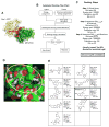
Figure 1
Figure 1. Virtual screening of p67phox inhibitors from the ZINC and UC DDC small molecule libraries
(A) The structure of the Rac1-p67phox complex is shown. Rac1 is shown in yellow, whereas p67phox is shown in green, respectively. Arginine residues 38 and 102 of p67phox, which surround the site within the interaction interface that was targeted by virtual screening, are shown using red spheres. Rac1 residues 25–27 (Thr-Ans-Ala motif in Switch I) that are buried upon complex formation and are sandwiched by Arg38 and Arg102 are shown in magenta. Arg38 is directly involved in interface formation. (B) The flow chart reports the strategy that was implemented for automated docking. Autodock 4 and related scripts were utilized to assign charges and perform docking energy calculations. (C) The main docking parameters adopted in the three different phases of the screening are displayed. The molecular geometry, cluster convergence, hydrogen bonding and lowest predicted docking energy criteria have been used to select the best 50% of candidates coming out from the third screening step. (D) Molecular surface representation of p67phox in complex with a predicted inhibitor is displayed. The region comprising p67phox Arginine 102 and 38 is defined by our grids and is highlighted in yellow. Docked Energy: −8.77Kcal/mol, Binding energy: −8.90 Kcal/mol, Ki 2.9×10⁻⁷ M−1. (E) A summary of the candidate inhibitors specific for p67phox; their Lipinski parameters and predicted Ki are reported on the side of each molecule.
Figure 2
Figure 2. Binding affinity and specificity of Phox-I1 to p67phox
(A) Using microscale thermophoresis, p67phox recombinant protein (1–200) was able to bind Phox-I1 with an Kd of ~100nM. (B) Similar to A, the ability of Phox-I1 to bind a recombinant mutant of p67phox at the site critical for Rac1-GTP binding, p67R38Q, was tested by microscale thermophoresis. (C) Experiment described in B was repeated with a random p67phox mutation, R188A. (D) Constitutively active Rac1V12 mutant protein binds p67phox with an Kd of ~40 nM using microscale thermophoresis. (E) However, Rac1 wild type protein (predominantly in GDP- bound state) cannot bind p67phox using microscale thermophoresis. (F) Competitive binding of p67phox with Phox-I1 or vehicle control, followed by titration of RacV12 protein using above methods. (G) Phox-I1 is unable to bind RacV12 recombinant protein via above technique.
Figure 3
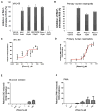
Figure 3. fMLP- stimulated ROS production is abrogated by Phox-I1 in human HL-60 cells and primary murine neutrophils
(A) Ability of Phox-I1 to inhibit ROS production in fMLP- stimulated differentiated HL-60 cells as compared to standard ROS inhibitors was assessed by H2-DCFDA staining and FACS analysis. Levels of ROS production in non-fMLP treated controls were subtracted from all samples, data was then normalized to fMLP- stimulated vehicle treated control. (B) Experiment described in A was repeated with primary murine neutrophils. (C) As described in A, HL-60 cells were treated with various concentrations of Phox-I1 and an IC50 curve was generated. (D) Dose response of fMLP-induced ROS production to Phox-I1 by primary human neutrophils. Levels of ROS production in non-fMLP or fMLP-stimulated human neutrophils were assayed by the luminol chemiluminescence method in increasing concentrations of Phox-I1. Data was normalized to fMLP- stimulated vehicle treated control. (E) Effect of Phox-I on glucose oxidase-generated ROS. (F) Effect of Phox-I1 on PMA induced ROS production in human neutrophils assayed by the luminol chemiluminescence method.
Figure 4
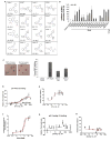
Figure 4. Phox-I1-analog analysis yields compounds with improved or similar cellular ROS inhibitory activity
(A) List of analogs derived from a search of the UCDDC and ZINC compound libraries for Phox-I1-like structures with medicinal chemistry optimized features. (B) H2-DCFDA staining in fMLP-stimulated dHL-60 cells treated with Phox-I1 analogs. Analog 4, 10 and 16 (Phox-I2) all display improved ROS inhibition over Phox1. (C) Freshly isolated primary murine neutrophils were stimulated with fMLP to initiate ROS production, cells were then treated with DMSO control, DPI, Phox-I1, or Phox-I2 and a Nitroblue tetrazolium (NBT) assay was performed and imaged (left panel). Blue stain is superoxide anion, pink stain is neutrophil nucleus. Cells displaying ROS production were quantified from the images and non-fMLP treated ROS levels were subtracted prior to normalization to vehicle control treated sample (right panel). (D) Using microscale thermophoresis, p67phox protein binds to Phox-I2 with high affinity. (E) IC50 for Phox-I2 was assayed by H2-DCFDA ROS production method in dHL-60 cells. (F) Phox-I2 dosage response of ROS production in human neutrophils assayed by luminol chemiluminescence. (G) As a negative control, Analog 13 was unable to bind p67phox. (H) Anolag 13 showed no cellular ROS inhibitory activity in HL-60 cells.
Figure 5
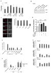
Figure 5. Phox-I1 and Phox-I2 show undetectable toxicity and site effects
(A) Apoptosis analysis by FACS of HL-60 cells treated with compound or vehicle control for 2 hours prior to 7-AAD and Annexin V staining. (B) HL-60 cells from A were harvested and lysates were immunoblotted for levels of pPAK, and actin was used as a control for loading. (C) F-actin reorganization in freshly isolated fMLP-stimulated primary murine neutrophils was analyzed. Representative images (left panel) and quantification (right panel) are displayed. Treatment with Analog 13 is included as a “dead analog” that possesses no intrinsic ROS inhibitory activity, and nocodazole is included as a positive control for actin disruption. Cells were exposed to a 10μM dose of Phox-I1, Phox-I2, and Analog 13, and 200nM nocodazole. (D) The effect of Phox-I on NOX4 mediated ROS production was tested in primary murine neutrophils transfected with a NOX4 expressing plasmid. 10 uM Phox-I1 was applied to the cells for 30 min prior to ROS assay by luminol chemiluminescence in the presence of HRP. The fMLP-stimulated ROS activity in the presence or absence of 10 uM Phox-I1 was measured in parallel. (E) The antioxidant abilities of these lead compounds were tested by prestimulating dHL-60 cells with fMLP for 30 minutes prior to treatment with Phox-I1 or Phox-I2. Levels of superoxide were analyzed by DCFDA assay and FACS. NAC, apocyanin, DPI, and NSC23766 treated cells served as controls. (F) For affinity assay, DMSO-differentiated HL-60 cells were treated with standard effective dose of indicated compound for 2 hours, washed, and allowed to recover in normal media for 4 hours or 2 hours prior to fMLP stimulation and DCFDA ROS production assay by FACS analysis. (G) For stability assay, DMSO-differentiated HL-60 cells were treated with 20μM dose of compound for the indicated time period prior to fMLP stimulation and DCFDA ROS production assay by FACS analysis. 30 minute and 18 hour time periods are not displayed because they revealed no ROS inhibition.
Figure 6
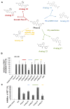
Figure 6. Medicinal chemistry optimization of Phox-I2 allows for the replacement of potentially toxic nitro groups
(A) Compounds with similar structures to Phox-I2 were synthesized and broken down into 4 different categories; 1. NO2 position scan, 2. NO2 substitution, 3. Addition of an aromatic ring (rendering it similar to Phox1), 4. Aromatic Phox2. (B) DCFDA FACS analysis was performed using differentiated HL-60 cells treated for 2 hours with compounds from A prior to stimulation with fMLP. (C) Freshly isolated primary murine neutrophils were stimulated with fMLP to initiate ROS production, cells were then treated with DMSO control, DPI, Phox-I2, Analogs 20, 21, 22, 23. A Nitroblue tetrazolium assay was performed and imaged in order to quantitate superoxide inhibition. Non-fMLP treated ROS levels were subtracted prior to normalization to vehicle control treated sample.

Comment in

  • Exploiting effectors of Rac GTPase.
    Jo H, Luo HR. Jo H, et al. Chem Biol. 2012 Feb 24;19(2):169-71. doi: 10.1016/j.chembiol.2012.02.001. Chem Biol. 2012. PMID: 22365599 Free PMC article.

Similar articles

Cited by

References

    1. Abo A, Pick E, Hall A, Totty N, Teahan CG, Segal AW. Activation of the NADPH oxidase involves the small GTP-binding protein p21rac1. Nature. 1991;353:668–670. - PubMed
    1. Ahmed S, Prigmore E, Govind S, Veryard C, Kozma R, Wientjes FB, Segal AW, Lim L. Cryptic Rac-binding and p21(Cdc42Hs/Rac)-activated kinase phosphorylation sites of NADPH oxidase component p67(phox) J Biol Chem. 1998;273:15693–15701. - PubMed
    1. Aldieri E, Riganti C, Polimeni M, Gazzano E, Lussiana C, Campia I, Ghigo D. Classical inhibitors of NOX NAD(P)H oxidases are not specific. Curr Drug Metab. 2008;9:686–696. - PubMed
    1. Armitage ME, Wingler K, Schmidt HH, La M. Translating the oxidative stress hypothesis into the clinic: NOX versus NOS. J Mol Med. 2009;87:1071–1076. - PMC - PubMed
    1. Bedard K, Krause KH. The NOX family of ROS-generating NADPH oxidases: physiology and pathophysiology. Physiol Rev. 2007;87:245–313. - PubMed

Publication types

MeSH terms