. 2012:4:61.
doi: 10.3389/fnmol.2011.00061. Epub 2012 Jan 10.

Fibroblast growth factor 22 contributes to the development of retinal nerve terminals in the dorsal lateral geniculate nucleus

Affiliations

Fibroblast growth factor 22 contributes to the development of retinal nerve terminals in the dorsal lateral geniculate nucleus

Rishabh Singh et al. Front Mol Neurosci. 2012.

Abstract

At least three forms of signaling between pre- and postsynaptic partners are necessary during synapse formation. First, "targeting" signals instruct presynaptic axons to recognize and adhere to the correct portion of a postsynaptic target cell. Second, trans-synaptic "organizing" signals induce differentiation in their synaptic partner so that each side of the synapse is specialized for synaptic transmission. Finally, in many regions of the nervous system an excess of synapses are initially formed, therefore "refinement" signals must either stabilize or destabilize the synapse to reinforce or eliminate connections, respectively. Because of both their importance in processing visual information and their accessibility, retinogeniculate synapses have served as a model for studying synaptic development. Molecular signals that drive retinogeniculate "targeting" and "refinement" have been identified, however, little is known about what "organizing" cues are necessary for the differentiation of retinal axons into presynaptic terminals. To identify such "organizing" cues, we used microarray analysis to assess whether any target-derived "synaptic organizers" were enriched in the mouse dorsal lateral geniculate nucleus (dLGN) during retinogeniculate synapse formation. One candidate "organizing" molecule enriched in perinatal dLGN was FGF22, a secreted cue that induces the formation of excitatory nerve terminals in muscle, hippocampus, and cerebellum. In FGF22 knockout mice, the development of retinal terminals in dLGN was impaired. Thus, FGF22 is an important "organizing" cue for the timely development of retinogeniculate synapses.


Figures

Figure 1
Figure 1
Synaptic development in mouse dLGN. (A–D) VGluT2-immunoreactivity in coronal sections of dLGN at P3, P7, P14, P21. dLGN are encircled by white dots. Arrows indicate the intergeniculate leaflet (IGL). Tissue orientation is indicated in (A): D, dorsal; L, lateral; M, medial; V, ventral. (E–H) High magnification of (A–D) [from areas similar to that shown by the white box in (A)] show the transformation of immature and small VGluT2-positive retinal terminals into large, morphologically distinct RLPs. (E′–H′) show the development of Gad67 inhibitory synapses in the postnatal dLGN. (E″–H″) show merged images of retinal terminals (red) and inhibitory terminals (green). Arrows in (G,H) highlight presumptive VGluT2-expressing RLPs [which are shown in higher magnification in the insets in (G,H)]. All images were acquired with identical settings on a confocal microscope. Scale bar in (D) = 200 μm for (A–D), in (H″) = 25 μm for (E–H″), and in the inset in (H″) = 5 μm for all insets.
Figure 2
Figure 2
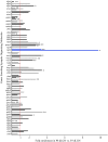
Changes in the expression of synaptic organizing molecules in the postnatal dLGN. Relative mRNA expression levels of known families of synaptic organizing molecules in P8 dLGN was compared to that at P3 by Agilent microarray. Bar graphs represent fold enrichment (or decrease) in P8 dLGN vs. P3 dLGN. The red line represents no change in gene expression between these ages. Bar color is alternated between gray and black for each adjacent gene for ease of viewing, with the exception of blue bars representing mRNA expressional levels for fgf22, the synaptic organizer showing the greatest enrichment. The expression of many genes is shown with multiple bars: in these cases each bar represents data from a unique probe set in the array. *Denote data that are statistically significant with p < 0.05. **Denote data that are statistically significant with p < 0.01. Expression of 11 synaptic organizers was enriched in P8 samples (nlgn3, sparc, sparcl1, fgf22, wnt5a, wnt7a, wnt7b, bmp4, lrrtm1, ephb3, ephb4) whereas seven were significantly down-regulated at P8 (nlgn1, nrxn1, cadm1, cbln2, ptprf, epha7, efna2).
Figure 3
Figure 3
Increases in fgf22 mRNA and FGF22 protein levels in dLGN coincide with the maturation of retinal terminals. (A) The developmental up-regulation of fgf22 mRNA from P2 to P14 in dLGN was examined by quantitative RT-PCR (qPCR). Data from P3, P8, and P14 were compared against that at P2. All data was normalized to actin mRNA levels. Data are shown ± SEM; n > 4. (B) Western blots demonstrate an increase in FGF22 protein from P3 to P8 in dLGN protein extracts. Levels of actin were used as loading controls.
Figure 4
Figure 4
The FGF22 receptor FGFR2 is expressed by classes of retinal ganglion cells that innervate dLGN. (A) Confocal analysis of immunostained P14 wild-type retinal cross-sections revealed the presence of FGFR2 in the ganglion cell layer (gcl) and nerve fiber layer (nfl). Retinal layers were identified by nuclear-labeling with DAPI. (B,C) Single optical slices from confocal analyses of retinal cross-sections immunostained for FGFR2 and either Calr (B) or Brn3a (C). Nuclei were labeled with DAPI. inl, inner nuclear layer; ipl, inner plexiform layer; gcl, ganglion cell layer; nfl, nerve fiber layer. Scale bar in (A) = 50 μm and in (B) = 20 μm for (B,C).
Figure 5
Figure 5
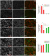
Deletion of FGF22 impairs the formation and maturation of retinal terminals in the dLGN. VGluT2- and GAD67-immunoreactivity in coronal sections of control (Ctl) and fgf22−/− mutant (KO) dLGN at P7 (A,B), P14 (D,E), and P21 (G,H). Differences in the percent area of immunoreactivity in each image were quantified for each age and genotype (C,F,I). At P7 fewer VGluT2-immunoreactive puncta were observed in mutant dLGN, leading to a statistically significant reduction in the area of immunoreactivity (A–C). At P14, VGluT2-immunoreactive (VGluT2-IR) terminals appeared less mature and the area that mutant terminals occupied in each field of view was significantly lower than in controls (D–F). By P21 no differences were observed in VGluT2–IHC in dLGN of mutants and controls (G–I). At all ages GAD67-immunoreactivity (GAD67-IR) appeared similar in fgf22−/− and control dLGN. For (C,F,I) data shown are ±SEM; n = 4 mice. *Differs from age-matched controls at p < 0.05 by Student’s t-test. **Differs from age-matched controls at p < 0.01 by Student’s t-test. Scale bar in (H) = 15 μm.
Figure 6
Figure 6
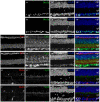
Normal morphological development of the retina in mice lacking FGF22. Confocal images of retinal cross-sections from P14 fgf22−/− mutant mice (B,D,F) and littermate controls (A,C,E). (A–D) Classes of dLGN-projecting RGCs were labeled by Brn3a–IHC (A,B) and Calr–IHC (C,D). The number and distribution of Brn3a- and Calr-expressing RGCs appeared similar in control and fgf22−/−retina. (C–F) Labeling of synaptic layers with anti-synaptophysin (Syn) (C,D) and synaptotagmin2 (Syt2) (E,F) revealed no remarkable differences in the density of synapses or laminar arrangement of the retina between mutants and controls. Likewise labeling with anti-Syt2, anti-Calr, and anti-melanopsin (Meln) revealed that sublaminar specificity was indistinguishable in controls and mice lacking FGF22 (D,F) and controls (C,E). Arrows in (E,F) show that dendrites from Meln-expressing RGCs correctly target the inner most region of the IPL in the absence of FGF22. In all sections nuclei were labeled with DAPI. inl, inner nuclear layer; ipl, inner plexiform layer; gcl, ganglion cell layer. Scale bar = 100 μm.
Figure 7
Figure 7
Deletion of FGF22 does not delay targeting of dLGN-projecting RGC axons. Calr–IHC was used to assess the development of dLGN-projecting RGC axons in P7 control [Ctl; (A)] and fgf22−/− mutant [KO; (B)] dLGN. dLGN are encircled by white dots. (C) The area of Calr-IR in control and mutant P7 dLGN was measured in ImageJ. No significant differences were detected in the percent area of dLGN innervated by Calr-expressing RGCs in P7 controls and mutants. Data are shown ± SEM; n = 4 mice. Scale bar = 125 μm.

Similar articles

Cited by

References

    1. Badea T. C., Cahill H., Ecker J., Hattar S., Nathans J. (2009). Distinct roles of transcription factors brn3a and brn3b in controlling the development, morphology, and function of retinal ganglion cells. Neuron 61, 852–86410.1016/j.neuron.2009.01.020 - DOI - PMC - PubMed
    1. Balice-Gordon R. J., Lichtman J. W. (1993). In vivo observations of pre- and postsynaptic changes during the transition from multiple to single innervation at developing neuromuscular junctions. J. Neurosci. 13, 834–855 - PMC - PubMed
    1. Bickford M. E., Slusarczyk A., Dilger E. K., Krahe T. E., Kucuk C., Guido W. (2010). Synaptic development of the mouse dorsal lateral geniculate nucleus. J. Comp. Neurol. 518, 622–63510.1002/cne.22223 - DOI - PMC - PubMed
    1. Bjartmar L., Huberman A. D., Ullian E. M., Renteria R. C., Liu X., Xu W., Prezioso J., Susman M. W., Stellwagen D., Stokes C. C., Cho R., Worley P., Malenka R. C., Ball S., Peachey N. S., Copenhagen D., Chapman B., Nakamoto M., Barres B. A., Perin M. S. (2006). Neuronal pentraxins mediate synaptic refinement in the developing visual system. J. Neurosci. 26, 6269–628110.1523/JNEUROSCI.4212-05.2006 - DOI - PMC - PubMed
    1. Catalina E., Tomassini S., Dal Monte M., Bosco L., Casini G. (2009). Localization patterns of fibroblast growth factor 1 and its receptors FGFR1 and FGFR2 in postnatal mouse retina. Cell Tissue Res. 336, 423–43810.1007/s00441-009-0787-9 - DOI - PubMed

LinkOut - more resources