. 2012 Mar 1;26(5):445-60.
doi: 10.1101/gad.182477.111. Epub 2012 Feb 22.

The splicing regulator Rbfox2 is required for both cerebellar development and mature motor function

Affiliations

The splicing regulator Rbfox2 is required for both cerebellar development and mature motor function

Lauren T Gehman et al. Genes Dev. .

Abstract

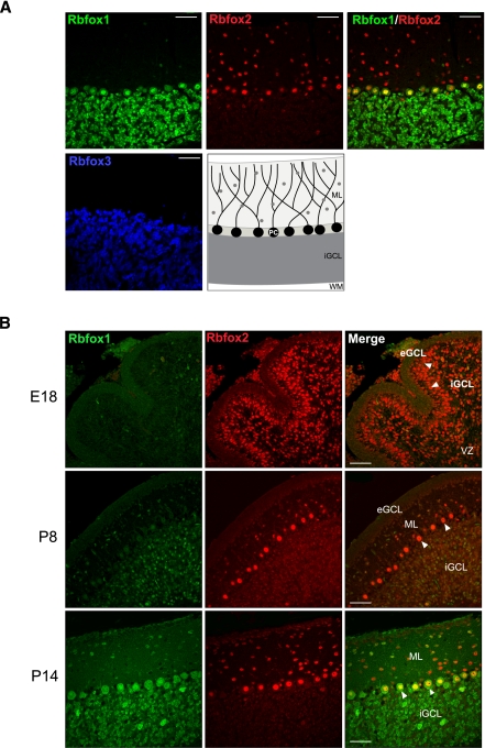
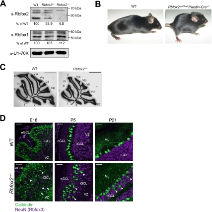
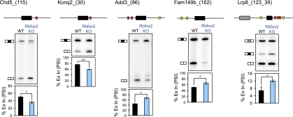
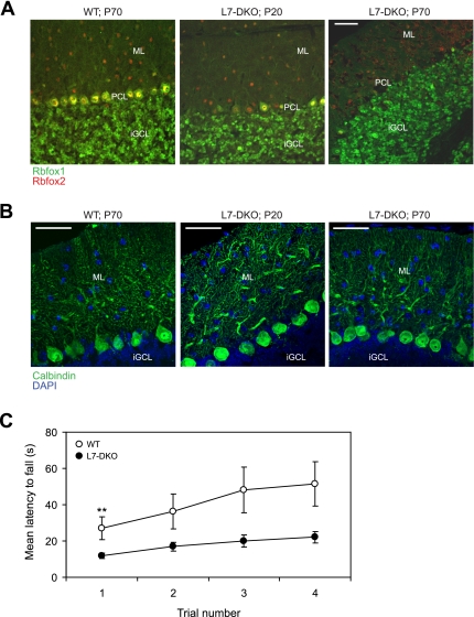
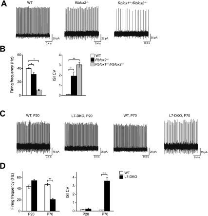
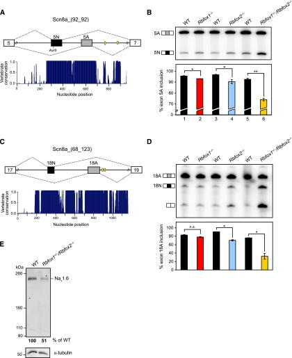
The Rbfox proteins (Rbfox1, Rbfox2, and Rbfox3) regulate the alternative splicing of many important neuronal transcripts and have been implicated in a variety of neurological disorders. However, their roles in brain development and function are not well understood, in part due to redundancy in their activities. Here we show that, unlike Rbfox1 deletion, the CNS-specific deletion of Rbfox2 disrupts cerebellar development. Genome-wide analysis of Rbfox2(-/-) brain RNA identifies numerous splicing changes altering proteins important both for brain development and mature neuronal function. To separate developmental defects from alterations in the physiology of mature cells, Rbfox1 and Rbfox2 were deleted from mature Purkinje cells, resulting in highly irregular firing. Notably, the Scn8a mRNA encoding the Na(v)1.6 sodium channel, a key mediator of Purkinje cell pacemaking, is improperly spliced in RbFox2 and Rbfox1 mutant brains, leading to highly reduced protein expression. Thus, Rbfox2 protein controls a post-transcriptional program required for proper brain development. Rbfox2 is subsequently required with Rbfox1 to maintain mature neuronal physiology, specifically Purkinje cell pacemaking, through their shared control of sodium channel transcript splicing.


Figures

Figure 1.
Figure 1.
The Rbfox proteins show differing patterns of expression in the wild-type cerebellum. (A) Confocal immunofluorescence microscopy on sagittal sections of wild-type (WT) adult cerebellar cortex probed for Rbfox1 (green), Rbfox2 (red), and Rbfox3 (blue) expression; overlayed Rbfox1 and Rbfox2 images are shown in the fourth panel. The fifth panel shows a schematic of the cerebellar cortex; gray circles represent inhibitory interneurons (basket and stellate cells). (B) Confocal immunofluorescence microscopy on sagittal sections of wild-type cerebelli at E18, P8, and P14 probed for Rbfox1 and Rbfox2 expression; overlayed images are shown in the far-right panels, and arrowheads point to Purkinje cells. (ML) Molecular layer; (PC) Purkinje cell; (iGCL) inner granule cell layer; (WM) white matter; (eGCL) external granule cell layer; (VZ) ventricular zone. Bars, 50 μm.
Figure 2.
Figure 2.
Rbfox2loxP/loxP/Nestin-Cre+/– mice are prone to hydrocephalus and possess abnormal cerebellar morphology. (A) Immunoblot analysis of Rbfox2 and Rbfox1 in nuclear lysates isolated from wild-type (WT), Rbfox2+/–, and Rbfox2−/− brains. U1-70K was used as a loading control for total nuclear protein. Below each gel is the amount of Rbfox2 or Rbfox1 protein in each sample as a percentage of wild type, normalized by U1-70K expression. Note that the Rbfox1 and Rbfox2 genes produce multiple protein isoforms that react with the antibodies. The top band in the Rbfox2 panel is nonspecific and was not used in quantification of Rbfox2 levels. (B) Wild-type and Rbfox2loxP/loxP/Nestin-Cre+/– mice at 2 mo of age. (C) Representative Nissl stains of wild-type and Rbfox2−/− cerebelli at 1 mo. Bar, 1 mm. (D) Confocal immunofluorescence microscopy on sagittal sections of wild-type and Rbfox2−/− cerebelli probed for Calbindin (green) and Rbfox3 (also known as NeuN; purple) expression at E18, P5, and P21. Bar, 50 μm. Arrowheads point to ectopic Purkinje cells. (eGCL) External granule cell layer; (iGCL) internal granule cell layer; (VZ) ventricular zone; (ML) molecular layer.
Figure 3.
Figure 3.
The Rbfox2−/− brain exhibits splicing changes of exons with adjacent Rbfox-binding sites. Representative denaturing gel electrophoresis of RT–PCR products for Rbfox2-dependent exons. Above each gel is a schematic indicating the alternative exon (horizontal black boxes) and the location of (U)GCAUG binding sites (red and yellow boxes) in the flanking introns (thin horizontal lines). Red boxes indicate (U)GCAUG sites conserved across multiple vertebrate species (Phastcons score >0.5). Shown below the gel is a graph quantifying the mean percentage of alternative exon inclusion (percent spliced in, PSI) in wild-type (WT; black bars) and Rbfox2−/− (blue bars) brains. Error bars represent SEM; n = 3. (*) P < 0.05; (**) P < 0.005; (n.s.) not significant by paired, one-tailed Student's t-test. Exact P-values are shown in Table 1.
Figure 4.
Figure 4.
At P70, L7-Cre DKO mice no longer express Rbfox1 and Rbfox2 in Purkinje cells and exhibit impaired motor function. (A,B) Confocal immunofluorescence microscopy on sagittal sections of wild-type (WT) cerebellum (left panel), L7-Cre DKO cerebellum at P20 (middle panel), and L7-Cre DKO cerebellum at P70 (right panel). (A) Overlayed images of sections probed for Rbfox1 (green) and Rbfox2 (red) expression. (B) Overlayed Z-stack projections of sections probed for Calbindin (green), counterstained with DAPI (blue). (ML) Molecular layer; (PCL) Purkinje cell layer; (iGCL) internal granule cell layer. Bars, 50 μm. (C) Quantification of wild-type and L7-DKO performance on the rotarod test; error bars represent SEM. Statistical significance was calculated by Wilcoxon rank sum test (nonparametric). (**) P = 0.0032; (n.s.) not significantly different between wild type and L7-DKO. n = 27 wild-type and 25 L7-DKO animals.
Figure 5.
Figure 5.
Rbfox1+/loxP/Rbfox2loxP/loxP/Nestin-Cre+/– and L7-DKO mice show highly irregular Purkinje cell electrophysiology. (A) Representative contiguous segments of an extracellular recording from a single Purkinje cell (PC) in the various Rbfox/Nestin knockouts. (B) Pooled data for Purkinje cell mean firing frequency and coefficient of variation of interspike intervals (ISI CV) in the various Rbfox/Nestin knockouts. n = 99, 89, 100, and 139 cells for wild type (WT), Rbfox2−/−, Rbfox1−/−/Rbfox2+/–, and Rbfo1+/–/Rbfox2−/−, respectively. (C) Representative contiguous segments of an extracellular recording from a single Purkinje cell in wild-type and L7-DKO mice at age P20 or P70. (D) Pooled data for Purkinje cell mean firing frequency and ISI CV in wild-type and L7-DKO mice. n = 43, 63, 39, and 71 cells for wild-type P20, L7-DKO P20, wild-type P70, and L7-DKO P70, respectively. Error bars, SEM. Statistical significance was calculated by ANOVA testing, followed by post-hoc Tukey paired comparisons with Bonferroni correction for multiple comparisons. (*) P < 0.005; (**) P < 2 × 10⁻⁶.
Figure 6.
Figure 6.
The Rbfox1+/–/Rbfox2−/− cerebellum exhibits strong changes in splicing of two pairs of mutually exclusive exons of Scn8a, the transcript encoding the Nav1.6 sodium channel. (A,C) Schematics showing the two pairs of Scn8a mutually exclusive exons, 5N/5A (A) and 18N/18A (C); horizontal black bars represent the upstream “N” (neonatal) exon, and horizontal gray bars represent the downstream “A” (adult) exon. Thin horizontal lines represent the intervening intron and the promixal 300 nt of the adjacent introns. Yellow boxes represent (U)GCAUG motifs. A histogram displaying the degree of conservation of this region among 30 vertebrate species, as determined by phastCons, is shown below each schematic. A score of 1 indicates 100% identity among all species at that nucleotide position. A distance scale in nucleotides is shown below the histogram. (B,D) Denaturing gel electrophoresis of Scn8a 5N/5A (B) and 18N/18A (D) RT–PCR products in cerebellar samples from mice of the listed genotypes. The exon A-included and exon N-included bands are indicated. Graphs of the mean inclusion percentage of exons 5A and 18A are shown below the gels. Error bars, SEM; n = 3. (*) P < 0.05; (**) P < 0.005 by paired, one-tailed Student's t-test. (E) Immunoblot analysis of Nav1.6 protein in membrane fractions isolated from wild-type and Rbfox1+/–/Rbfox2−/− cerebelli. α-Tubulin was used as a loading control for total membrane protein. Below the gel is the amount of Nav1.6 protein in each sample as a percentage of wild type, normalized by α-tubulin expression.

Similar articles

Cited by

References

    1. Auweter SD, Fasan R, Reymond L, Underwood JG, Black DL, Pitsch S, Allain FH 2006. Molecular basis of RNA recognition by the human alternative splicing factor Fox-1. EMBO J 25: 163–173 - PMC - PubMed
    1. Baraniak AP, Chen JR, Garcia-Blanco MA 2006. Fox-2 mediates epithelial cell-specific fibroblast growth factor receptor 2 exon choice. Mol Cell Biol 26: 1209–1222 - PMC - PubMed
    1. Barnby G, Abbott A, Sykes N, Morris A, Weeks DE, Mott R, Lamb J, Bailey AJ, Monaco AP 2005. Candidate-gene screening and association analysis at the autism-susceptibility locus on chromosome 16p: Evidence of association at GRIN2A and ABAT. Am J Hum Genet 76: 950–966 - PMC - PubMed
    1. Barski JJ, Dethleffsen K, Meyer M 2000. Cre recombinase expression in cerebellar Purkinje cells. Genesis 28: 93–98 - PubMed
    1. Bhalla K, Phillips HA, Crawford J, McKenzie OL, Mulley JC, Eyre H, Gardner AE, Kremmidiotis G, Callen DF 2004. The de novo chromosome 16 translocations of two patients with abnormal phenotypes (mental retardation and epilepsy) disrupt the A2BP1 gene. J Hum Genet 49: 308–311 - PubMed

Publication types