. 2011:1:1.
doi: 10.1038/srep00001. Epub 2011 Jun 14.

Interplay between NS3 protease and human La protein regulates translation-replication switch of Hepatitis C virus

Affiliations

Interplay between NS3 protease and human La protein regulates translation-replication switch of Hepatitis C virus

Upasana Ray et al. Sci Rep. 2011.

Abstract

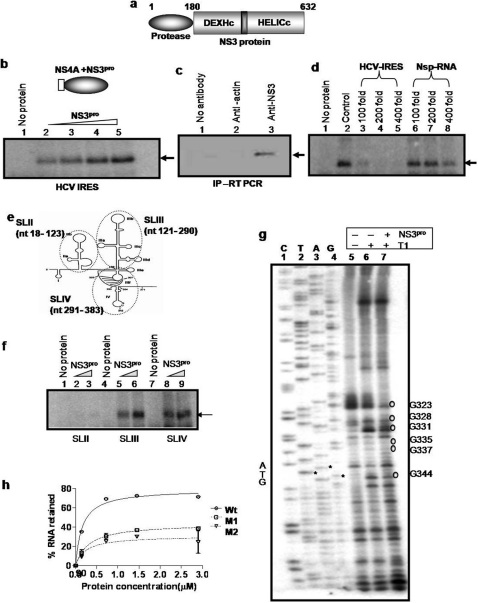
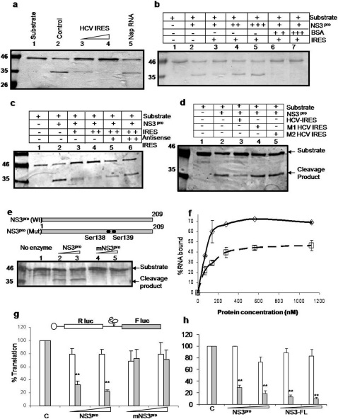
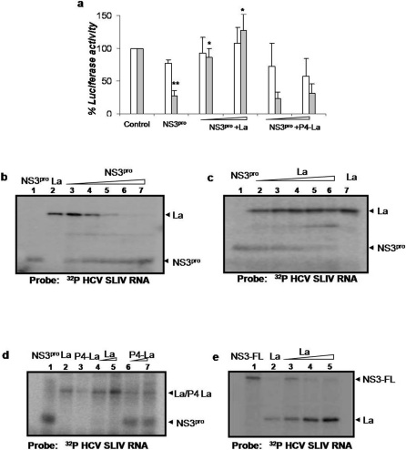
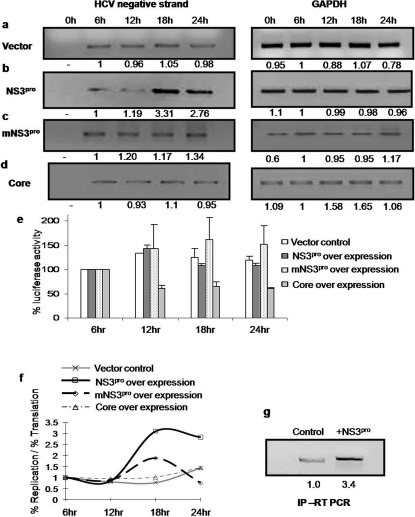
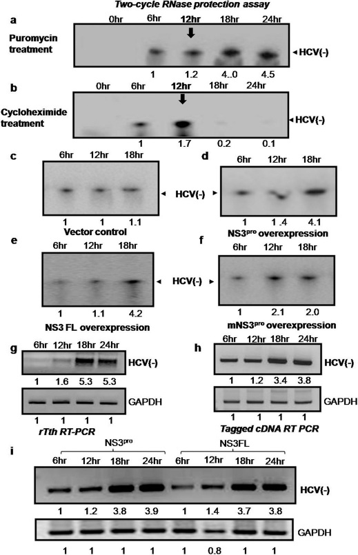
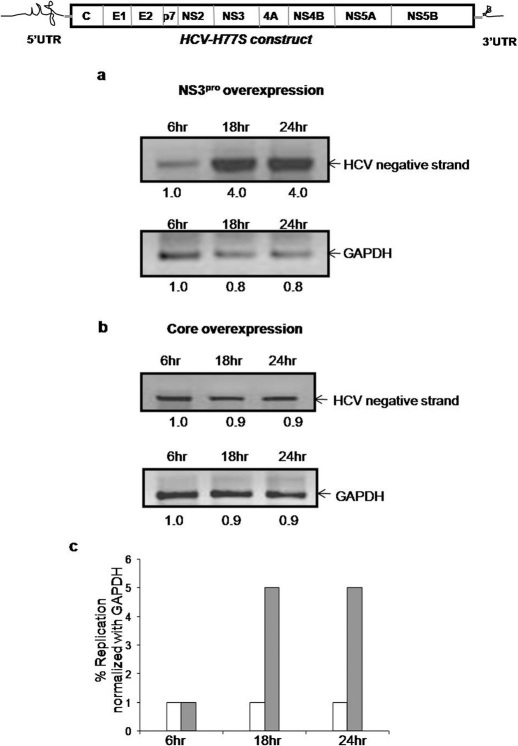
HCV NS3 protein plays a central role in viral polyprotein processing and RNA replication. We demonstrate that the NS3 protease (NS3(pro)) domain alone can specifically bind to HCV-IRES RNA, predominantly in the SLIV region. The cleavage activity of the NS3 protease domain is reduced upon HCV-RNA binding. More importantly, NS3(pro) binding to the SLIV hinders the interaction of La protein, a cellular IRES-trans acting factor required for HCV IRES-mediated translation, resulting in inhibition of HCV-IRES activity. Although overexpression of both NS3(pro) as well as the full length NS3 protein decreased the level of HCV IRES mediated translation, replication of HCV replicon RNA was enhanced significantly. These observations suggest that the NS3(pro) binding to HCV IRES reduces translation in favor of RNA replication. The competition between the host factor (La) and the viral protein (NS3) for binding to HCV IRES might regulate the molecular switch from translation to replication of HCV.


Figures

Figure 1
Figure 1. NS3pro specifically binds to HCV IRES RNA.
Panel a: Schematic representation of the domain organization of HCV NS3 protein. Panel b: UV-crosslinking. [α32P]UTP labeled HCV IRES RNA was UV cross linked with increasing concentrations (0.1, 0.2, 0.4 and 0.8 µM) of NS3pro (lane 2 –5). Lane 1 shows only probe control. Schematic representation of the NS3pro protein is shown above the panel. Panel c: Immuno-pulldown assay and RT-PCR. NS3-RNA complex was immunoprecipitated from HCV replicon bearing cells using anti-NS3 antibody. RNA was isolated and RT-PCR was performed for HCV IRES (lane 3). No antibody (lane 1) and anti-actin antibodies (lane 2) were used as negative controls for the pull down. Panel d: Competition UV-crosslinking. [α32P]UTP labeled HCV IRES RNA was UV cross linked with NS3pro in the absence (lane 2) and presence of molar excess of unlabeled HCV IRES RNA (lanes 3–5) or a nonspecific RNA (lanes 6–8). Lane 1 shows only probe control. Panel e: Schematic representation of HCV IRES RNA showing the stem and loop (SL) regions . The domains that are used as SLII, III and IV in the study are encircled and indicated. Panel f: UV-crosslinking. [α32P]UTP labeled HCV SLII (lane 2–3); SLIII (lane 5–6) and SLIV (lane 8–9) RNAs were UV cross linked to increasing concentrations (0.2 and 0.4µM) of NS3pro. Lanes 1, 4 and 7 represents only probe controls for SLII, SLIII and SLIV respectively. Panel g: RNase T1 Foot-printing assay. Binding reactions of in vitro transcribed HCV IRES RNA were carried out in absence (lane 6) or presence (lane 7) of NS3 protease. The RNA was then digested with RNase T1. RNA was reverse-transcribed with an end labeled primer. The cDNA was resolved in along with a reference sequencing reaction (lanes 1–4). Lane 5 represents the no T1 control. Panel h: Filter-binding assay. [α32P]UTP labeled HCV IRES RNA or mutant IRES RNA (M1 and M2) was bound to increasing concentrations of NS3pro.
Figure 2
Figure 2. Effect of HCV RNA binding on the catalytic activity of NS3 protease.
Panel a: Recombinant NS3 protease was allowed to form complex with increasing amount of unlabeled HCV IRES RNA (lanes 3–4). Lane 2 shows the control without HCV IRES RNA. NS5A/5B protein was used as the substrate for NS3pro. Lane 1 shows substrate alone. Non specific RNA has been used (lane 5) to test for the specificity of the reaction. Panel b: Cleavage assay was carried out in the presence of HCV IRES RNA. The cleavage inhibition (lane 3) caused by the RNA was rescued by addition of excess amount of NS3pro (lanes 4–5). BSA was used as a negative control (lanes 6–7). Panel c: Cleavage assay was carried out in the presence of HCV IRES RNA. The cleavage inhibition (lane 3–4) caused by the RNA was rescued by addition of increasing concentrations of antisense RNA (lanes 5–6). Panel d: Cleavage assay in presence of the two mutant HCV IRES RNAs (M1 and M2). Panel e: Cleavage assay using increasing concentrations of wild type NS3pro (lanes 2 and 3) and mutant NS3pro (lanes 4 and 5). Panel f: Filter-binding assay. [α32P]UTP labeled HCV IRES RNA was bound to increasing concentrations of NS3pro and mutant NS3pro. Panel g: In vitro transcribed capped bicistronic RNA (shown above panel) was in vitro translated in the absence or presence of increasing concentrations (0.2 and 0.4µM) of either NS3pro or mutant NS3pro. Luciferase assay was performed. Percent luciferase activities corresponding to Rluc (white bar) and Fluc (grey bar) values were plotted against the protein concentrations. Values which significantly differ from control (P value<0.001) are indicated as asterisks. Panel h: In vitro transcribed capped bicistronic RNA was in vitro translated in the absence or presence of increasing concentrations of either NS3pro (0.2 and 0.4µM) or full length NS3 (0.2 and 0.4µM). Luciferase assay was performed. Percent luciferase activities corresponding to Rluc (white bar) and Fluc (grey bar) values were plotted against the protein concentrations.
Figure 3
Figure 3. Panel a: In vitro transcribed capped bicistronic RNA was in vitro translated in the absence or presence of NS3pro.
Luciferase assay was performed. Percent luciferase activities corresponding to Rluc (white bar) and Fluc (grey bar) values were plotted against the protein concentrations. The translation inhibition was rescued by adding equimolar and also 2 fold molar excess of wild type La protein. P4-La mutant was used as a negative control. Values which significantly differ from control are indicated as asterisk. ** indicates P value<0.001 and * indicates P value<0.01. Panel b: UV-crosslinking assay: [α32P] UTP labeled HCV SLIV RNA was UV crosslinked with La protein (0.2 µM) in the presence of equimolar, 2 fold, 4 fold, 8 fold and 10 fold molar excess of purified HCV NS3pro (lanes 3–7). The position of NS3pro and La protein is indicated. Lane 1 and 2 represents binding due to NS3pro and La protein alone respectively. Panel c: UV-crosslinking assay: [α32P]UTP labeled HCV SLIV RNA was UV cross linked to NS3pro (0.2 µM) in the presence of equimolar, 2 fold, 4 fold, 8 fold and 10 fold molar excess La protein (lanes 2–6). Lane 1 and 7 represents binding due to NS3pro and La protein alone respectively. Panel d: UV-crosslinking assay: [α32P]UTP labeled HCV SLIV RNA was UV cross linked to NS3pro (0.2µM) in the presence of equimolar or 2 fold molar excess of either wild type La protein (lanes 4 and 5) or P4-La (lanes 6 and 7). Lanes 1, 2 and 3 represent binding due to NS3pro and wild type La protein and P4-La alone respectively. Panel e: UV-crosslinking assay: [α32P] UTP labeled HCV SLIV RNA was UV crosslinked with the purified recombinant full-length NS3 protein (0.3µM) in the presence of equimolar, 2 fold, and 4 fold molar excess of the La protein (lanes 3–5). The position of NS3 and La protein is indicated. Lane 1 and 2 represents binding due to NS3-FL (0.3µM) and La protein (0.3µM) alone respectively.
Figure 4
Figure 4. Effect of NS3pro over expression on HCV RNA replication versus translation.
Huh7 cells were cotransfected with of pSGR JFH1/Luc RNA and construct expressing NS3pro, or vector or mutant NS3pro or core. RNA was isolated at different time points and HCV RNA was quantitated using semi quantitative RT PCR (left side of Panel a, b, c and d). GAPDH was used as internal control (right hand side of Panel a, b, c and d ). Panel e: For the experiments performed in fig. 4a–d, luciferase assay was carried out to detect translation levels. The percentage luciferase activities were plotted for each reaction at different time intervals taking 6 hour time point as 100 (control). Panel f: The ratio of percentage of replication to translation levels in vector control cells was compared with cells overexpressing NS3pro plasmid or the other controls and graphically represented by plotting ratio of percentage replication versus translation on Y axis and time on X axis. Panel g: RNA protein complex was pulled down at 18th hour time point from cells cotransfected with pSGR JFH1/Luc RNA and construct expressing NS3pro using anti-NS5B antibody followed by RT-PCR for HCV IRES.
Figure 5
Figure 5. Panel a: Huh 7 cells were transfected with pSGR JFH1 luc RNA.
12hours after transfection, cells were treated with puromycin. At different time points total cellular RNA was isolated and was subjected to two cycle RNase protection assay. Negative strand RNA was probed using radiolabelled positive strand HCV IRES RNA followed by RNAse digestion. Protected fragment was detected by running on 4% polyacrylamide-8M urea gel. The arrow (below 12hr) indicates the time of addition of puromycin. Panel b: Huh 7 cells were transfected with pSGR JFH1 luc RNA. 12hours after transfection, cells were treated with cycloheximide. At different time points total cellular RNA was isolated and was subjected to two cycle RNase protection assay. Negative strand RNA was probed using radiolabelled positive strand HCV IRES RNA followed by RNAse digestion. Protected fragment was detected by running on 4% polyacrylamide-8M urea gel. The arrow (below 12hr) indicates the time of addition of cycloheximide. Panel c–f: Two cycle RNase protection assay was performed under similar experimental set up as in fig.4 for vector control, NS3pro, NS3 full length and mutant. The total RNA was self hybridized followed by RNase treatment. Negative strand RNA was probed using radiolabelled positive strand HCV IRES RNA followed by RNAse digestion. Protected fragment was detected by running on 4% polyacrylamide-8M urea gel. Panel g: Under similar experimental set up as in fig. 4b, instead of semiquantitative RT-PCR, negative strand was detected using thermostable RT-PCR. Panel h–i: Under similar experimental set up as in fig. 4b, negative strand was detected using a tagged cDNA. Here, cDNA was made using a HCV primer that was tagged with a long non HCV nucleotide tag. This was followed by first round of PCR using tag only primer and HCV reverse primer. A second cycle nested PCR was performed using 50 times diluted first PCR product by a second set of internal primers. Similarly, the above assay was performed under over expression of NS3pro as well as NS3 full length protein as indicated (panel i).
Figure 6
Figure 6. Panel a: Huh7 cells were cotransfected with H77S RNA (schematic representation shown above panel) and the construct encoding NS3pro.
RNA was isolated at different time points and HCV RNA was quantitated using semi quantitative RT PCR. GAPDH was used as internal control. Panel b: Huh7 cells were cotransfected with H77S RNA and construct encoding HCV core. RNA was isolated at different time points and HCV RNA was quantitated using semi quantitative RT PCR. GAPDH was used as internal control. Panel c: Graph plotted taking percentage replication of HCV RNA normalized with that of GAPDH amplified on Y axis and time on X axis. White bar indicates overexpression of HCV core and grey bar indicates that of NS3pro

Similar articles

Cited by

References

    1. Barbato G. et al. The solution structure of the N-terminal proteinase domain of the hepatitis C virus (HCV) NS3 protein provides new insights into its activation and catalytic mechanism. J. Mol. Biol. 289, 371–384 (1999). - PubMed
    1. Beran R. K. F., Serebrov V. & Pyle A. M. The serine protease domain of hepatitis C viral NS3 activates RNA helicase activity by promoting the binding of RNA substrate. J. Biol. Chem. 282, 34913–34920 (2007). - PubMed
    1. Frick D. N., Rypma R. S., Lam A. M. I. & Gu B. The nonstructural protein 3 protease/helicase requires an intact protease domain to unwind duplex RNA efficiently. J. Biol. Chem. 279, 1269–1280 (2004). - PMC - PubMed
    1. Herold J. & Andino R. Poliovirus RNA replication requires genome circularization through a protein–protein bridge. Mol. Cell 7, 581–591 (2001). - PMC - PubMed
    1. Ali N., Pruijn G. J. M., Kenan D. J., Keene J. D. & Siddiqqui A. Human La antigen is required for the Hepatitis C virus internal ribosome entry site mediated translation. J. Biol. Chem. 275, 27531–27540 (2000). - PubMed

Publication types

MeSH terms